#frog oviducts
Text
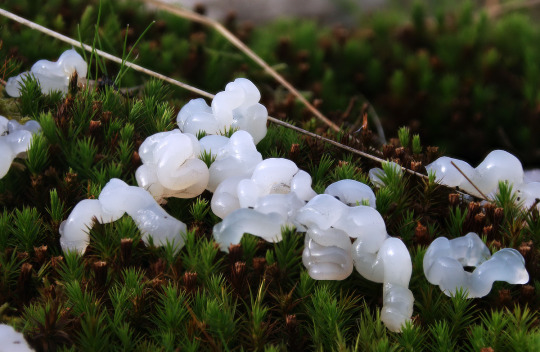
Some super-fresh star slime found on top of a wall yesterday.
734 notes
·
View notes
Text
Pacific Rim kaiju biology funtimes!
The Newmann server looked at screencaps of the kaiju anatomical chart from Newt's drift flashback and @newtgottlieb figured out that the info came from a male frog anatomy chart. I'm absolutely losing my mind over here; "kaiju are really big frogs" was not a discovery I expected. 🤣
On a slightly more serious note, my guess is that somebody yoinked the info from a frog anatomy chart during production because the kaiju are described as "amphibious."
For the curious, the labels on the chart are:
Dorsal aorta
Fat body
Testes
Renal veins
Vestigial oviduct
Cloaca
Postcaval vein
Vasa efferentia
Adrenal (should have read "Adrenal gland")
Kidney
Ureter
Bladder
16 notes
·
View notes
Note
First i must apologize I have a tendency to forget that tumblr - even when we're all 'on' - is not live interaction and additionally you can't actually read my mind 😅
This was in reaction to seeing your ... delightful?? .. new frog. In such a state. And I got nervous 😋🦀💖

Annon... I´m gonna cry... I love you♡♡♡ you´re making me do happy jumps♡
And I understand fully what you mean about "reading minds" if you can call it that, we´re all there at times!
I hope this is you trying to get me to talk about my wonderful frog Kaj because that´s what I´m gonna do know XD
-
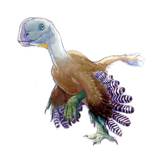
This is Kaj - named after my second favorite show as a child - he´s 25 cm long, and he´s a treefrog, there didn´t stand which one but my best guess is a red eyed tree frog, he just doesn't have the blue stripes, he could also be a Agalychnis moreletii (Morelet's tree frog) but he doesn't have black eyes and Morelet´s are known for their black eyes (a nickname of theirs is black eyed frog).
He could also be an American green tree frog, but his feet are a bit too pigmented, and back a bit to light for that, although the eyes would match there.
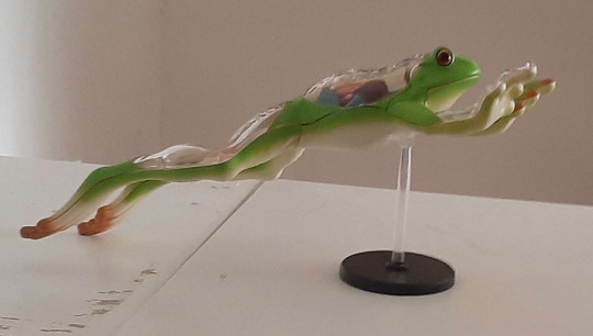

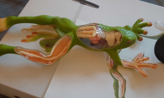
Actually he´s a woman as he has an ovary and oviduct (the blue stuff you can see below his urostyle) but I didn´t know that until I unpacked him, where I already had named him
Going with the idea he´s a Morelet (as that´s what he looks most like) he would be around 60mm (f-54 m-60/64)
Red eyed tree frog gets around 6/7 cm (f-7,5 m-5)
All in conclusion he´s a tree frog. Which kind of one? Karl.
And no, the crabs need not fear, he´s much too small to be of any danger in the wild, and my Kaj is made of plastic so he doesn't really jump or eat, unfortunately.
Although some crabs might want to watch out for the Goliath frog... (world's biggest frog at around 32 cm and 3.3 kilograms)
#im always so nervous if my english is good enough and if i have misunderstood something because of it#no art just talk#kaj the frog#frog#frogs#i dont know how to replay when people gives me compliments#they make me so happy and i cant even get it down in words so i end up usually just giving them a thank you but i mean so much more#if any better frogs experts can help me out please do#this is just a take out of my own knowledge#ive always had an oppositions with reptile and amphibians#mostly because my dad talked about how he grew up with turtles fish reptiles and frogs#but the frogs just kicked extra hard this last month and they have been filling almost every minut of my days in some way#ask
7 notes
·
View notes
Note
Frogs don’t have penises they have cloaca and sperm is ejected from the cloaca directly onto the eggs as the female lays them. The ovaries of the female frog are beside the kidneys and the eggs pass down a pair of oviducts and through the cloaca to the exterior.
Thank you for that wonderful biology lesson nonnie
Still doesn't prove whether Kermit and Miss Piggy can have kids

0 notes
Text
Frog dissection diagram worksheet

Observe several frogs to see the difference between males and females. Male frogs are also usually smaller than female frogs. A male frog usually has thick pads on its “thumbs,” which is one external difference between the sexes, as shown in the diagram below. To determine the frog’s sex, look at the hand digits, or fingers, on its forelegs. Put on safety goggles, gloves, and a lab apron.safety goggles, gloves, and a lab apron.In this lab, you will dissect a frog in order to observe the external and internal structures of frog anatomy. Name the organs that make up various systems of the frog.Describe the appearance of various organs found in the frog.When the extensor of that body part contracts, the part straightens. When a flexor of a leg or other body part contracts, that part is bent. Voluntary muscles, which are those over which the frog has control, occur in pairs of flexors and extensors. The frog’s skeletal and muscular systems consist of its framework of bones and joints, to which nearly all the voluntary muscles of the body are attached. The central nervous system of the frog consists of the brain, which is enclosed in the skull, and the spinal cord, which is enclosed in the backbone. The ovaries produce eggs, or female sex cells, which move through oviducts into the uteri, then through the cloaca outside the body. The testes produce sperm, or male sex cells, which move through sperm ducts, tubes that carry sperm into the cloaca, from which the sperm move outside the body. Those of the female system are the ovaries, oviducts, uteri, and cloaca. The organs of the male reproductive system are the testes, sperm ducts, and cloac a. Connected to each kidney is a ureter, a tube through which urine passes into the urinary bladder, a sac that stores urine until it passes out of the body through the cloaca. The kidneys are organs that excrete urine. The urinary system consists of the frog’s kidneys, ureters, bladder, and cloaca. Blood from both atria goes into the ventricle and then is pumped into the arteries, which are blood vessels that carry blood away from the heart. Veins from different parts of the body enter the right and left atria. Blood is carried to the heart in vessels called veins. The heart has two receiving chambers, or atria, and one sending chamber, or ventricle. The circulatory system consists of the heart, blood vessels, and blood. The walls of the lungs are filled with capillaries, which are microscopic blood vessels through which materials pass into and out of the blood. The respiratory system consists of the nostrils and the larynx, which opens into two lungs, hollow sacs with thin walls. Indigestible materials pass through the large intestine and then into the cloaca, the common exit chamber of the digestive, excretory, and reproductive systems. The contents of the common bile duct flow into the small intestine, where most of the digestion and absorption of food into the bloodstream takes place. Bile flows into a tube called the common bile duct, into which pancreatic juice, a digestive juice from the pancreas, also flows. Bile is a digestive juice made by the liver and stored in the gallbladder. From the esophagus, swallowed food moves into the stomach and then into the small intestine. The digestive system consists of the organs of the digestive tract, or food tube, and the digestive glands. In the pharynx, there are several openings: one into the esophagus, the tube into which food is swallowed one into the glottis, through which air enters the larynx, or voice box and two into the Eustachian tubes, which connect the pharynx to the ear. Also inside the mouth behind the tongue is the pharynx, or throat. Inside the mouth are two internal nares, or openings into the nostrils two vomerine teeth in the middle of the roof of the mouth and two maxillary teeth at the sides of the mouth. The third lid, called the nictitating membrane, is transparent. On the outside of the frog’s head are two external nares, or nostrils two tympani, or eardrums and two eyes, each of which has three lids. As members of the class Amphibia, frogs may live some of their adult lives on land, but they must return to water to reproduce.
0 notes
Text
Grey Foam Nesting Frog,(Chiromantis xerampelina)
Also known as the Grey Tree frog as these frogs are arboreal (they live in trees), the foam ball is the nest of the Foam Nest Frog - the nests are formed as females secrete a fluid from the oviduct, which the froth into a foam, before laying eggs inside, males will join by adding sperm into the nest.
The Foam dries and hardens protecting the eggs from drought or predators,tadpoles hatch between 4 to 5 days - dropping into the water where they continue their life sickles.

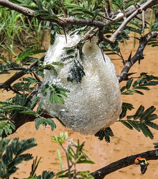

#safari#nature#wildlifephotography#underafricanskies#buyelaeafrica#wildlife#fieldguide#thisisafrica#facts#conservation#returntoafrica#amphibians
2 notes
·
View notes
Text
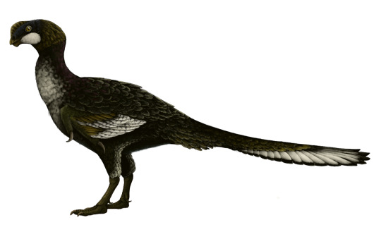
Microvenator celer
By Ripley Cook
Etymology: Small Hunter
First Described By: Ostrom, 1970
Classification: Dinosauromorpha, Dinosauriformes, Dracohors, Dinosauria, Saurischia, Eusaurischia, Theropoda, Neotheropoda, Averostra, Tetanurae, Orionides, Avetheropoda, Coelurosauria, Tyrannoraptora, Maniraptoromorpha, Maniraptoriformes, Maniraptora, Pennaraptora, Oviraptorosauria, Caenagnathoidea, Caenagnathidae
Status: Extinct
Time and Place: Between 115 and 105 million years ago, from the Aptian to the Albian ages of the Early Cretaceous

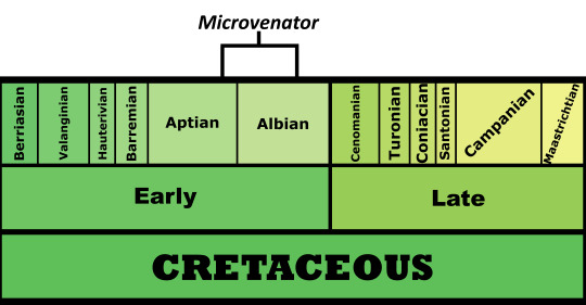
Microvenator is known from the Cloverly Formation of Montana and Wyoming; specifically the Little Sheep and Himes members of the Formation

Physical Description: Microvenator was an Oviraptorosaur - aka, a Chickenparrot - one closely related to the later Anzu from the Hell Creek formation at the end of the Cretaceous. Still, the main remains it is known from are a juvenile, which actually lead to its name - prior to further study, we thought the juvenile was as big as it got, and thus named it for its small size. As such, Microvenator probably got much bigger than the original estimates - while the juvenile was about 1.2 meters long, an adult would probably be around 3 meters long.
As a chickenparrot, Microvenator would have been fairly squat in body, and it had a shorter tail than your average dinosaur - but, as an earlier member of the group, it didn’t have a pygostyle like some of the later species. It had long arms and moderate length legs, and was covered in feathers - especially notable wings on the arms and a tail fan on the tail. It probably had a large beak, as well, but little of the skull is known.
Diet: As an Oviraptorosaur, Microvenator probably had an omnivorous diet, eating a wide variety of food, especially plants.
By Scott Reid
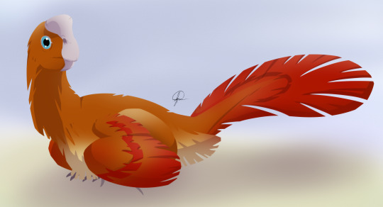
Behavior: Though Microvenator isn’t particularly well-known, it probably behaved similarly to its close relatives, including in taking care of its young. Oviraptorosaurs are known to have been great parents, creating large nests with eggs laid around the edge, laid from a single oviduct. Microvenator would have then sat in the center of the nest, and used its wings to keep the eggs warm - much like how birds do so today. Microvenator laid ovular, elongated eggs, which were probably turquoise or teal in color.
Microvenator was a feathered, warm-blooded animal, and thus very active in its environment and with its behavior; using its feathers to display to other members of the species for sex and communication - and its large wings and tail fan would have been excellent for these activities. As an omnivore, it would have been an opportunistic animal as it looked around its environment for food.
Ecosystem: The Cloverly Formation was one of many distinctive “middle” Cretaceous environments in Western North America, a transition between the iconic Late Jurassic and Late Cretaceous faunas of the area. Here, the Western Interior Seaway was growing in Microvenator’s backyard, which was mainly a floodplain and river system with extensive flooding that buried many animals at a time. These plains were forested, covered with many conifers and cycads.
By José Carlos Cortés
The earlier environment, the Little Sheep ecosystem, was much muddier and less associated with the sea than later times. Here, Microvenator lived with the ankylosaur Sauropelta, the ornithopod Tenontosaurus, the small fast-moving herbivore Zephyrosaurus, and the medium-sized theropod Deinonychus. There was an unnamed sauropod as well, as well as lungfish, sharks, fish, frogs, salamanders, crocodilians, and turtles - and a lot of types of mammals and lizards, which would have been decent food for Microvenator in a pinch.
The later Himes ecosystem was sandier, and more associated with the sea as it grew into North America. Microvenator still lived alongside Deinonychus, Sauropelta, and Tenontosaurus, but also the large sauropod Sauroposeidon and the poorly known Rugocaudai; a new nodosaur, Tatankacephalus, and the large predator Acrocanthosaurus which would have been a major threat to Microvenator. There also may have been a proto-bird present in the ecosystem, along with many kinds of lizards, frogs, salamanders, crocodilians, turtles, fish, sharks, and mammals.
Other: Microvenator is the oldest known Oviraptorosaur from North America!
~ By Meig Dickson
Sources under the Cut
Mackovicky, Peter J., Sues, Hans-Dieter. (1998). "Anatomy and phylogenetic relationships of the Theropod Dinosaur Microvenator celer from the Lower Cretaceous of Montana” American Museum Novitates. Number 3240, 27pp. 27 August 1998.
Maxwell, W. D. 1993. Neonate dinosaur remains and dinosaur eggshell from the Lower Cretaceous Cloverly Formation, Montana. Journal of Vertebrate Paleontology 13(3, suppl.):49A
Norell, M.A., Gaffney, E.S., and Dingus, L. 1995. Discovering Dinosaurs in the American Museum of Natural History. Alfred A. Knopf, Inc.:New York, 204 p.
Oreska, M. P. J., M. T. Carrano, and K. M. Dzikiewicz. 2013. Vertebrate paleontology of the Cloverly Formation (Lower Cretaceous), I: faunal composition, biogeographic relationships, and sampling. Journal of Vertebrate Paleontology 33(2):264-292 [
Ostrom, J. H. 1970. Stratigraphy and paleontology of the Cloverly Formation (Lower Cretaceous) of the Bighorn Basin area, Wyoming and Montana. Peabody Museum Bulletin 35:1-234
Sato, T., Y. Cheng, X. Wu, D. K. Zelenitsky, Y. Hsaiao. 2005. A pair of shelled eggs inside a female dinosaur. Science 308 (5720): 375.
Varricchio, D. J. 2001. Late Cretaceous oviraptorosaur (Theropoda) dinosaurs from Montana. pp. 42–57 in D. H. Tanke and K. Carpenter (eds.), Mesozoic Vertebrate Life. Indiana University Press, Indianapolis, Indiana.
Wiemann, J., T.-R. Yang, P. N. Sander, M. Schneider, M. Engeser, S. Kath-Schorr, C. E. Müller, P. M. Sander. 2017. Dinosaur origin of egg color: oviraptors laid blue-green eggs. PeerJ 5: e3706.
#Microvenator#Microvenator celer#Dinosaur#Oviraptorosaur#Bird#Birds#Dinosaurs#Terrestrial Tuesday#Palaeoblr#Birblr#Feathered Dinosaurs#Prehistoric life#Paleontology#Prehistory#Omnivore#North America#Cretaceous#Factfile#biology#a dinosaur a day#a-dinosaur-a-day#dinosaur of the day#dinosaur-of-the-day#science#nature
183 notes
·
View notes
Link
Scientists Grow Mice Embryos in a Mechanical Womb The mouse embryos looked perfectly normal. All their organs were developing as expected, along with their limbs and circulatory and nervous systems. Their tiny hearts were beating at a normal 170 beats per minute. But these embryos were not growing in a mother mouse. They were developed inside an artificial uterus, the first time such a feat has been accomplished, scientists reported on Wednesday. The experiments, at the Weizmann Institute of Science in Israel, were meant to help scientists understand how mammals develop and how gene mutations, nutrients and environmental conditions may affect the fetus. But the work may one day raise profound questions about whether other animals, even humans, should or could be cultured outside a living womb. In a study published in the journal Nature, Dr. Jacob Hanna described removing embryos from the uteruses of mice at five days of gestation and growing them for six more days in artificial wombs. At that point, the embryos were about halfway through their development; full gestation is about 20 days. A human at this stage of development would be called a fetus. To date, Dr. Hanna and his colleagues have grown more than 1,000 embryos in this way. “It really is a remarkable achievement,” said Paul Tesar, a developmental biologist at Case Western Reserve University School of Medicine. Alexander Meissner, director of genome regulation at the Max Planck Institute for Molecular Genetics in Berlin, said that “getting this far is amazing” and that the study was “a major milestone.” But the research has already progressed beyond what the investigators described in the paper. In an interview, Dr. Hanna said he and his colleagues had taken fertilized eggs from the oviducts of female mice just after fertilization — at Day 0 of development — and had grown them in the artificial uterus for 11 days. Until now, researchers were able to fertilize eggs from mammals in the laboratory and grow them for only a short time. The embryos needed a living womb. “Placental mammals develop locked away in the uterus,” Dr. Tesar said. That prevented scientists from answering fundamental questions about the earliest stages of development. “The holy grail of developmental biology is to understand how a single cell, a fertilized egg, can make all of the specific cell types in the human body and grow into 40 trillion cells,” Dr. Tesar said. “Since the beginning of time, researchers have been trying to develop ways to answer this question.” The only way to study the development of tissues and organs was to turn to species like worms, frogs and flies that do not need a uterus, or to remove embryos from the uteruses of experimental animals at varying times, providing glimpses of development more like snapshots than video. What was needed was a way to get inside the uterus, watching and tweaking development in mammals as it happened. For Dr. Hanna, that meant developing an artificial uterus. He spent seven years developing a two-part system that includes incubators, nutrients and a ventilation system. The mice embryos are placed in glass vials inside incubators, where they float in a special nutrient fluid. The vials are attached to a wheel that slowly spins so the embryos do not attach to the wall, where they would become deformed and die. The incubators are connected to a ventilation machine that provides oxygen and carbon dioxide to the embryos, controlling the concentration of those gasses, as well as the gas pressure and flow rate. At Day 11 of development — more than halfway through a mouse pregnancy — Dr. Hanna and his colleagues examined the embryos, only the size of apple seeds, and compared them to those developing in the uteruses of living mice. The lab embryos were identical, the scientists found. By that time, though, the lab-grown embryos had become too large to survive without a blood supply. They had a placenta and a yolk sack, but the nutrient solution that fed them through diffusion was no longer sufficient. Getting past that hurdle is the next goal, Dr. Hanna said in an interview. He is considering using an enriched nutrient solution or an artificial blood supply that connects to the embryos’ placentas. In the meantime, experiments beckon. The ability to keep embryos alive and developing halfway through pregnancy “is a gold mine for us,” Dr. Hanna said. The artificial womb may allow researchers to learn more about why pregnancies end in miscarriages or why fertilized eggs fail to implant. It opens a new window onto how gene mutations or deletions affect fetal development. Researchers may be able to watch individual cells migrate to their ultimate destinations. The work is “a breakthrough,” said Magdalena Zernicka-Goetz, professor of biology and biological engineering at Caltech. It “opens the door to a new age of studying development in the experimental mouse model.” A recent development provides another opportunity. Researchers have directly created mouse embryos from mouse fibroblasts — connective tissue cells — making early embryos without starting with a fertilized egg. Combine that development with Dr. Hanna’s work, and “now you don’t need mice to study mouse embryo development,” Dr. Meissner said. Scientists may be able to make all the embryos they need from connective tissue. If scientists could make embryos without fertilizing eggs and could study their development without a uterus, Dr. Meissner said, “you can get away from embryo destruction.” There would be no need to fertilize mouse eggs only to destroy them in the course of study. But the work might eventually extend beyond mice. Two other papers published in Nature on Wednesday report on attempts that edge near creating early human embryos in this way. Of course, Dr. Meissner said, creation of human embryos is years away — if it is permitted at all. And for now, international regulations prohibit studying human embryos beyond 14 days of fertilization. In the future, Dr. Tesar said, “it is not unreasonable that we might have the capacity to develop a human embryo from fertilization to birth entirely outside the uterus.” Of course, even the suggestion of this science fiction scenario is bound to horrify many. But it is early days, with no assurance human fetuses could ever develop entirely outside the womb. Even assuming they could, Dr. Tesar noted, “whether that is appropriate is a question for ethicists, regulators and society.” Source link Orbem News #Embryos #grow #mechanical #Mice #Scientists #Womb
0 notes
Photo

Star jelly (also called astromyxin, astral jelly) is a gelatinous substance sometimes found on grass or even on branches of trees. According to folklore, it is deposited on the Earth during meteor showers. Star jelly is described as a translucent or grayish-white gelatin that tends to evaporate shortly after having "fallen." There have been reports of star jelly for centuries. John of Gaddesden(1280–1361), for example, mentions stella terrae (Latin for 'star of the earth' or 'earth-star') in his medical writings, describing it as "a certain mucilaginous substance lying upon the earth" and suggesting that it might be used to treat abscesses. A fourteenth-century Latin medical glossary has an entry for uligo, described as "a certain fatty substance emitted from the earth, that is commonly called 'a star which has fallen.'" Similarly, an English-Latin dictionary from around 1440 has an entry for "sterre slyme" with the Latin equivalent given as assub (a rendering of Arabic ash-shuhub, also used in medieval Latin as a term for a "falling" or "shooting" star). The Oxford English Dictionary lists a large number of other names for the substance, with references dating back to the circa-1440 English-Latin dictionary entry mentioned above: star-fallen, star-falling, star-jelly, star-shot, star-slime, star-slough, star-slubber, and star-slutch. Slightly related, the slime mold Enteridium lycoperdon is called "caca de luna" (moon's excrement) by the locals in the state of Veracruz in Mexico. A long article in the paranormal magazine Fate declared star jelly to be of extraterrestrial origin, calling it "cellular organic matter" which exists as "prestellar molecular clouds" which float through space. In The Book of British Amphibians and Reptiles (page 138), author M. Smith states that star jelly is most likely formed from the glands in the oviducts of frogs and toads. Birds and mammals will eat the animals but not the oviducts which, when they come into contact with moisture, swell and distort leaving a vast pile of jellylike substance sometimes also referred to as otter jelly. #destroytheday https://www.instagram.com/p/B_kRCWxBPLO/?igshid=cmmoowub3cyw
0 notes
Text
Molecules, Vol. 23, Pages 1384: Comparative Proteomic Analysis of Rana chensinensis Oviduct
As one of most important traditional Chinese medicine resources, the oviduct of female Rana chensinensis (Chinese brown frog) was widely used in the treatment of asthenia after sickness or delivery, deficiency in vigor, palpitation, and insomnia. Unlike other vertebrates, the oviduct of Rana chensinensis oviduct significantly expands during prehibernation, in contrast to the breeding period. To explain this phenomenon at the molecular level, the protein expression profiles of Rana chensinensis oviduct during the breeding period and prehibernation were observed using isobaric tags for relative and absolute quantitation (iTRAQ) technique. Then, all identified proteins were used to obtain gene ontology (GO) annotation. Ultimately, KEGG (Kyoto Encyclopedia of Genes and Genomes) enrichment analysis was performed to predict the pathway on differentially expressed proteins (DEPs). A total of 4479 proteins were identified, and 312 of them presented different expression profiling between prehibernation and breeding period. Compared with prehibernation group, 86 proteins were upregulated, and 226 proteins were downregulated in breeding period. After KEGG enrichment analysis, 163 DEPs were involved in 6 pathways, which were lysosome, #RNA transport, glycosaminoglycan degradation, extracellular matrix (ECM)–receptor interaction, metabolic pathways and focal adhesion. This is the first report on the protein profiling of Rana chensinensis oviduct during the breeding period and prehibernation. Results show that this distinctive physiological phenomenon of Rana chensinensis oviduct was mainly involved in ECM–receptor interaction, metabolic pathways, and focal adhesion. http://bit.ly/2M5gjc1 #MDPI
0 notes
Text
Development - the cells inside of you
The fertilized egg, travels through the oviduct and before even the implantation onto the urine tract, it has turned from one enormous cell to blastocyst - as the rapid mitotic divisions take place. When the rate of these divisions slows down, our ball of cells will become a gastrula (from a blastula) as the cells rearrange themselves and start organization into the three germ layers: ectoderm, mesoderm and endoderm. Let's start with the first one. What is this ectoderm layer? Is it the layer form which epidermis cells will arise (skin cells) and your nervous system. The neural tissue is induced after the organizer, autonomous cells which determine neurulation and other events, this is a very important structure as the experiments have shown that if we transplant the organizer from the blastopore of a frog into another embryo, an additional embryo will form. So it has the capability to induce formation of a second individual! (Which will be connected to the first). It's job is tremendous as it will induce the formation of the nervous system and from gastrula, we will move on to the stage called neurula. Here, neurulation will take place which is the folding process of the neural plate into neural tube. It is called neural plate because at first it is just part of the embryo, flat-like top (anterior) structure. But when a node of mesodermal cells (middle layer) tell the ectodermal cells above them that they are not going to become epidermis but instead form the nervous system, it all changes.
SO when this rod of mesodermal cells, or notochord, induces the formation of neural tube, first the cells above it will elongate and then begin to fold, forming a tube-like structure. This happens because of the cytoskeleton of the cells which can allow them to constrict their "necks" and thus acquire the mechanical force to pull themselves together and then elongate. Neural crest cells play a part here because they are part of the neural plate and when the folding begins, they will elevate themselves to form a "circle" (the to be neural tube) below them and thus close it.
It is just one out of millions amazing processes that contribute to who you are - don't ever doubt your body. It is incredible.
#incredible#curious#biology#ectoderm#germ layers#development#student#learning#love#amazing body#aware#cells#induction#communication#crest cells#neural cells#neural plate#neural tube#formation
1 note
·
View note
Video
youtube
This is a video of caecilian from the BBC's Life in Cold Blood documentary series.
Caecilians (/sɪˈsɪliən/; New Latin for "blind ones") are a group of limbless, serpentine amphibians. They mostly live hidden in the ground and in stream substrates, making them the least familiar order of amphibians. They are mostly distributed in the tropics of South and Central America, Africa, and southern Asia. Their diet consists of earthworms and small subterranean creatures.
The order completely lack limbs, making the smaller species resemble worms, while the larger species, with lengths up to 1.5 m (5 ft), resemble snakes. Their tails are short or absent, and their cloacae are near the ends of their bodies Their skin is smooth and usually dark, but some species are colourful. The skin also has numerous ring-shaped folds, or annuli, that partially encircle the body, giving them a segmented appearance. Like some other living amphibians, the skin contains glands that secrete a toxin to deter predators.
Caecilians' vision is limited to dark-light perception and their anatomy is highly adapted for a burrowing lifestyle. In water or very loose mud,they swim in an eel-like fashion. As an example the family Typhlonectidae are aquatic, and the largest of their kind. The representatives of this family have a fleshy fin running along the rear section of their bodies, which enhances propulsion in water
They are the only order of amphibians to use internal insemination exclusively (although most salamanders have internal fertilization and the tailed frog in the US uses a tail-like appendage for internal insemination in its fast-flowing water environment). The male caecilians have a long tube-like organ, the phallodeum, which is inserted into the cloaca of the female for two to three hours. About 25% of the species are oviparous (egg-laying); the eggs are guarded by the female. For some species, the young caecilians are already metamorphosed when they hatch; others hatch as larvae. The larvae are not fully aquatic, but spend the daytime in the soil near the water.
About 75% of caecilians are viviparous, meaning they give birth to already-developed offspring. The foetus is fed inside the female with cells lining the oviduct, which they eat with special scraping teeth.
The egg-laying species Boulengerula taitana feeds its young by developing an outer layer of skin, high in fat and other nutrients, which the young peel off with modified teeth. This allows them to grow by up to 10 times their own weight in a week. The skin is consumed every three days, the time it takes for a new layer to grow, and the young have only been observed to eat it at night. It was formerly thought that the juveniles subsisted only on a liquid secretion from their mothers
0 notes
Photo
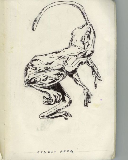
13 of 31 #inktober Forest Frog. There's a particular piece of the frog that is used for traditional chinese medicine. The oviduct (the little bit that connects the eggs to the uterus) is used to heal weakness after illness or delivery, night sweat, cough and hematemesis due to tuberculosis. #traditionalchinesemedicine #tcm #ink #frog #animal #inktober2018 #doodle #drawing https://www.instagram.com/p/Bo-RhO4nFdU/?utm_source=ig_tumblr_share&igshid=exgjh3uplbtw
0 notes
Text
Funny kitten videos
kitten videos best of the best right here right now. Watch them and enjoy your soul. You will laugh with this cute kittens. More silly cat videos for you right here. See the cutest cats or cats and have a wonderful time . Kittens are some of the great domestic mammals. They are so sweet that you almost could eat them. So funny kitten videos and so adorable. If you love kittens than come here. This is the place for youpeople like you. Want more thinks like this? There is no problem. We have plenty of it. Just click the button. Cute cats in adorable videos like nowhere before. We gained all the funniest videos from the world just for you. Nice lens. It was nice fun seeing all the alternatives on the market. Nice for making early schooling studying material. The baby will study fast, making you proud and glad! And simply like the Historic Egyptians, we worship them simply slightly as well. Jack Russells 9/10 instances are once more one other little piranha. Elephant foot watsebaskets and umbrella stands are in poor style, I feel. Are they warm sufficient?
The gonads are linked to oviducts which are related to outdoors either by means of cloaca or vagina or an intromittent organ. They know when we are comfortable and can inform when we aren't. Funny kitten videos
youtube
Rides, reveals, parades and themed restaurants are amongst essentially the most thrilling typical options of Animal Kingdom. Attention-grabbing and this is give more info in regards to the exotic animal to me and my buddies . How It works journal has studied the science behind cute animal faces and revealed their allure is right down to our evolutionary need to take care of and defend our own kids.
We consider that the correction, whether a puzzle piece is positioned in the proper or unsuitable place, needs to be extra emphasized in order to assist youngsters perceive they've bought it fallacious or proper. Some of the lesser identified familiars included frogs, toads, lizards, blackbirds, snakes, insects and more. The truth that horses love to run round ensures that they will exercise by themselves. A fox can burrow below your property's foundation or beneath stone walls and trigger structural harm. Nearly information can be used for business. No Marco, you feel sorry for yourself because you haven't any phrases of substance to defend your emotional response to my words but have a futile need to bring me down.
0 notes
Last Seen Blogs

aiqc
AIQC

fatgermanbabe
fatgermanbabe

sexiforever
Senza titolo

law-of-neutrality
I am known by many names

critical-methodology
ENGL 6600: Critical Methodology