#fundus examination
Text
Eye Fundus Camera: Revolutionizing Ophthalmic Imaging
Introduction to Eye Fundus Camera
Eye fundus cameras, also known as retinal cameras, are specialized devices used to capture images of the back of the eye, including the retina, optic disc, macula, and blood vessels. These images, referred to as fundus photographs, play a crucial role in diagnosing and monitoring various eye conditions, such as diabetic retinopathy, glaucoma, macular degeneration, and hypertensive retinopathy.
Importance of Eye Fundus Examination
A comprehensive eye examination typically involves the evaluation of the eye's posterior segment, which includes the retina and optic nerve head. Fundus examination allows ophthalmologists and optometrists to assess the health of these structures, detect abnormalities, and monitor disease progression over time.
Types of Eye Fundus Cameras
Traditional Fundus Cameras
Traditional fundus cameras are stationary devices typically found in ophthalmology clinics and hospitals. They offer high-resolution imaging capabilities and are suitable for detailed retinal examinations.
Handheld Fundus Cameras
Handheld fundus cameras provide flexibility and portability, allowing eye care professionals to capture fundus images outside of traditional clinical settings. These devices are particularly useful for screening programs and telemedicine applications.
Smartphone Fundus Cameras
Smartphone fundus cameras leverage the power of mobile technology to transform smartphones into cost-effective fundus imaging devices. With the use of specialized adapters or attachments, these cameras enable primary care providers and even patients themselves to capture fundus images conveniently.
How Eye Fundus Cameras Work
Fundus cameras utilize various imaging techniques, such as digital photography, scanning laser ophthalmoscopy, and optical coherence tomography, to capture detailed images of the eye's posterior segment. These images are then analyzed by eye care professionals to detect abnormalities and guide treatment decisions.
Benefits of Using Eye Fundus Cameras
Early detection of eye diseases
Monitoring disease progression
Guiding treatment decisions
Facilitating patient education
Enhancing collaboration among healthcare providers
Applications in Ophthalmology
Eye fundus cameras are indispensable tools in ophthalmic practice, with applications including:
Diabetic retinopathy screening
Glaucoma management
Age-related macular degeneration evaluation
Retinopathy of prematurity assessment
Optic nerve head analysis
Challenges and Limitations
Despite their numerous benefits, eye fundus cameras face certain challenges, such as:
Cost of equipment and maintenance
Training requirements for operators
Limited access in underserved areas
Image interpretation variability
Future Trends in Eye Fundus Imaging Technology
Advancements in imaging technology, such as artificial intelligence and telemedicine, are poised to revolutionize eye fundus imaging by improving accuracy, efficiency, and accessibility.
Comparison with Other Imaging Techniques
Compared to other imaging modalities like optical coherence tomography and fluorescein angiography, fundus photography offers a non-invasive and cost-effective approach to visualizing the retina and optic nerve.
Considerations for Purchasing an Eye Fundus Camera
When selecting an eye fundus camera, factors to consider include image quality, ease of use, compatibility with existing systems, technical support, and cost-effectiveness.
Tips for Efficient Use of Eye Fundus Cameras
Ensure proper patient preparation and positioning
Adjust camera settings for optimal image quality
Use appropriate imaging techniques for different eye conditions
Regularly calibrate and maintain equipment
Training and Certification for Eye Fundus Imaging
Eye care professionals should undergo specialized training and certification programs to acquire the necessary skills for operating and interpreting fundus images accurately.
Costs Associated with Eye Fundus Imaging
The initial investment in purchasing an eye fundus camera may vary depending on the type and features of the device. Additional costs include maintenance, accessories, and ongoing training.
Case Studies and Success Stories
Numerous studies have demonstrated the clinical utility and cost-effectiveness of eye fundus cameras in various healthcare settings, highlighting their role in improving patient outcomes and reducing healthcare costs.
Conclusion
Eye fundus cameras have transformed the landscape of ophthalmic imaging, enabling early detection, accurate diagnosis, and personalized management of eye diseases. As technology continues to evolve, these devices will play an increasingly vital role in preserving vision and enhancing patient care.
FAQs (Frequently Asked Questions)
Are eye fundus cameras safe for all patients?
Yes, fundus cameras are non-invasive and safe for most patients, including children and pregnant women.
Can fundus cameras detect all eye diseases?
Fundus cameras can detect a wide range of eye conditions, but they may not capture certain abnormalities that require specialized imaging techniques.
Is dilating the pupils necessary for fundus photography?
Dilating the pupils may be necessary to obtain clear fundus images, especially in cases where a detailed examination is required.
Are smartphone fundus cameras as accurate as traditional devices?
While smartphone fundus cameras offer convenience and accessibility, they may not provide the same level of image quality and diagnostic accuracy as traditional devices in all cases.
How often should fundus examinations be performed?
The frequency of fundus examinations depends on various factors, including age, medical history, and risk factors for eye diseases. It is best to consult with an eye care professional for personalized recommendations.
#eyecare#fundus examination#health#medical care#eye health#eyes#choroida#choroid#medical devices#medical equipment
0 notes
Text
Fundus Examination
People with type 1 and type 2 diabetes are at a heightened risk for eye complications and peripheral neuropathy.
You may have heard that diabetes causes eye problems and may lead to blindness. People with diabetes do have a higher risk of blindness than people without diabetes. But most people who have diabetes have nothing more than minor eye disorders over time.
With regular checkups, you can keep minor problems minor. And, if you do develop a major problem, some treatments often work well if you begin them right away.
1 note
·
View note
Text
Fundus Camera: An Essential Equipment for Eye Examination

What is a Fundus Camera?
A fundus camera, also known as an ophthalmoscope or retinal camera, is a specialized low-power microscope used to afford high-quality imaging of the internal tissues of the living human eye, known as the fundus. The fundus refers to the interior surface of the eye opposite the lens, including the retina, optic disc, and posterior pole of the eye. Fundus cameras allow eye care professionals to non-invasively examine and document the health of the retina, optic disc and macula.
Parts
A standard fundus camera consists of the following key components:
- Illumination System: Provides controlled light to illuminate the fundus via the pupil. Koehler illumination is commonly used, allowing homogenous lighting of the fundus. Halogen or Xenon lamps are commonly used light sources.
- Optical System: Consists of lenses that magnify the light reflected from the fundus for visualization and imaging purposes. Typical magnification ranges from 15x to 60x. Wide-angle and ultra-wide angle lens options are available.
- Eye Piece: The eyepiece allows the examiner to view the magnified fundus image. Binocular eyepieces provide stereoscopic viewing.
- Digital Camera Sensor: Replaces traditional film in modern digital Fundus Camera. Charged coupled device (CCD) or complementary metal-oxide-semiconductor (CMOS) digital sensors capture fundus images which can be stored, analyzed and shared digitally.
- Shields/Filters: Polarization filters reduce glare from light reflections within the eye. Alignment lights and targets help with proper centration of the fundus image.
- Stand or Slit Lamp Attachment: It may sit on a stand alone or attach to an existing slit lamp biomicroscope, allowing integration with other examination equipment.
Uses and Applications of Fundus Photography
Fundus photography allows detailed examination and documentation of retinal pathologies, which is invaluable for diagnosing and monitoring many common eye diseases. Some key applications include:
- Diabetic Retinopathy Screening: Photographing the retina allows detection of microaneurysms, hemorrhages, cottonwool spots, hard exudates and neovascularization indicative of diabetic eye disease.
Optimal Fundus Photography Techniques
Careful technique is needed to obtain high quality fundus photos enabling accurate clinical evaluation:
- Pupil Dilation: Most fundus details are only visible after dilating the pupil with eyedrops to at least 6 mm. This improves illumination levels reaching the fundus.
- Centration: Centering the fundus camera lens on the pupil ensures the retina is evenly illuminated and the entire posterior pole is in view. Targeting lights assist alignment.
Get more insights on Fundus Camera
About Author:
Ravina Pandya, Content Writer, has a strong foothold in the market research industry. She specializes in writing well-researched articles from different industries, including food and beverages, information and technology, healthcare, chemical and materials, etc. (https://www.linkedin.com/in/ravina-pandya-1a3984191)
#Fundus Camera#Retinal Imaging#Eye Examination#Ophthalmology#Fundus Photography#Retina#Ocular Health#Fundus Imaging#Eye Care#Diabetic Retinopathy Screening
0 notes
Text
Early this morning, one crew took us for a Government boat, and treated us accordingly. We saw in this a way of smoothing matters, so at Fundu, where the Bistritza runs into the Sereth, we got a Roumanian flag which we now fly conspicuously. With every boat which we have over-hauled since then this trick has succeeded; we have had every deference shown to us, and not once any objection to whatever we chose to ask or do.
This whole "impersonating officials" venture feels extra funny because they just stumbled into it basically first thing. I guess their plan up till that point was just to rely on the social lubricant of Arthur's charm and money and maybe the intimidation factor of Jonathan's fierce glare, but getting to actually examine other boats in any detail seems like a big ask.
62 notes
·
View notes
Text
So.... I got diagnosed with idiopatic intracranial hypertension (IIH) just a few hours ago. We have no idea what made me develop it but we are working on trying to find out. This could explain a lot of my symptoms that have made it difficult to make art.
Funny thing is that I've had these symptoms for years but didn't think I'd be taken seriously by doctors so I never bothered until I went to get a routine eye examination for an eye glass prescription and the optician found something weird in my fundus images 🤪
27 notes
·
View notes
Video
undefined
tumblr
Asteroid hyalosis or human snow globe?
A 54-year-old woman with type 2 diabetes mellitus presented to the ophthalmology clinic for a routine dilated fundus examination. Slit-lamp examination of the left eye revealed multiple sparkling, white, globular, refractile, dotlike opacities in the vitreous cavity. The opacities moved with ocular movement and returned to their original position after movement stopped. The vitreous of the right eye appeared normal. The visual acuity was 20/20 in each eye, and the fundus in each eye was normal.
Asteroid hyalosis is a benign condition that is often unilateral, and is associated with older age. The asteroid bodies are composed of calcium, phosphorus, and various phospholipids. Given the benign nature of this condition and this patient’s normal visual acuity, no treatment was recommended.
Source: NEJM case report (doi: 10.1056/NEJMicm1712355)
294 notes
·
View notes
Text
PART 10
of the dfk audiobook translation
@cnka
Direktorin Kreuzkamm: „I hereby hand over the production for the closing celebration to you.“
Narrator (Nichtraucher):
Given her overflowing time planner…
Direktorin Kreuzkamm: „Have fun and toi, toi, toi!“
[Note: is „Toi, toi, toi“ a thing in other countries?? Its basically a way to wish luck in the theatre. Like „break a leg“]
Narrator (Nichtraucher):
…Frau Kreuzkamm counted on Jo and the self-responsibilty of the students.
Narrator (audiobook):
Jo steps in front of the group.
Jo, overly enthusiastic and cheerful: „Let’s get started!"
Narrator (audiobook):
Ruda, however, doesn’t play along with it.
Ruda: „You can do that on your own.“
Jo: „Come on, guys, it’s gonna be cool! Who's with me?“

Apart from Jo's friends, only Egerland raises his hand, but Sebi slaps it down again and pulls him with her.
Narrator (audiobook):
The Externs leave the hall as one, and even the majority of the Internals have other plans.
Internal: „Sorry, Jo, we’re not getting involved in that.“
Other Internal: „Too much stress for us…“
Everyone leaves, muttering among each other („No, no thank you“)
Jo: „Are you serious?!“
Narrator (audiobook):
Apart from our four friends, all the other Internals seem to be serious about it as well, because Martina, Jo, Matze and Uli are the only ones left.
Jo: „Ugh, it can’t work with this amount of people!“
Martina: „It has to work! If I get a reprimand, I won’t be taken in.“
Matze: „Ask me, my father always punches me in the face when I get another one!“
Uli: „…It would be my first one…“

Jo: „Guys. I have an idea.“
Narrator (audiobook):
Jo brings her friends back to the clearing and proudly points to the train wagon.

Jo: „Okay, guys, this is the flying classroom! We’ll put a nose on the front. A wing here. And a rocket engine in the back. We get the stuff from the theatre fundus, and then we produce a film that we’ll show at the closing celebration!“
Martina, lighting up: „A film?“
Jo, excitedly: „Yes!! And-“
Narrator (audiobook):
Jo falls silent. From the inside of the train wagon comes piano music.
Jo: „Do you hear that?“
Narrator (audiobook):
She sneaks to the wagon and looks through one of the windows. She sees a man sit there and play with a cigarette in his mouth. All of a sudden, he abruptly turns his head and looks directly into her eyes.


Narrator (audiobook):
Jo frantically comes running back to her friends.
Jo: „Shit, guys! Do you hear that? There’s someone in there! He saw me!“
The door of the train wagon slams open. The kids gasp.
Narrator (audiobook):
Suddenly, the door of the wagon opens. At first, the Internals only hear a voice.
Nichtraucher: „Are you trying to break in here?“
Matze: „Co-come on, let’s scram!“
Narrator (audiobook):
But Martina forges ahead.
Martina: „Excuse us! We got lost.“
Narrator (audiobook):
The smoking man -approximately Herr Bökh’s age- steps out of the door and examines the Internals closely.
Jo: „Uh- What- What are you doing here?“
Nichtraucher: „That’s really none of your business.“

Martina, with emphasis: „Jo, come on!“
Jo: „No, wait- Do you sleep in there?“
Nichtraucher: „… This is private property here.“
Narrator (audiobook):
But Jo doesn’t let up and persistently asks further.
Jo: „What was that song? Who is it from?“
Nichtraucher: „…“
Nichtraucher: „From me.“
Jo: „Do you give lessons?“
Nichtraucher: „Piano?“

Jo: „No, for smoking. Of course Piano.“

Nichtraucher: „No, I don’t.“


Nichtraucher: „Ok, and now off you go, you little squirts!“
He turns around to go back inside.
Jo: „No, stop, wait! Uh… We’re four Internals from the boarding school Sigismund. And we want to shoot a movie for the closing celebration. Because we have trouble with the Externs and… Doesn’t matter, you can’t explain that. In any case, we wanted to ask if we could shoot on your property.“
The Nichtraucher pauses when Jo mentions the boarding school and turns around.
He listens silently and seems to be thinking.


Narrator (audiobook):
Robert, that’s what the man is called, who, as previously mentioned, is also the author of this story, seems to be surprised by this blunt question. He takes a long drag and looks Jo in the eye.

Jo: „Please?“

#das fliegende klassenzimmer#dfk 2023 audiobook translation#das fliegende klassenzimmer 2023#dfk#how its just casually brought up that matze’s dad physically abuses him and then they never mention it again 👍#mine#also that the narrator emphasizes that the nichtraucher is „approximately herr bökh’s age“#really smooth very sneaky
7 notes
·
View notes
Text
Tagging: @weekofobitine
Day 6 prompts used: Senate, archive, intrigue
Bonus prompts used: none

Idle curiosity was never his weakness, he respected other people's boundaries, realizing that he himself wasn't completely honest all the time, preferring to keep some things secret even from those closest to him. And studying Satine's personal datapad wasn't something he had ever wanted to do without her personal permission. And yet… Yet when his finger, once again touching the kyber crystal, was too close to the border of the touch screen, and the last document had viewed was again opened, his gaze unconsciously slid along the straight lines of tables of symbols and numbers.
After Kadavo the Jedi Archives became the most frequent place where Obi-Wan spent the little time that he was on Coruscant between the Order’s regular missions, but the mission under the guise of Rako Hardeen turned the most frequent place into the only one. Being a huge accumulation of information, the Jedi Archives attracted him in his youth too, now, however, it wasn't so much a craving for new knowledge but an attempt to find peace in the silence of the majestic library for his soul, torn to shreds by recent events. Trying to distract his mind from painful memories, he studied all the holodiscs, all the holocrons that he had access to, including some historical sources in Mando'a, and therefore he was able to translate now some individual phrases and get an overall picture and general meaning of the documents stored on the datapad.
It were medical reports. Multiple a blood, a plasma, a bone marrow tests. The ultrasound diagnostics. The tone and height of the uterine fundus, a fetal position, a heartbeat… The datapad stored all information about examinations carried out during pregnancy, as well as birth and postpartum data about the child and the mother. His updated knowledge of Mando'a was still not enough to understand whether the dynamics of certain indicators were positive or not, and the notes still weren't fully understood - its often exceeding the results themselves in volume. But when the graphs, tables and photographs passed where something over a hundred, he got a test, which a Jedi of his status was required to know in all known languages of the Galaxy - an analysis of the content of Midi-chlorian in the blood.
The name of the creature being examined, written on Basic, made he shudder.
Korkie.
Continue read on ao3
#Obitine Week 2023#Obitine Week#Star Wars#The Clone Wars#Obi Wan Kenobi#Satine Kryze#Duchess Satine#Korkie Kryze#Obitine#Obi Wan x Satine#Obi x Satine#Obi Wan/Satine#Star Wars fanfiction#Obitine fanfiction#Obitine fanfic#Russian fanfiction#ao3 fanfic
2 notes
·
View notes
Text
I'm mad at the ophtalmologist I saw last year because I did a dilated fundus examination for the first time and the first thing the doctor told me was that I have an "incredibly beautiful retina" and then she DIDN'T SHOW ME PICS OF IT
#so now I have to live wondering what an 'incredibly beautiful retina' even looks like#bee tries to talk
8 notes
·
View notes
Text
Persistence of myelinated nerve fibers
Persistence of myelinated nerve fibers is a condition where myelin, which typically sheaths the optic nerve, extends abnormally into the retinal nerve fiber layer. This can appear as white, opaque patches on fundus examination, often with feathery edges. Myelinated nerve fibers can sometimes obscure underlying retinal structures and are generally benign but may be associated with visual field…
0 notes
Text
Retinal Fundus Camera: Revolutionizing Eye Health
Imagine a device capable of peering into the depths of your eyes, revealing early signs of potentially sight-threatening diseases. Enter the retinal fundus camera, a groundbreaking tool in ophthalmology that has revolutionized the way eye health is monitored and managed. In this article, we delve into the intricacies of retinal fundus cameras, exploring their history, functionality, applications, and future prospects.
Introduction to Retinal Fundus Camera
At its core, a retinal fundus camera is a specialized imaging device used to capture detailed images of the back of the eye, including the retina, optic disc, macula, and blood vessels. These images, known as fundus photographs, provide invaluable insights into the health of the eye, aiding in the diagnosis and management of various ocular conditions.
History and Evolution of Retinal Fundus Imaging
The roots of retinal fundus imaging can be traced back to the late 19th century when ophthalmologists began experimenting with rudimentary devices to visualize the posterior segment of the eye. Over the decades, advancements in optics, electronics, and imaging technology have led to the development of highly sophisticated fundus cameras capable of capturing high-resolution images with remarkable clarity.
Importance of Retinal Fundus Imaging in Eye Health
Early Detection of Eye Diseases
One of the primary advantages of retinal fundus imaging is its ability to detect subtle changes in the retina that may indicate the presence of various eye diseases, such as diabetic retinopathy, age-related macular degeneration (AMD), and glaucoma, at their earliest stages.
Monitoring Disease Progression
Retinal fundus cameras play a crucial role in monitoring the progression of ocular conditions over time, allowing healthcare providers to assess the effectiveness of treatments and intervene promptly if necessary.
Types of Retinal Fundus Cameras
Traditional Fundus Cameras
Traditional fundus cameras are stationary devices typically found in ophthalmology clinics and hospitals. They consist of complex optical systems capable of capturing high-resolution images of the retina.
Handheld Fundus Cameras
Handheld fundus cameras offer portability and versatility, making them ideal for use in remote or underserved areas where access to traditional imaging equipment may be limited.
Smartphone-Based Fundus Cameras
With the advent of smartphone technology, it is now possible to transform a regular smartphone into a portable fundus camera using specially designed attachments or accessories.
How a Retinal Fundus Camera Works
Fundus imaging involves the capture of digital photographs of the retina, which is the light-sensitive tissue lining the back of the eye. The process typically begins with the dilation of the pupil to provide a clear view of the retina. The camera then emits a flash of light, illuminating the back of the eye, while a specialized lens focuses and captures the resulting image.
Applications of Retinal Fundus Cameras
Diabetic Retinopathy Screening
Diabetic retinopathy is a leading cause of vision loss among individuals with diabetes. Retinal fundus cameras are instrumental in screening for diabetic retinopathy by detecting early signs of retinal damage, such as microaneurysms, hemorrhages, and exudates.
Age-Related Macular Degeneration Diagnosis
Age-related macular degeneration is a progressive eye disease that affects the macula, the central part of the retina responsible for sharp, central vision. Fundus imaging is crucial for diagnosing and monitoring the progression of AMD, enabling timely intervention to preserve vision.
Glaucoma Detection
Glaucoma is a group of eye conditions characterized by damage to the optic nerve, often resulting in irreversible vision loss. Retinal fundus cameras help in identifying optic nerve abnormalities and monitoring changes in the optic disc that may indicate the presence of glaucoma.
Retinopathy of Prematurity Screening
Retinopathy of prematurity is a potentially blinding condition that affects premature infants. Fundus cameras are used to screen premature babies for signs of retinopathy, allowing for early intervention to prevent vision loss.
Advantages of Retinal Fundus Imaging
Non-Invasive Procedure
Retinal fundus imaging is a non-invasive procedure that does not require direct contact with the eye, making it safe and comfortable for patients of all ages.
High-Quality Images
Modern fundus cameras are capable of capturing high-resolution images with exceptional detail, allowing for accurate diagnosis and treatment planning.
Early Disease Detection
By detecting subtle changes in the retina at an early stage, retinal fundus imaging enables healthcare providers to initiate timely interventions and prevent vision loss.
Challenges and Limitations
Despite its numerous advantages, retinal fundus imaging is not without its challenges and limitations.
Accessibility Issues
In many parts of the world, access to retinal fundus cameras may be limited, particularly in rural or underserved areas where healthcare infrastructure is lacking.
Cost
The initial cost of acquiring and maintaining retinal fundus imaging equipment can be prohibitive for smaller clinics or healthcare facilities with limited budgets.
Operator Skill Requirement
Interpreting fundus images requires specialized training and expertise, highlighting the importance of ensuring that healthcare providers receive adequate education and training in fundus photography.
Future Trends in Retinal Fundus Imaging
Artificial Intelligence Integration
Advancements in artificial intelligence are poised to revolutionize retinal fundus imaging, enabling automated analysis of fundus photographs for the early detection of eye diseases.
Miniaturization and Portability
The development of compact and portable fundus cameras is opening up new possibilities for remote screening and telemedicine, particularly in resource-limited settings.
Telemedicine Applications
Retinal fundus imaging has the potential to transform the delivery of eye care by facilitating remote consultations and telemedicine services, allowing patients to access specialized care from the comfort of their homes.
Conclusion
Retinal fundus cameras represent a paradigm shift in the field of ophthalmology, offering a non-invasive and effective means of assessing and monitoring eye health. As technology continues to advance, the role of retinal fundus imaging in preventing vision loss and preserving sight will only continue to grow.
FAQs
Are retinal fundus cameras safe?
Yes, retinal fundus imaging is a non-invasive procedure that poses minimal risk to patients.
Can retinal fundus cameras detect all eye diseases?
While retinal fundus cameras are highly effective for detecting many eye diseases, they may not be able to identify certain conditions that require specialized testing or imaging techniques.
How often should someone undergo retinal fundus imaging?
The frequency of retinal fundus imaging depends on various factors, including age, medical history, and risk factors for eye diseases. It is best to consult with an eye care professional to determine an appropriate screening schedule.
Are retinal fundus cameras only used by ophthalmologists?
While retinal fundus cameras are commonly used by ophthalmologists, they may also be utilized by optometrists and other eye care professionals trained in fundus photography.
What should I expect during a retinal fundus imaging procedure?
During a retinal fundus imaging procedure, your eyes may be dilated with eye drops to provide a clear view of the retina. You will then be asked to focus on a target while the camera captures images of the back of your eye.
#eyecare#eyes#health#medical care#medical devices#eye health#choroida#choroid#fundus examination#medical equipment
0 notes
Text
Fundus Examination | Samyak Eye Care Clinic
Fundus Examination
People with type 1 and type 2 diabetes are at a heightened risk for eye complications and peripheral neuropathy.
You may have heard that diabetes causes eye problems and may lead to blindness. People with diabetes do have a higher risk of blindness than people without diabetes. But most people who have diabetes have nothing more than minor eye disorders over time.
With regular checkups, you can keep minor problems minor. And, if you do develop a major problem, some treatments often work well if you begin them right away.
How the eye works
Understanding what happens in eye disorders, helps to understand how the eye works. The eye is covered with a tough outer membrane. The covering in front is clear and curved. This curved area is the cornea, which focuses light while protecting the eye.
After light passes through the cornea, it travels through a space called the anterior chamber (which is filled with a protective fluid called the aqueous humor), through the pupil (which is a hole in the iris, the colored part of the eye), and then through a lens that performs more focusing. Finally, light passes through another fluid-filled chamber in the center of the eye (the vitreous) and strikes the back of the eye, the retina.
The retina records the images focused on it and converts those images into electrical signals, which the brain receives and decodes.
One part of the retina is specialized for seeing fine detail. This tiny area of extra-sharp vision is called the macula. Blood vessels in and behind the retina nourish the macula.
Glaucoma
People with diabetes are more likely to suffer from glaucoma than people without diabetes. The longer someone has had diabetes, the more common glaucoma is. Risk also increases with age.
Glaucoma occurs when pressure builds up in the eye. The pressure pinches the blood vessels that carry blood to the retina and optic nerve. Vision is gradually lost because the retina and nerves are damaged.
There are several treatments for glaucoma. Some use drugs to reduce pressure in the eye, while others involve surgery.
Cataract
Many people without diabetes get cataracts, but people with diabetes are more likely to develop this eye condition. People with diabetes also tend to get cataracts at a younger age and have them progress faster. With cataracts, the eye’s clear lens clouds, blocking sight.
To help deal with mild cataracts, you may need to wear sunglasses more often and use glare-control lenses in your glasses. For cataracts that interfere greatly with vision, doctors usually remove the lens of the eye and replace it with a new artificial lens. In people with diabetes, retinopathy can get worse after the removal of the lens, and glaucoma may start to develop.
Retinopathy
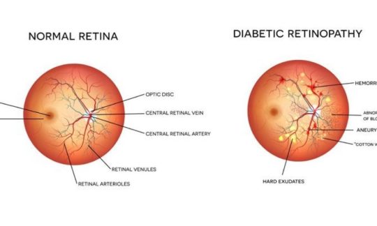
Diabetic retinopathy is a general term for all disorders of the retina caused by diabetes. There are two major types of retinopathy: nonproliferative and proliferative.
Nonproliferative retinopathy
In nonproliferative retinopathy, the most common form of retinopathy is capillaries in the back of the eye balloon and forming pouches. Nonproliferative retinopathy can move through three stages (mild, moderate, and severe), as more and more blood vessels become blocked.
Macular edema
Although retinopathy does not usually cause vision loss at this stage, the capillary walls may lose their ability to control the passage of substances between the blood and the retina. Fluid can leak into the part of the eye where focusing occurs, the macula. When the macula swells with fluid, a condition called macula edema, vision blurs and can be lost entirely. Although nonproliferative retinopathy usually does not require treatment, macular edema must be treated, but fortunately, treatment is usually effective at stopping and sometimes reversing vision loss.
Proliferative retinopathy
In some people, retinopathy progresses after several years to a more serious form called proliferative retinopathy. In this form, the blood vessels are so damaged they close off. In response, new blood vessels start growing in the retina. These new vessels are weak and can leak blood, blocking vision. The new blood vessels can also cause scar tissue to grow. After the scar tissue shrinks, it can distort the retina or pull it out of place, a condition called retinal detachment.
How is retinopathy treated?
Huge strides have been made in the treatment of diabetic retinopathy. Treatments such as scatter photocoagulation, focal photocoagulation, and vitrectomy prevent blindness in most people. The sooner retinopathy is diagnosed, the more likely these treatments will be successful. The best results occur when sight is still normal.
In photocoagulation, the eye care professional makes tiny burns on the retina with a special laser. These burns seal the blood vessels and stop them from growing and leaking.
In scatter photocoagulation (also called panretinal photocoagulation), the eye care professional makes hundreds of burns in a polka-dot pattern on two or more occasions. Scatter photocoagulation reduces the risk of blindness from vitreous hemorrhage or detachment of the retina, but it only works before bleeding or detachment has progressed very far. This treatment is also used for some kinds of glaucoma.
Side effects of scatter photocoagulation are usually minor. They include several days of blurred vision after each treatment and possible loss of side (peripheral) vision.
In focal photocoagulation, the eye cares professional aims the laser precisely at leaking blood vessels in the macula. This procedure does not cure blurry vision caused by macular edema, but it does keep it from getting worse.
When the retina has already detached or a lot of blood has leaked into the eye, photocoagulation is no longer useful. The next option is vitrectomy, which is surgery to remove scar tissue and cloudy fluid from inside the eye. The earlier the operation occurs, the more likely it is to be successful. When the goal of the operation is to remove blood from the eye, it usually works. Reattaching a retina to the eye is much harder and works in only about half the cases.
There are two types of treatment for macular edema: focal laser therapy that slows the leakage of fluid, and medications that can be injected into the eye that slow the growth of new blood vessels and reduce the leakage of fluid into the macula.
A newer retinopathy treatment involves injecting medication directly into the eye. The injection contains a drug that blocks the activity of vascular endothelial growth factor (VEGF). This hormone promotes the growth of new blood vessels and plays a key role in retinopathy by promoting the growth of weak, leaky blood vessels. Anti-VEGF drugs put a stop to problem vessels, improving vision in people with retinopathy. In many cases, these treatments have to be repeated every few months (sometimes every month) to decrease the inflammation in the eye.
There are also some other new treatments with substances that are put into the back of the eye to help it heal. All of these advances in eye care have made a big difference in helping people’s eyes. Prevention is always first, but if damage happens, it can be treated.
Am I at risk for retinopathy?
Several factors influence whether you get retinopathy:
blood sugar control
blood pressure levels
how long you have had diabetes
genes
The longer you’ve had diabetes, the more likely you are to have retinopathy. Almost everyone with type 1 diabetes will eventually have nonproliferative retinopathy. And most people with type 2 diabetes will also get it. But the retinopathy that destroys vision, proliferative retinopathy, is far less common.
People who keep their blood sugar levels closer to normal are less likely to have retinopathy or to have milder forms.
Your retina can be badly damaged before you notice any vision change. Most people with nonproliferative retinopathy have no symptoms. Even with proliferative retinopathy, the more dangerous form, people sometimes have no symptoms until it is too late to treat them. For this reason, you should have your eyes examined regularly by an eye care professional.
Visit the Focus on Diabetes site for more information regarding eye health and diabetes.
high blood pressure and other disorders, because many diseases can have an impact on vision and eye health
1 note
·
View note
Text
Eye Testing Equipment Market : Technology Advancements, Industry Insights, Trends And Forecast 2033
The Eye Testing Equipment Global Market Report 2024 by The Business Research Company provides market overview across 60+ geographies in the seven regions - Asia-Pacific, Western Europe, Eastern Europe, North America, South America, the Middle East, and Africa, encompassing 27 major global industries. The report presents a comprehensive analysis over a ten-year historic period (2010-2021) and extends its insights into a ten-year forecast period (2023-2033).

Learn More On The Eye Testing Equipment Market:
https://www.thebusinessresearchcompany.com/report/eye-testing-equipment-global-market-report
According to The Business Research Company’s Eye Testing Equipment Global Market Report 2024, The eye testing equipment market size is expected to see strong growth in the next few years. It will grow to $4.41 billion in 2028 at a compound annual growth rate (CAGR) of 7.8%. The growth in the forecast period can be attributed to the increasing adoption of telemedicine, growing demand for portable and handheld devices, rising prevalence of diabetes and related eye disorders, increasing focus on preventive healthcare, and an expanding middle-class population. Major trends in the forecast period include the integration of AI and machine learning in diagnostics, the increasing use of virtual reality in eye examinations, the rise in demand for personalized eye care solutions, the development of advanced imaging technologies, and growing collaboration between tech companies and healthcare providers.
The increasing prevalence of eye-related disorders is expected to propel the growth of the eye-testing equipment market going forward. Eye-related disorders refer to a range of conditions affecting the eyes, including diseases such as glaucoma, cataracts, macular degeneration, and refractive errors such as myopia and hyperopia. The increase in the number of patients with eye-related disorders is due to factors such as aging populations, lifestyle changes, environmental factors, and lack in diagnostic capabilities leading to delay in problem detection. Eye testing equipment is used for early detection, accurate diagnosis, and effective monitoring of eye-related conditions such as glaucoma, cataracts, and macular degeneration. For instance, in January 2024, according to the Centers for Disease Control and Prevention, a US-based science and data-driven service organization that protects the public's health, over 3.4 million Americans aged 40 years and older were blind or visually impaired, which is expected to double by 2030. Therefore, the increasing prevalence of eye-related disorders will drive the growth of the eye-testing equipment market.
Get A Free Sample Of The Report (Includes Graphs And Tables):
https://www.thebusinessresearchcompany.com/sample.aspx?id=17147&type=smp
The eye testing equipment market covered in this report is segmented –
1) By Device: Slit Lamp, Biometer, Perimeter, Tonometer, Optical Coherence Tomography (OCT), Fundus Camera, Autorefractor And Keratometer, Other Devices
2) By Application: General Examination, Glaucoma, Cataract, Other Applications
3) By End User: Hospital, Eye Clinic, Optometry Academic Institute
Major companies operating in the eye-testing equipment market are developing advanced eye-testing devices, such as handheld artificial intelligence fundus cameras, to enhance diagnostic accuracy and streamline screening processes. A handheld fundus camera enhanced with AI technology is used for capturing detailed images of the eye's fundus to assist in diagnosing eye conditions. For instance, in May 2024, Optomed USA, a US-based medical technology company, launched Optomed Aurora AEYE. It is a handheld AI-powered fundus camera specifically designed to quickly detect diabetic retinopathy beyond mild stages, enabling prompt eye screenings in primary care settings. This device simplifies the screening process with its user-friendly design, needing only one image per eye and delivering results within a minute.
The eye testing equipment market report table of contents includes:
1. Executive Summary
2. Eye Testing Equipment Market Characteristics
3. Eye Testing Equipment Market Trends And Strategies
4. Eye Testing Equipment Market - Macro Economic Scenario
5. Global Eye Testing Equipment Market Size and Growth
.............
32. Global Eye Testing Equipment Market Competitive Benchmarking
33. Global Eye Testing Equipment Market Competitive Dashboard
34. Key Mergers And Acquisitions In The Eye Testing Equipment Market
35. Eye Testing Equipment Market Future Outlook and Potential Analysis
36. Appendix
Contact Us:
The Business Research Company
Europe: +44 207 1930 708
Asia: +91 88972 63534
Americas: +1 315 623 0293
Email: [email protected]
Follow Us On:
LinkedIn: https://in.linkedin.com/company/the-business-research-company
Twitter: https://twitter.com/tbrc_info
Facebook: https://www.facebook.com/TheBusinessResearchCompany
YouTube: https://www.youtube.com/channel/UC24_fI0rV8cR5DxlCpgmyFQ
Blog: https://blog.tbrc.info/
Healthcare Blog: https://healthcareresearchreports.com/
Global Market Model: https://www.thebusinessresearchcompany.com/global-market-model
0 notes
Text
Finding the Best Eye Hospital in Rawalpindi for Medical Retina Needs
Introduction:
When it comes to eye care, especially concerning delicate conditions like medical retina issues, finding the Best eye hospital in Rawalpindi is crucial. Your eyes deserve the best care, and this article will guide you on how to identify the top facilities in Rawalpindi that specialize in medical retina treatments.

Understanding Medical Retina Care:
The medical retina refers to the branch of ophthalmology that deals with diagnosing and treating diseases of the retina, such as diabetic retinopathy, age-related macular degeneration, and retinal vein occlusion. These conditions require specialized attention and advanced treatment methods, which can only be provided by the Best eye hospital in Rawalpindi.
Why Choose the Best Eye Hospital in Rawalpindi?
Rawalpindi is home to several eye care facilities, but not all are equipped to handle complex medical retina cases. The Best eye hospital in Rawalpindi will have:
Experienced Retina Specialists: Look for hospitals with board-certified ophthalmologists who specialize in medical retina care. Their expertise is vital for accurate diagnosis and effective treatment.
Advanced Diagnostic Equipment: The Best eye hospital in Rawalpindi will be equipped with state-of-the-art diagnostic tools, such as Optical Coherence Tomography (OCT) and Fundus Fluorescein Angiography (FFA), to thoroughly examine the retina.
Comprehensive Treatment Options: Whether it’s laser therapy, anti-VEGF injections, or vitrectomy surgery, the Best eye hospital in Rawalpindi should offer a full range of treatments tailored to your specific medical retina condition.
What to Look for in an Eye Hospital for Medical Retina Needs:
Choosing the Best eye hospital in Rawalpindi for your medical retina needs involves several considerations:
Reputation and Patient Reviews:
Start by researching the hospital’s reputation. Patient reviews and testimonials can provide insights into the quality of care provided. The Best eye hospital in Rawalpindi will have a track record of successful medical retina treatments and positive patient outcomes.
Qualified Medical Team:
Ensure that the hospital has a team of qualified ophthalmologists, including specialists in medical retina. The expertise of the medical team plays a significant role in determining the success of your treatment.
Technology and Equipment:
The Best eye hospital in Rawalpindi will invest in the latest technology to diagnose and treat medical retina conditions. Make sure the hospital is up-to-date with the latest advancements in eye care.
Personalized Care:
Your medical retina condition is unique, and so should be your treatment. The Best eye hospital in Rawalpindi will provide personalized care plans tailored to your specific needs, ensuring the best possible outcomes.
Conclusion:
Finding the Best eye hospital in Rawalpindi for medical retina needs is essential for preserving your vision and ensuring your eye health is in the best hands. By considering factors like the hospital’s reputation, the qualifications of the medical team, the technology available, and the level of personalized care, you can make an informed decision that will lead to better health outcomes.
Take the time to research and choose the Best eye hospital in Rawalpindi for your medical retina care, as this decision can make all the difference in the quality of your vision and overall well-being.
0 notes
Text
Ophthalmoscopes: A Vital Medical Device for Eye Examinations

The history of the ophthalmoscope dates back to the 1850s when Hermann von Helmholtz, a German physician and physicist, invented the first basic version to examine the interior of the human eye. Since then, the device has undergone many innovations and advancements. In 1890, American ophthalmologist Henry Gradle developed an improved version of the direct device which allowed for better retinal examinations. In the early 1900s, indirect ones became available which enabled eye inspection without contact with the patient's eyeball making examinations more comfortable. In the following decades, power sources were introduced to provide brighter illumination for viewing the fundus and photo capabilities were integrated to document findings. Modern ones today are highly portable, lightweight digital devices offering advanced imaging, magnification and illumination functions.
Workings
Ophthalmoscopes utilize a combination of lenses and light sources to view the interior structures of the eye. Direct ones hold a lens near the patient's eye which reflects and magnifies the retinal image while the examiner views through an eyepiece. Indirect scopes project an inverted magnified image of the retina onto a lens placed on the patient's forehead, allowing the examiner to observe from a short distance. Both types focus a small bright light beam on the retina using lenses or an optic fiber bundle. Dioptic lenses enable viewing of both the illuminated retina and immediate surroundings simultaneously. Powered ones contain a battery that powers high-intensity LEDs or halogen bulbs. Digital models have cameras to photograph the retina electronically.
Applications in Medical Screening and Diagnosis
Routine eye examinations conventionally involve visualizing the retina, optic disc and macula using an ophthalmoscope. This helps detect signs of numerous common conditions. Diabetic retinopathy screenings rely heavily on retinal visualization via ophthalmoscopy. Changes indicative of the disease such as microaneurysms, hemorrhages and exudates can be spotted. These are indispensable for hypertensive retinopathy evaluation where they reveal arteriolar narrowing, silver wiring of vessels and optic disc edema. Papilledema, a swelling of the optic disc, is a key sign often picked up on ophthalmoscopy in conditions like brain tumors. Retinal detachments, tumors and occlusions can also be promptly diagnosed with these instruments. Ophthalmoscopic findings are regularly recorded to monitor progression or response to treatment over time.
Applications in Research and Telemedicine
Ophthalmoscopy makes significant contributions beyond clinical practice. Research in ocular diseases extensively utilizes information gathered through standardized retinal examinations using scopes. Digital retinal images obtained are stored in databases for large population studies analyzing risk factors. Technology partnerships have enabled “eyecams” that can tele-ophthalmoscopically transmit retinal photos from remote locations to expert reading centers, expanding access to specialty eye care. The US military uses tele-ophthalmoscopy to remotely screen troops for eye and systemic issues like hypertension. These are pivotal research tools, aiding discovery of pathological changes, disease mechanisms and treatment responses. Advancing scope technology itself drives retinal imaging research contributing to medicine.
0 notes
Text
Enhancing Proficiency in Eye Care with Ophthalmology EMR Software

Ophthalmology is a specialized branch of medicine that deals with the diagnosis, treatment, and prevention of eye disorders. Given the intricate nature of this field, ophthalmologists require an Electronic Medical Records (EMR) system tailored to meet their specific needs. Ophthalmology EMR software can significantly enhance the efficiency of clinical workflows, improve patient care, and streamline administrative tasks. Among the leading providers in this space is 1st Providers Choice, known for their comprehensive and customizable EMR solutions.
Specialized Templates and Forms
Ophthalmology involves a wide range of procedures and examinations, from routine eye exams to complex surgeries. An efficient Ophthalmology EMR system must offer dedicated templates and forms that cater to these numerous requirements. These templates should cover comprehensive eye exams, visual acuity tests, slit-lamp exams, and more. Customizable templates allow ophthalmologists to efficiently document patient encounters, ensuring that all relevant data is captured accurately.
Imaging and Diagnostic Integrations
Imaging is a cornerstone of ophthalmology, with tools like Optical Coherence Tomography (OCT), fundus photography, and visual field testing playing critical roles in diagnosis and treatment planning. A robust EMR system should integrate seamlessly with these imaging devices, allowing for easy import, storage, and viewing of images directly within the patient’s record. This integration facilitates quick access to diagnostic data and aids in more accurate and timely clinical decision-making.
E-Prescribing and Medication Management
Efficient management of medications is essential in ophthalmology, where treatments often involve various eye drops, oral medications, and injections. An EMR system should include e-prescribing capabilities, allowing ophthalmologists to send prescriptions electronically to pharmacies. Additionally, features such as medication reconciliation and alerts for potential drug interactions enhance patient safety and streamline the prescribing process. That is why, it is always suggested to choose software from 1st Providers Choice. Being a leading provider of EMR EHR software, we will guide you every possible way to manage an efficient healthcare practice.
Patient Education and Engagement
Educating patients about their eye conditions and treatment options is crucial for successful outcomes. An Ophthalmology EMR software should include patient education resources that can be easily shared during consultations. These might include informational brochures, videos, and personalized care plans accessible through a patient portal. Engaged and informed patients are more likely to adhere to treatment plans and make necessary lifestyle adjustments.
Workflow Automation
Ophthalmology practices handle a high volume of patients and complex administrative tasks. Workflow automation features in an EMR system can significantly reduce the administrative burden. Automated appointment reminders, billing processes, and follow-up schedules ensure that nothing falls through the cracks, allowing providers to focus more on patient care.
We at 1st Providers Choice, offer you an innovative Ophthalmology EMR software that encompasses essential features. By investing in a comprehensive EMR system, ophthalmologists can enhance their clinical efficiency and patient care standards, ultimately advancing the quality and effectiveness of eye care services. You can also check our Physical Therapy Practice Management Software for better efficiency.
#Ophthalmology EMR Software#Ophthalmology EMR#EMR EHR software#Physical Therapy Practice Management Software
0 notes
Last Seen Blogs

brieffreakbonkgoth
Untitled

incorrectmarvelquotess
missing my babe nat romanoff, end tweet

thealphareporter
The Alpha Reporter

the-silver-peahen-residence
The silver rose peahen Mansion

incorrect-wynonnaearp-quotes
Totally Accurate Wynonna Earp