#light microscopy
Explore tagged Tumblr posts
Visit Tumblr Blog
Explore Tumblr blogs with no restrictions, modern design and the best experience.
Last Seen Tumblr Blogs


Fun Fact
The total number of visits Tumblr.com received during January 2021 is 327 million.
Text
Freshwater Microalgal Diversity and Culturing Conditions in Karak, PakistanFreshwater Microalgal Diversity and Culturing Conditions in Karak, Pakistan

Abstract
Algae are photosynthetic and predominantly aquatic organisms that produce up to half of the oxygen in Earth’s atmosphere. In this study, the algal flora of district Karak has been isolated, identified, and explored for diversity based on its optimum in-vitro culturing and microscopic technique. Microalgal samples were collected from freshwater bodies of ecologically diverse sites of district Karak. The microalgae samples were collected from February to April (spring season) and August to September (summer season) in 2020-2022. In the aseptic environment, three different types of media (BBM, MBBM, and BG-11) were used to evaluate microalgal growth parameters. The fluctuation in temperature, pH, water density, and nutrient availability varies with species distribution; however, BBM media was shown to be more optimal and standard than others for algae cultivation. A total of 33 microalgae strains were investigated that belong to 4 classes, 10 orders, 12 families, and 17 genera. Among them 5 species were cyanobacteria, 16 species were green microalgae, and 12 species from Diatoms; in which Bacillariaceae was the dominant family with 6 species and their contribution was 19%. The 2nd most dominant families were Scenedesmaceae, Volvocaceae, and Desmidiaceae with each 4 species respectively and their contribution was 12%. The other families Oscillatoriaceae followed by Chlorococcaceae (9%) while some families represented only two species (6%) that were Fragilariaceae, Pinnulariaceae and Nostocaceae, and Gomphonemataceae, Naviculaceae, Chaetophoraceae were one (3%) species each. These species belonged to 17 genera and 12 families; three key categories of microalgae (Cyanobacteria, Green Algae, and Diatoms) were reported in this study area. This study’s scope is to examine the scientific studies of microalgae diversity from various habitats of fresh water and investigate the optimal culture conditions for these algae growth which is essential for multiple applications. Hence, the present taxonomic findings demonstrate that District Karak is a rich source of microalgae biomes unexplored till now.
Introduction
The word algae are derivative of the epithet “alga” in Latin & Greek, “phykos” means seaweed; which has a lack of defined body parts system like plant (Sahoo and Seckbach, 2015; Oyewumi and Olukunle, 2017; Selvaraj et al., 2021). Microalgae are a planktonic vast and diverse group of microscopic unicellular may be prokaryotic or eukaryotic organisms (Masojídek et al., 2013; Ghani et al., 2020; Puchkova et al., 2021). Blue-green algae also known as Cyanobacteria (Gramnegative bacteria) are among the most ancient photosynthetic microorganism due to the existence of a bluish pigment “Phycocyanin” in them (Singh et al., 2016; Barinova et al., 2018; Selvaraj et al., 2021). They are oxygen-evolving microbes by photosynthesis process to get their energy; ranges from unicellular structures to colonial, branched, and un-branched filaments, non-motile and widely distributed organisms on Earth (Klm et al., 2011; Shakir et al., 2014; Tragin et al., 2016;). Blue-green algae play a crucial role as primary producers in aquatic ecosystems with distinctive characteristics that provide lodging to varying environments (Raghuwanshi et al., 2011; Halder, 2016; Narchonai et al., 2019). They have massive phylogenetic diversity, as ancestors of plants from billion years of evolutionary history and often developed extremely habitats (Singh et al., 2013; Barinova et al., 2018; Arsad et al., 2022). Microalgae are photosynthetic microbes that have generated rapid interest in applied research with multifunctional applications in the modern era (Abdelaziz et al., 2013; Alam et al., 2019; Ramos et al., 2021).
Microalgae are assorted organisms with numerous prospective traits like cell organization, plastids, biochemical composition, morphological features, and habitat as well (Cheng, 2011; Wali et al., 2017; Elisabeth et al., 2021). They cover about 2, 00,000 – 8, 00,000 existing species, but currently, less than 5% of them are well-described (Guiry, 2012; Watanabe and Lewis, 2017; Puchkova et al., 2021).
The wide variability of microalgae in terms of photosynthetic pigment compositions "photosynthates" (storage polysaccharide), and the plastid structures are almost unexploited natural resources (Sahoo and Seckbach, 2015; Narchonai et al., 2019; Selvaraj et al., 2021). Based on plastid structures Algal world is divided into 10 groups, consisting of (Cyanobacteria, Glaucophyte, Rhodophyte (red algae), Chlorophyte (green algae), Haptophyte, Heterokontophyte, Dryptophyte, Dinophyte, Chlorarachnid, and Euglenid).
Microalgae are widely distributed in natural habitats almost in all ecosystems; they can grow in such habitats as sedimentary, deserts, soil, wall, stone, epiphytic, hot-spring water, salty lake, snow, freshwater, or seawater as well as in moist areas that have been adapted to extreme environments (Raghuwanshi et al., 2011; Hopes and Mock, 2015; Alam et al., 2019).
The physical and chemical parameters are essential for the standard medium and selections of suitable strains for the growth of microalgae; they can be grown in various bioreactors in the field, agar, liquid media, glycerol, cryoprotectant, and various organic wastes (Singh et al., 2016; Rimsha et al., 2020; Kamboj et al., 2022). Temperature is a conditional factor for algae growth and development of greenalgae grow up at 47℃, Diatoms develop up to 60℃, and Thermal blue-green algae at 74℃. There are several standard culture media are present for microalgae culturing; been reported that Light intensity, pH, and nutrient composition is capable in artificial habitats related to scientific experiments and environmental habitats (Chader et al., 2011; Hokmollahi et al., 2016; Jabeen et al., 2021). Furthermore, microalgae can also maintain a unique relationship with other microorganisms in certain habitats observed naturally and artificially perform as symbiotic mutualism to support each other’s life (Abdelaziz et al., 2013; Watanabe and Lewis, 2017).
Freshwater algae were previously studied from various ecological zones of Pakistan including Naz and Hasan (2004) observed from the northern area, by Munir et al. (2012) from Kallar Kahar lake of salt range, by Naveed et al. (2011) from Contra District Karak, by Khalid et al. (2014) from Taxila, by Ali et al. (2015), from Malakand, by Khan et al. (2017), from Tehsil Landi Kotal, by Suhaib et al. (2017) from Dir lower and later selected spots of district Peshawar were explored for algal communities by Imtiaz et al. (2018) as well as by Ullah et al. (2021) from District Mardan. In the past microalgae were classified based on morpho-anatomical characteristics by “Harvey” into four groups (Shakir et al., 2014, Ten et al., 2016), while presently according to “Lee” either prokaryote “cyanophyta” is one division or eukaryote based on chloroplast membrane (Suhaib et al., 2017; Shah et al., 2019; Tabassum et al., 2021).
Algae have an enormous importance and essential marker usually in the food chain, green energy, wastewater remediation, CO2 cycling, toxic molecule assimilation, and biodegradability to life on the planet. Microalgae are photosynthetic autotrophic organisms with rapid growth that hold great promise as significant sources for new products and other roles (Ayubli and Valeem, 2019; Umen, 2020; Ramos et al., 2021). Currently, biochemicals, pharmaceuticals, medicines, and biomass production for nutrients are the main commercial products produced by green microalgae. The souk for microalgae use is still developing, and new regions will be exploited.
The freshwater algae of district Karak have been studied poorly in the past and the baseline of algal flora is needed for the exploration of their potential. Hence, the present study scope & aims to examine the scientific studies on the diversity of microalgae found in freshwater environments and investigated the optimal conditions for growing these microalgae in culture, which is important for various applications. Furthermore, the present study was making a checklist of species diversity based on morphology and cytology.
Source : Freshwater Microalgal Diversity and Culturing Conditions in Karak, Pakistan | InformativeBD
0 notes
Text
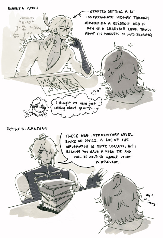
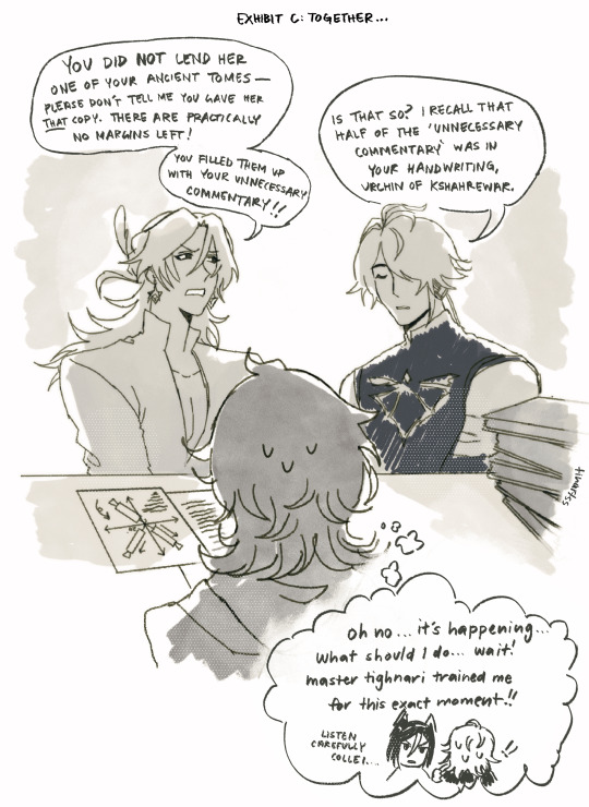
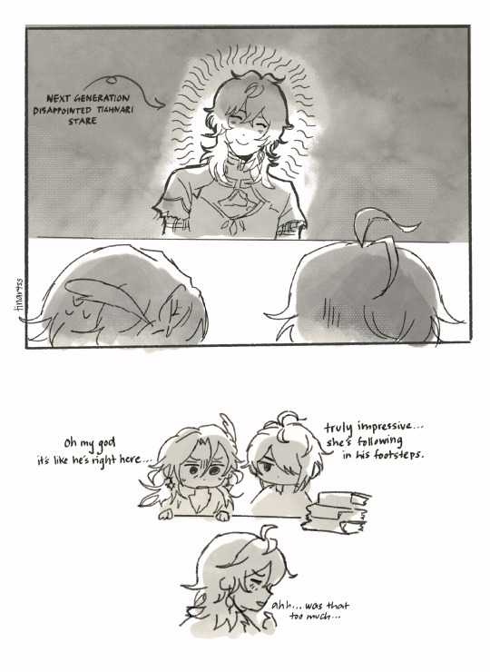
when you ask your uncles for help with your physics homework but they’re both geniuses (and also like the sound of each other’s voices way too much)
#collei#haikaveh#kavetham#alhaitham#kaveh#genshin impact#projecting btw im taking intro physics + light microscopy methods this sem and i was like woah…hkvthm ref….
4K notes
·
View notes
Text


i've been mixing flesh, wax, chemicals and dyes for months to obtain these images
#first one look like a skull??#flesh wizard#i modified them for esthetical purpose only here ahah#but maybe its gonna be my first paper omgg#ik its an artblog but can we agree that this count??#fluorescence#science#cells#biology#immunofluorescence#microscopy#medecine#medblr#pathology#skull#retrowave#synthpop#synthwave#neon light#microscope
274 notes
·
View notes
Text
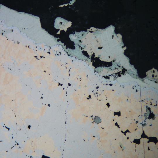
Nickeline (pink/peach) and Maucherite (pinkish grey) with a rim of Gersdorffite (bluish grey) overgrown by intergrown Parkerite (light grey, twinning) and Hauchecornite (dull pink), then overgrown by Matildite (darker grey, rough texture).
Locality: Easter-Duffy Deposit, East Arm Region, Greater Slave Lake, NWT, Canada
Reflected Plane Polarized Light, 200X Magnification
Quick laundry list of minerals most people will have never heard of!
#geology#rocks#minerals#mineralogy#mineral#canada minerals#earth#science#reflected light#microscopy#economic geology#parkerite#hauchecornite#matildite#gersdorffite#nickeline#maucherite#easter duffy#nwt#canada#greater slave lake#rare minerals
41 notes
·
View notes
Text

Nervous system of a juvenile sea star (Patiria miniata) about 1 cm wide. Labeled with an antibody against acetylated tubulin after optical clearing, and captured using a color-coded Z-projection.
By Laurent Formery (USA).
Light Microscopy Awards
#laurent formery#photographer#united states#light microscopy awards#micro photography#juvenile sea star#patiria miniata#nature
100 notes
·
View notes
Text

An opaque matrix usually makes for a pretty boring and uninformative thin section, unless you can polish it! This is a reflected light view of a partially metamorphosed iron sandstone. I love the way you can see the grains shifting!
10 notes
·
View notes
Text

A single atom can change the directional profile of the light emitted in scanning tunneling microscopes
Researchers from Madrid explain a phenomenon that allows the direction of light emission to be controlled at the atomic scale. The paper provides a detailed explanation of how the profile of the light collected in a scanning tunneling microscope (STM) experiments changes when the tip is placed on an atomic step. The properties of light in the far field are determined by what happens in the near field. The manipulation of light at the nanometer scale, below its wavelength, can be carried out in STM microscopes because the electromagnetic field is extremely confined between two metal nanostructures, the tip of the microscope and the sample, both separated by a typical distance of 1 nanometer. This configuration is called a nanocavity. If an element is introduced into this nanocavity, such as an atomic defect, the system becomes a picocavity and has unique properties. It has been observed that, by introducing atomic steps into the nanocavities, it is possible to modify the direction of light emission in the experiments. This phenomenon, which researchers had previously observed, lacked a scientific explanation until now.
Read more.
13 notes
·
View notes
Text


Some miracles of my life. 🫧
#art#personal tumblr#aesthetic#photography#photos of nature#photos#thailand#koh samui#photographers on tumblr#lights#microscopy#glitter
8 notes
·
View notes
Text
Delving in Detail
Live imaging using light sheet microscopy allows the depths of multicellular systems like organoids to be interrogated at the single cell level
Read the published research article here
Video from work by Franziska Moos & Simon Suppinger and colleagues
Friedrich Miescher Institute for Biomedical Research, Basel, Switzerland
Image originally published with a Creative Commons Attribution 4.0 International (CC BY 4.0)
Published in Nature Methods, March 2024
You can also follow BPoD on Instagram, Twitter and Facebook
11 notes
·
View notes
Text
A Journey into the World of Microscopy: From Humble Beginnings to High-Tech Magnification
The science of looking into the hidden invisible Microscopy has transformed our understanding of the world around us. It can explore the universe beyond the reach of our naked eyes, with complex cellular structures, red blood cells, viruses and other viruses and microorganisms taking on amazing perspectives
The history of the microscope is a fascinating story of human curiosity, scientific genius, and relentless exploration. From the humble beginnings of simple magnifying glasses to the sophistication of modern electronic microscopes, the invention of microscopes has shaped our understanding of the microscopic world
In the 1600s, Dutch opticians such as Hans and Zachary Janssen are credited with inventing the first microscope. Known for this hybrid microscope, many lenses were used to magnify objects up to 30 times.At the end of the 17th century, Antony van Leeuwenhoek, Dutch draper some changed our perception of thumbnails. Armed with a well-made single-lens microscope, and explored the hidden reaches of nature. In 1674, Leeuwenhoek discovered microorganisms in lake water, which he aptly named “animalcules”. His discovery laid the foundations of biology and inspired generations of scientists. This incredible feat allowed him to uncover a hidden universe – the first sightings of bacteria, red blood cells, and other microorganisms.
Formation of the scientific environment (17th-19th centuries): Leeuwenhoek’s discoveries boosted scientific research. Robert Hooke, an English scientist, established these developments. In 1665, his book "Micrographia" recorded his observations with a compound microscope. Notably, the term "cell" was coined by Hooke when he examined cork tissue, laying the foundation for cell biology.Microscope systems flourished throughout the 18th and 19th centuries Joseph Lister and other scientists addressed the limitations of the early lenses, introducing improvements that reduced image distortion.
Beyond the Limits of Light: The Beginning of the New Age (19th-20th century): As the 19th century progressed, the limitations of optical microscopy became apparent and scientists yearned for a tool which can go deeper into cells. This research culminated in the development of the electron microscope in the 1930s. The 20th century was revolutionary with the invention of the electron microscope. Unlike light microscopes, which use visible light, electron microscopes use electron beams to achieve much higher magnification.Formation of the scientific environment (17th-19th centuries): Leeuwenhoek’s discoveries boosted scientific research. Robert Hooke, an English scientist, established these developments. In 1665, his book "Micrographia" recorded his observations with a compound microscope. Notably, the term "cell" was coined by Hooke when he examined cork tissue, laying the foundation for cell biology.Microscope systems flourished throughout the 18th and 19th centuries Joseph Lister and other scientists addressed the limitations of the early lenses, introducing improvements that reduced image distortion.
Beyond the Limits of Light: The Beginning of the New Age (19th-20th century): As the 19th century progressed, the limitations of optical microscopy became apparent and scientists yearned for a tool which can go deeper into cells. This research culminated in the development of the electron microscope in the 1930s. The 20th century was revolutionary with the invention of the electron microscope. Unlike light microscopes, which use visible light, electron microscopes use electron beams to achieve much higher magnification.
In the 1930s, German experts Max Knoll and Ernst Ruska made the first electron microscope. This tool let us see tiny things like cells and even atoms by using electron beams, not light, getting images many times bigger. This cool invention showed us the tiny parts inside cells, viruses, and stuff too small to see before. The 1900s brought even more cool microscopes. New kinds like phase-contrast and confocal microscopy let scientists look at live cells without using stuff that could hurt them. Now, the world of looking at tiny things is getting even better. Today, we have high-tech microscopes that use computers and lasers. These let us see and even change tiny things in ways we never could before.
Modern Microscopy's Diverse Arsenal - Today, the field of microscopy boasts a diverse range of specialized instruments, each tailored to address specific scientific needs. Here's a glimpse into some remarkable examples:
Scanning Electron Microscope (SEM): Imagine a high-tech camera that captures images using a beam of electrons instead of light. That's the essence of a SEM. By scanning the surface of a sample with a focused electron beam, SEMs generate detailed information about its topography and composition. This makes them ideal for studying the intricate structures of materials like insect wings, microchips, and even pollen grains.
Transmission Electron Microscope (TEM): While SEMs provide exceptional surface detail, TEMs take us a step further. They function by transmitting a beam of electrons through a very thin sample, allowing us to observe its internal structure. TEMs are the go-to instruments for visualizing the intricate world of viruses, organelles within cells, and macromolecules like proteins.
Confocal Microscopy: Ever wished to focus on a specific layer within a thick biological sample and blur out the rest? Confocal microscopy makes this possible. It utilizes a laser beam to precisely illuminate a chosen plane within the sample, effectively eliminating information from out-of-focus regions. This allows researchers to create sharp, three-dimensional images of cells, tissues, and even small organisms.
Atomic Force Microscopy (AFM): This technique takes a completely different approach, venturing into the realm of physical interaction. AFM employs a tiny cantilever, akin to a microscopic feeler, to physically scan the surface of a sample. By measuring the minute forces between the cantilever and the sample's surface, AFM can map its topography at an atomic level. This provides invaluable insights into the properties of materials at an unimaginable scale, making it crucial for research in fields like nanotechnology and surface science.
Fluorescence Microscopy: Imagine illuminating a sample with specific wavelengths of light and observing it glowing in response. That's the essence of fluorescence microscopy. This technique utilizes fluorescent molecules or tags that bind to specific structures within a cell or tissue. When excited by light, these tags emit their own light, highlighting the target structures with remarkable clarity. This allows researchers to visualize specific proteins, DNA, or even pathogens within biological samples.
Super-resolution Microscopy (SRM): Overcoming the limitations imposed by the wavelength of light, SRM techniques like STED (Stimulated Emission Depletion) and PALM (Photoactivated Localization Microscopy) achieve resolutions surpassing the diffraction limit. This allows researchers to visualize structures as small as 20 nanometers, enabling the observation of intricate cellular machinery and the dynamics of individual molecules within living cells.
Cryo-Electron Microscopy (Cryo-EM): This powerful technique takes a snapshot of biological samples in their near-life state. Samples are rapidly frozen at ultra-low temperatures, preserving their native structure and minimizing damage caused by traditional fixation methods. Cryo-EM has been instrumental in determining the three-dimensional structures of complex molecules like proteins and viruses, providing crucial insights into their function and potential drug targets.
Correlative Microscopy: Combining the strengths of multiple microscopy techniques, correlative microscopy offers a comprehensive view of biological samples. For instance, researchers can utilize fluorescence microscopy to identify specific structures within a cell and then switch to electron microscopy to examine those structures in high detail. This integrated approach provides a deeper understanding of cellular processes and their underlying mechanisms.
Light Sheet Microscopy (LSM): Imagine illuminating a thin slice of a sample within a living organism. LSM achieves this feat by focusing a laser beam into a thin sheet of light, minimizing photobleaching and phototoxicity – damaging effects caused by prolonged exposure to light. This allows researchers to observe dynamic processes within living organisms over extended periods, providing valuable insights into cellular behavior and development.
Expansion Microscopy (ExM): This innovative technique physically expands biological samples by several folds while preserving their structural integrity. This expansion allows for better resolution and visualization of intricate cellular structures that would otherwise be difficult to distinguish using traditional microscopy methods. ExM holds immense potential for studying the organization and function of organelles within cells.
Scanning Near-Field Optical Microscopy (SNOM): This innovative technique pushes the boundaries of resolution by utilizing a tiny probe that interacts with the sample at an extremely close range. SNOM can not only image the surface features of a sample with exceptional detail but also probe its optical properties at the nanoscale. This opens doors for research in areas like material science and photonics, allowing scientists to study the behavior of light at the interface between materials.
X-ray Microscopy: Stepping outside the realm of light and electrons, X-ray microscopy offers unique capabilities. By utilizing high-energy X-rays, this technique can penetrate deep into samples, making it ideal for studying the internal structure of dense materials like bones and minerals. Additionally, it allows for the visualization of elements within a sample, providing valuable information about their distribution and composition.
From revealing the building blocks of life to aiding in the development of new medicines, the microscope has played an undeniable role in shaping our scientific understanding. As technology continues to evolve, one can only imagine the future breakthroughs this remarkable invention holds in unveiling the secrets of our universe, both seen and unseen. These advancements hold the potential to revolutionize our understanding of biological processes, develop new materials with extraordinary properties, and ultimately pave the way for breakthroughs in medicine, nanotechnology, and countless other fields. As we continue to refine and develop novel microscopy techniques and the future holds immense promise for further groundbreaking discoveries that will undoubtedly revolutionize our perception of the world around us.
#science sculpt#life science#science#molecular biology#biology#biotechnology#artists on tumblr#microscopy#microscope#Scanning Electron Microscope#Transmission Electron Microscope#Confocal Microscopy#Atomic Force Microscopy#Fluorescence Microscopy#Expansion Microscopy#X-ray Microscopy#Super-resolution Microscopy#Light Sheet Microscopy#illustration#illustrator#illustrative art#education#educate yourself#techniques in biotechnology#scientific research#the glass scientists#scientific illustration#scientific advancements
7 notes
·
View notes
Link
1 note
·
View note
Text
Meet the Founders: Mohs
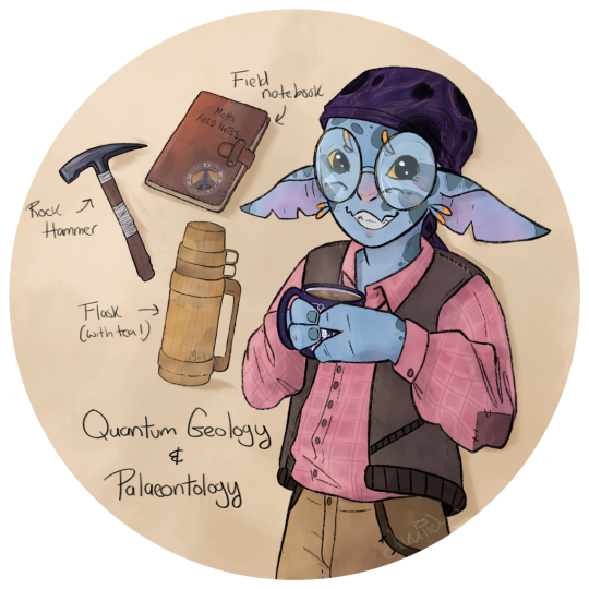
Outer Wilds Geological Survey Founder: Mohs, drawn by @dekkiidan Howdy! Thanks so much for stopping by the Outer Wilds Geological Survey database, it's always great to know there are folks out there interested in the amazing stories and secrets that our local, and not so local, geology can tell us!
Anyways, I'm Mohs - nice to meet you! I'm one of the founders of the survey alongside my good pal and esteemed colleague and pilot, Lari. Ah, yeah, most folks already know, I'm not a fan of flying . . . but that's why Lari is our pilot and not me. Besides, how can I solve the mysteries of the Quantum Shards with only the samples we have on Timber Hearth when there are more outcrops out there waiting to be analysed? Conclusion:- I can't! Unfortunately. And, you know, Lari hasn't crashed us! No yet, anyway.
Geology wise, my main areas of expertise lie in what most folks would call laboratory and technician work, so things like sample and specimen prep, sorting out thin sections for microscopy, and sawing large samples to get a nice clean surface for proper observational analysis. This is why you're more likely to find me in the museum prep. room than out in the field. Don't get me wrong, I love field work too, especially when Lari and I are wrangling a particularly tricky outcrop, or if there are fossils involved; but somebody has to ensure the survey sample and field note collections are properly organised, analysed and documented. And, I'm not sure if you've ever seen Lari or Hornfels' own collections, whether that be geological, astronomical or research based, but - well let's just say, I don't think they're the right hearthians for keeping things organised. Sorry Lari! Sorry Hornfels!
I'm also currently working on a research paper and presentation regarding Quantum Geology that has recently come along in leaps and bounds thanks to Hal and the hatchling's translation tool titled -
"Quantum Geology: A morphological mystery, or lost in translation?"
Because, you know, based on some of the latest translations, I really don't think the Nomai fully understood the complex nature of these curious shards, and I would love to crack the mystery of their formation and origins!
Ah, apologies, I got a little carried away there, heh! Well, if you ever have any questions, don't hesitate to get in touch with either of us, we'll do our best to shed some light on the mysteries and puzzles of the amazing geological history of our solar system! And if you have any questions about Quantum Geology, or even want to share theories, I'm all ears!
Thanks again for stopping by!
57 notes
·
View notes
Note
"don't leave" from the microscopy prompts place 🥺
"Don't leave." Her voice was soft when she called out to him. Usually larger than life, she looked so very small in her nest of blankets and tears.
A warm hand, devoid of the gold she should wear, reached out and he caught it firmly between his ringed ones. The jingle of his bracelets made her smile, even if only for a brief moment.
She was terrified. The terror crawled out of her face and devoured the light that clung to her.
Emmrich sat down on the bed again and kissed her hand. Soft. Intently. Keeping his eyes on hers.
Did she not know how loved she was? If he could have made himself leave at any moment in their shared story, it had fled many moments before.
Maybe it never had been there at all.
So he did the only thing he could think of to assure her. He unfastened his boots, shed away the layers of propriety, folding them neatly to rest on the chair and laid down next to her.
He held her tight, all night and whispered promises in her hair.
"I will never leave you, darling."
#Emmrook#Mikroprompts#emmrich x rook#Emmrich Volkarin#Rook#Look this is Siobhan to me but feel free to think of any Rook#dragon age the veilguard
24 notes
·
View notes
Text

Autofluorescence of the tip of the stamens of flowers at 405 nm, 488 nm, and 561 nm, emitting a greenish blue and emerald green color like peacock feathers.
By Mei Yu (China)
Light Microscopy Awards
#mei yu#photographer#china#light microscopy awards#micro photography#autofluorescence of the tip of the stamens of flowers#nature
52 notes
·
View notes
Text

An opaque matrix usually makes for a pretty boring and uninformative thin section, unless you can polish it! This is a reflected light view of a partially metamorphosed iron sandstone. I love the way you can see the grains shifting!
5 notes
·
View notes
Text

Native Silver (tarnished yellow) overgrown by Nickeline (pink) and Rammelsbergite (whitish grey rim on Nickeline) and Gersdorffite (light grey, euhedral crystals) and partially replaced by intergrown Acanthite (dull, scratched grey) and Stephanite (slightly lighter, "cleaner" grey)
Locality: Easter-Duffy Deposit, Greater Slave Lake, Northwest Territories, Canada
Plane Polarized Reflected Light, 50X Magnification
Have not had time to take new pictures of my newer rocks, as I have been super busy working on my MSc, so I thought I would upload some reflected light images from my thesis! If it seems like these are something folks want to see, I have loads more where this came from. Plus tumblr has a lack of reflected light microscopy images anyways. Feel free to reach out if you have any questions about this stuff!
#native silver#silver#nickeline#rammelsbergite#acanthite#stephanite#gersdorffite#nwt#canada#geology#mineralogy#earth#nature#science#reflected light#petrography#ore deposit#microscope#thin section#mineral#rock#fossils#minerals
99 notes
·
View notes