#neurohypophysis
Text
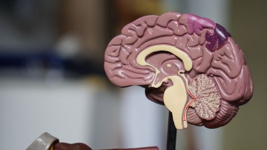
How does one identify the location of the pituitary gland in the brain?
The pituitary gland, also known as the hypophysis, is a small, pea-sized gland located at the base of the brain. It is often referred to as the “master gland” because it controls the function of other endocrine glands in the body. Despite its small size, the pituitary gland plays a critical role in maintaining a wide range of bodily functions, including growth and development, metabolism, and…
View On WordPress
#adenohypophysis#Anatomy#brain#computed tomography#diagnosis#endocrine system#Health#hormones#imaging techniques#magnetic resonance imaging#nasal endoscopy#neurohypophysis#pituitary disorders#Pituitary gland#symptoms#treatment
0 notes
Text
why didnt our brain made it easier to study it bro im having a stroke reading hypothalamus, neurohypophysis, vasopressin and nucleus supraoptic all in one sentence
11 notes
·
View notes
Text
0 notes
Quote
Many neuropeptides were originally identified as pituitary or gastrointestinal hormones
Probably the first neuropeptide to be identified was vasopressin, a nine-amino-acid peptide secreted by the nerve endings in the neural lobe of the pituitary. The source of the vasopressin is the magnocellular neurons of the hypothalamus, which send axons to the neurohypophysis, which is the site of release into the blood, in classic neurosecretory fashion. Like vasopressin, a number of gastrointestinal peptides, such as cholecystokinin (CCK), are also found at high concentrations in the nervous system. In the gastrointestinal (GI) system, CCK is secreted by the duodenum and governs the delivery of digestive enzymes and bile acids into the intestine. In contrast to vasopressin and CCK, the hypothalamic releasing factors are peptides released into a special portal blood system that bathes the anterior pituitary, controlling the secretion of pituitary hormones. In this system, “portal” means two successive capillary beds, one in the hypothalamus and one in the anterior pituitary. Substance P was first purified as a “sialogogic peptide,” causing salivation in a bioassay. Now substance P is recognized as a major bioactive peptide in many neuronal pathways, including pain signaling. Since there are so many peptides, this chapter focuses on the principles of how neuropeptides are synthesized, stored and released and how they act on the cells they regulate. Comparisons among peptides and smaller, “conventional” neurotransmitters will be emphasized. It is significant to note that the number of known neuropeptides far exceeds the number of classical neurotransmitters.
The Neuropeptides - Basic Neurochemistry - NCBI Bookshelf
0 notes
Link
Vasopressin is a nonapeptide that is produced in the paraventricular and supraoptic nuclei of the posterior hypothalamus as a prohormone. It travels down the supraoptic hypophyseal tract, attached to the carrier protein neurohypophysis, to the axonal terminals of magnocellular neurones in the posterior pituitary. Takes 1-2 hours for synthesis, delivery, and storage. 4 pg ml-1 are the normal plasma values. It is broken down by vasopressinases, which are present in the liver and kidney, and has a half-life of 10-35 minutes. Vasopressin impurities interact with oxytocin, V1, V2, and V3 type receptors (OTR).
Vascular smooth muscle in the systemic, splanchnic, renal, and coronary circulations contains V1 receptors. They can be found on platelets and myometrium as well.
The pituitary is the primary location of V3 receptors. They are Gq-coupled G-protein receptors, and when they are active, they raise intracellular calcium. They are believed to play a role in the production of ACTH and may function as a neurotransmitter or mediator in the consolidation or retrieval of memories as well as the control of body temperature. 1, 2
0 notes
Photo

A helpful mnemonic to remember the functions of the hypothalamus.
2 notes
·
View notes
Text
Human Hormones and their properties
The Endocrine System is the collection of glands and organs that produce hormones which helps in the chemical co-ordination and integration in the human body. The following image shows various glands and their hormones.

Properties of hormones
These are discharged by endocrine organ (biogenic in source).
Their discharges is delivered straightforwardly into blood (aside from nearby hormones for example gastrin).
These are conveyed to remotely find explicit organs, called target organ.
These have explicit physiological activity (excitatory or inhibatory). These co-ordinate diverse physical, mental and metabolic exercises and look after homeostasis.
The hormones have low sub-atomic weight for example ADH has an atomic load of 600–2000 daltons.
These demonstration in extremely low focus for example around10–10 molar.
Hormones are non antigenic.
These are generally fleeting. So have a no camulative impact.
Some hormones are snappy acting for example adrenalin, while some acting gradually for example ostrogen of ovary.
Some hormones emitted in dormant structure called Prohormone for example Favorable to insulin.
Hormones are explicit. They are transporters of explicit data to their particular objective organ. Just those objective cell react to a specific hormone for which they have receptors.
Pituitary Gland (Hypophysis)
(1) Pituitory is known as hypophysis cerebri, its name pituitary was given by vesalius.
(2) Muller's organ of amphioxus and subneural organ of hardmania is homologous to pituitary of vertebrates.
Parts and segment
(1) Adenohypophysis (Anterior projection)
Pars distalis
Pars tuberalis
Pars intermedia
(2) Neurohypophysis (Posterior projection)
Pars nervosa
Infundibulum
Hypothalamus
1. Position and Structure: Hypothalamus is the floor of diencephalon. It is shaped of masses of dark issue, called hypothalmic cores, containing neurosecretory cells. It is associated with front pituitary projection by blood vessels of hypophyseal entryway framework and with the back pituitary flap by axons of its neurons, both going through the pituitary tail.
2. Hormones of hypothalamus: Neurosecretory cells of hypothalamus emit neurohormones called delivering factors (RF) or restraining factors (IF). These neurohormones are conveyed by hypophyseal gateway framework to adenohypophysis (essential objective organ) and animate or restrain the arrival of trophic hormones from adenohypophysis. These neurohormones are proteinous in nature and shaped of 3 – 20 amino acids.
Thyroid organ
1. The name "thyroid" was presented by Thomas Wharton (1656).
2. It is gotten from Greek "Thyreos" a shield.
3. Location: This is the biggest endocrine organ of our body. It is situated in our neck upon the ventral part of larynx (sound box or Adam's apple) and a couple of foremost most tracheal rings. It is a dim earthy colored and H-molded bilobed organ.
4. Hormones of thyroid:
Thyroid organ secretes two iodinated hormones.
(a) Thyroxine: It is an iodine containing (6% iodine) amine hormone which is gotten from tyrosine amino corrosive. Chemically thyroxine is tetraidothyronine however likewise found as tri-iodothyronine. Emission of thyroxine is contrarily relative to the blood level of thyroxine (criticism system).
(b) Thyrocalcitonin (TCT): It is a long peptide hormone discharged by parafollicular by cells of thyroid organ (C-cells). It discharge is managed by expanded plasma level of calcium by criticism instrument.
5. Irregularities of thyroid organ
(a) Hypothyroidism: (Decreased segment of thyroxine from thyroid organ).
(b) Hypersecretion of thyroid hormones (Hyperthyroidism or thyrotoxicosis) : This may likewise be a hereditary imperfection, however for the most part it is given by interminable contaminations (flu, stiffness, tonsilitis, tuberculosis, measles, challenging hack, and so on.) pregnancy, admission of huge dosages of iodine, over-eating, and so forth. It results into an extensive increment in glucose and oxygen utilization by cells and the pace of oxidative digestion in the mitochondria.
Parathyroid organ
1. Hormones of parathyroid: Active hormone emitted by parathyroids is parathormone (PTH), additionally called Collip's Hormone (Phillips collip, 1925).
2. Irregularities of parathormones
(a) Hypoparathyroidism (Hyposecretion of parathormone)
(b) Hyperparathyroidism (Hypersecretion of parathormone)
Pancreas
1. Hormones of pancreas and their job:
(a) Insulin: Insulin manages how the body uses and stores glucose and fat.
(b) Glucagon: This is discharged by the alpha cells of islets of Langerhans. Its capacity is to hoist glucose level in blood when glucose is insufficient.
(c) Somatostatin and Pancreatic polypeptide: Modern physiologists have proposed that the d and F (PP) cells of pancreas separately discharge somatostatin (SS) and pancreatic polypeptide (PP). Somatostatin takes after the development hormone inhibitory hormone (GHIH) emitted by hypothalamus.
Thymus organ
1. Function of thymus organs
Thymus is haemopoietic, just as, an endocrine organ.
The significant capacity of thymus is to emit thymosin hormone, thymic humoral factor (THF), thymic factor (TF), thymopoietin.
Thymus is basic in neonatal (recently conceived) baby and postnatal kid for ordinary advancement of lymphoid organs and cell invulnerability.
Gonads
1. The gonads are the sex organs, the testicles and the ovary.
(i) Testes
The testis structure part of the male conceptive framework, and is where sperm and testosterone are created.
Elements of Testes
It animates the male conceptive framework to develop to full measure and become utilitarian.
It invigorates the arrangement of sperms (spermatogenesis) in the seminiferous tubules.
It additionally decides the male sexual conduct sex encourage, forceful conduct.
Under its impact protein anabolism increments.
(ii) Ovary
The ovary is a ductless conceptive organ in which the female regenerative cells are delivered.
2 notes
·
View notes
Text

🇺🇸 Patriotic Pituitary 🇺🇸
There's a little Red (acidophils) White (chromophobes) & Blue (basophils) in us all!
Happy 4th of July everyone!
i❤️histo
———
The pituitary gland (hypophysis) is the king of the endocrine glands! It is so important that it sits in its own throne on the floor of the skull called the sella turcica where is sends out hormones to control the entire body via the blood stream.
It has two distinct parts:
An anterior pituitary (adenohypophysis) which is glandular and colorful and a posterior pituitary (neurohypophysis) which is pale and subdued because it is composed of the axons of nerves that reside in the hypothalamus of the brain.
The patriotic adenohypophysis (shown here) constantly flies its red, white and blue flag. Cheering on the organs of the body by sending them signalling molecules.
The red cells are acidophils: they make Growth Hormone (to urge our bodies to grow) and Prolactin (to make sure milk is produced during pregnancy).
The white cells are chromophobes: they don’t have a clearly understood function in the pituitary.
The blue cells are basophils: they produce Thyroid Stimulating Hormone (TSH - which excites the secretory cells of the thyroid gland), Follicle Stimulating and Luteinizing Hormone (FSH, LH - these are important in reproductive processes in the body) and Adrenocorticoptrophic Hormone (ACTH - which excites the secretory cells of the adrenal cortex).
#histology#anatomy#pathology#science#4th of july#independence day#med school#med student#vet science#vet school#premed#med lab#nursing#nurse#dental student#dental school#dental#histotech#histopathology#endocrine#pituitary#ihearthisto
116 notes
·
View notes
Text
3-year Postdoc position
Institute of Molecular Biology & Biotechnology, Faculty of Biology
Application Deadline: 2022-08-31
3-year Postdoc position
We are seeking a highly motivated Postdoctoral fellow to join the Zebrafish developmental neurobiology research group led by Dr Savani Anbalagan at the Adam Mickiewicz University, Poznań, Poland.
In mammals, neurohypophysis (NH) is a major CNS neuroendocrine organ that regulates peripheral body functions ranging from milk let-down to water retention. How hypothalamic axons reach NH and create synapses to enable NH function is relatively understudied. The project will take advantage of the genetic amenability of zebrafish and optical transparency to understand how NH-derived signals (secreted factors and microRNAs) can regulate NH development. Using our recently developed CRISPR mutants and transgenic fish lines, we will address these questions. We will use advanced light microscopy techniques for characterizing the phenotypes.
Requirements:
· PhD in life science field awarded on or after 2016* or recently defended PhD thesis.
· High motivati...
See the full job description on jobRxiv: https://jobrxiv.org/job/institute-of-molecular-biology-biotechnology-faculty-of-biology-27778-3-year-postdoc-position/?feed_id=21135
#ScienceJobs #hiring #research #zebrafishjobs
0 notes
Text
Open Access journals on surgery- BJSTR Journal

Intranasal Spray of Oxytocin Might Improve Working Memory Performance in Alzheimer, Tracking with Diffusion Tensor Imaging by Fatemeh Bakouie* in Biomedical Journal of Scientific & Technical Research https://biomedres.us/fulltexts/BJSTR.MS.ID.001818.php
Alzheimer's disease (AD) is a neurodegenerative process; manifests as episodic memory loss and working memory impairment [1]. Prefrontal cortex plays a key role in working memory which depends on modulating transmitters, dopamine and gamma-aminobutyric acid (GABA) [2,3]. The neurohypophysial hormone, oxytocin acts on lactation, parturition, modulating stress responses, pain perception, learning and different aspects of social behavior. Investigators suggest that oxytocin has potential therapeutic effects on depressive disorders because it induces hippocampal neurogenesis- a process altered in depression [4]. In this regard intranasal oxytocin administration found to be the most effective way to access to the central nervous system [5]. Dopamine concentrations in Alzheimer patients decreased (about 18-27%) in temporal and hippocampal cortices [6]. In addition to dopamine, significant reduction in GABA level has been identified in AD [7]. A recent study suggested that administration of single oxytocin dose has improved "executive component" of working memory (component contributing in information maintenance plus manipulation) in schizophrenic patients [8]. One of the oxytocin analogues induces GABAergic transmission [9]. Prefrontal cortex dopamine amounts might multiply upon oxytocinergic stimulation.
For more Articles on Open Access journals on surgery Please Click Here
For bjstr
Follow on Twitter : https://twitter.com/Biomedres01
Follow on Blogger : https://biomedres01.blogspot.com/
Like Our Pins On : https://www.pinterest.com/biomedres/
#Journals on Medical drug and theraputics#Journals on Medical Casereports#Journals on Medical Microbiology#Journals on Biomedical Intervention#Journals on Biomedical Imaging
0 notes
Text
300+ TOP PHYSIOLOGY Objective Questions and Answers
PHYSIOLOGY Multiple Choice Questions :-
1. Damage to sensory area 1 of the cerebral cortex results in
a) Loss of perception of pain
b) Loss of tactile and two point discrimination
c) Loss of perception of touch
d) Loss of only tactile discrimination
Ans:b
2. Volume of CSF is about
a) 50ml
b) 100ml
c) 150 ml
d) 200 ml
Ans:c
3.Dissymmetria is seen in
a) Extra pyramidal lesions
b) Cerebellar lesions
c) Pyramidal lesions
d) Cortical lesions
Ans:b
4.Floculonodular lobe of cerebellum is concerned with
a) Equilibrium
b) Co-ordination
c) Baroreception
d) Chemoreception
Ans:a
5.In the postnatal period the greatest growth in the grey matter of the C.N.S is of a) Neuron cell number
b) Length of axon
c) Dendritic tree
d) Size of Perikaryon
Ans:c
6.CSF production per minute
a) 0.30-0.35 ml/min
b)0.5ml/min
c)2ml/min
d) 1 ml/min
Ans:a
7.Function of GABA on CNS is
a) Neuronal inhibition
b) Neuronal activation
c) Glial cell inhibition
d) Glial cell activation
Ans:a
8.The EEG rhythm having lowest frequency is:
a) Alpha
b) Beta
c) Delta
d) Theta
Ans:c
9.Medial geniculate body is concerned with
a) Hearing
b) Vision
c) Smell
d) Taste
Ans:a
10.True statement regarding CSF is
a)Daily production b)CSF analysis rules out active secretion as a cause of formation of CSF
c)It flows from III ventricle to the IV ventricle
d) Produced only by choroid plexus
Ans:c

PHYSIOLOGY MCQs
11. In cerebellar disease, all the statements are correct except
a) The Romberg's sign is positive
b) There is Adiodokokinesia
c) There is pendular knee jerk
d) There is involuntary tremor
Ans:d
12.Swallowing center is situated in
a) Midbrain
b) Pons
c) Medulla
d) Cerebellum
Ans:c
13.Hyper kinetic syndromes such as chorea and athetosis are usually associated with pathological changes in:
a) Motor areas of cerebral cortex
b) Anterior hypothalamus
c) Pathways for recurrent collateral inhibition in the spinal cord
d) Basal ganglia complex
Ans:d
14. An EEG:
a)Provides indication of intelligence
b)Tends to show waves of smaller amplitude during deep sleep than of alert state
c)Show waves with a lower frequency during intense thought than during sleep
d)Is bilaterally symmetrical
Ans:d
15.The following is true about brain metabolism except
a) Use fatty acid in starvation
b) In resting state 60% of total energy utilized
c) Ketone bodies are used in starvation
d) Has no energy store
Ans:a
16.Satiety center in hypothalamus is regulated by:
a) Gastric dilatation
b) Blood glucose levels
c) Blood insulin leve
d) All of the above
Ans:b
17. In hippocampus EEG waves are
a) Alpha wave
b) Beta wave
C) 65 mm of Hg
b)55mmofHg
Ans:d
18.EEG with spike and dome pattern is characteristic of epilepsy
a) Jacksonian
b) Grandmal
c) Petitmal
d) Temporal lobe
Ans:c
19.Buerger waves (alpha waves) of EEG have the rhythm per sec of:
a) 0-4
b)4-7
c)8-13
d) 13-30
Ans:c
20.All the following are more in CSF compared to plasma except
a)Mg
b)Cl
c)HC03
d) Glucose
Ans:d
21. During light sleep, the sleep spindles that appear have the frequency of:
a) 1 -2/sec
b) 6-12/sec
c) 14-16/sec
d) 21 -26/sec
Ans:c
22.Up to what systolic pressure is the brain capable of auto regulation
a) Dorsomedian nucleus of hypothalamus
b) Ventromedian nucleus of hypothalamus
c) Perifornical region
d) 75mmofHg
Ans:a
23. pH of CSF is:
a) 7.13
b) 7.23
c) 7.33
d) 7.40
Ans:b
24.Temporal lobe lesion causes:
a) Homonymous upper quadrantinopia
b) Homonymous lower quadrantinopia
c) Bitemporal hemianopia
d) Binasal hemianopia
Ans:a
25.Hyperphagia results from lesion of
a) Thalamus
b) Cerebral cortex
c) Ventromedial region of supra-optic nucleus
d) Ventrolateral region of supra-optic nucleus
Ans:c
26. Delta waves in EEG are seen in
a) Deep sleep
b) REM sleep
c) Awake with eyes open
d) Awake with eyes closed
Ans:a
27. Nightmares are seen in
a) REM sleep
b) NREM stage II
c) NREM stage III
d) NREM stage IV
Ans:a
28. Which of the following are true of median eminence
a) Portion of ventral hypothalamus
b) Hypothalamo hypophyseal vessels arise here
c) Outside the blood-brain barrier
d) All are correct
Ans:d
29. Unconscious kinesthetic sensations are carried by
a) Posterior columns
b) Ventral spinothalamic tract
c) Anterior spinothalamic tract
d) Lateral spinothalamic tract
Ans:a
30. Crossed extensor reflex is a
a) Withdrawal reflex
b) Postural reflex
c) Monosynaptic reflex
d) Sympathetic reflex
Ans:a
31.Renshaw cell inhibition is an example of
a) Feed-forward inhibition
b) Oscillating motor activity
c) Circuitry for bio feedback
d) All of the above
Ans:a
32.Arousal response is mediated by
a) Dorsal column
b) Reticulo activating system
c) Spinothalamic tract
d) Vestibulo cerebellar tract
Ans:b
33.All are seen in a spinal reflex except
a) Summation
b) Fatigue
c) Memory
d) Adaptation
Ans:b
34.First change to occur in the distal segment of cut nerves
a) Myelin degeneration
b) Axonal degeneration
c) Mitosis of Schwann cell
d) Sprouting
Ans:b
35.Maximum increase in pain threshold occurs in which phase of sleep
a) Phase I
b) Phase 3
c) Phase 4
d) REM sleep
Ans:d
PHYSIOLOGY Objective Questions with Answers
36.Below pressure, CSF absorption stops
a)60mmCSF
b)68mmCSF
c)80mmCSF
d)50cmCSF
Ans:b
37.Phagocytosis in the CNS is done by
a) Astrocytes
b) Schwann cells
c) Microglia
d) Oligocytes
Ans:c
38.The condition known as REM sleep is:
a)That point at which the individual becomes aware and alert
b)Referred to paradoxical sleep
c) Characterized by total lack of all muscular activity
d) Characterized by slow high voltage regular EEG activity
Ans:b
39.Gag reflex is mediated by cranial nerve:
a)VH
b)IX
c)X
d)XII
Ans:b
40.Hypertonia indicates
a) Upper motor neuron injury
b) Lower motor neuron injury
c) Cerebellar lesion
d) Autonomic imbalance
Ans:a
41.What is not true for respiration center?
a)Situated in the medulla and pons
b)Sends out regular bursts of impulses to expiratory muscles during quiet respiration
c)sends out regular impulses to expiratory muscles during quiet respiration
d)Is inhibited during swallowing and vomiting
Ans:c
42.In brain ischaemia, systemic blood pressure rises, this is called:
a) Monro-kellie doctrine
b) Cushing reflex
c) Auto regulation
d) White reaction
Ans:b
43.Vestibular fibers relay in
a) Vermis
b) Lateral geniculate body
c) Floculonodular lobe of cerebellum
d) Auditory cortex
Ans:c
44.Vomiting center is situated in the
a) Hypothalamus
b) Amygdala
c) Pons
d) Medulla
Ans:d
45. Broca's area
a) Is situated in temporal lobe
d) It is synonymous to Wernicke's area
c) Is an area of hearing
b) Is supplied by middle cerebral artery
Ans:c
46.Crude touch sensations are carried by
a) Lateral spinothalamic tract
b) Posterior columns
c) Ventral spinothalamic tract
d) P)tramidal tract
Ans:c
47.One of the following is a function of hypothalamus
a) Swallowing
b) Vomiting
c) Respiration
d) Homeostasis of temperature
Ans:d
48.Blood brain barrier is maximum permeable to:
a)Na+
b)K+
c) Chloride
d)CO,
Ans:d
49.The basic postural reflex is
a) Crossed extensor reflex
b) Golgi tendon reflex
c) Flexor reflex
d) Positive supporting reflex
Ans:c
50.All of the following manifestations are seen in cases of cerebellar damage in human beings except
a) Loss of non-declarative/reflexive memory
b) Loss of adjustment of ve9tibulo-ocular reflex
c)Static tremor and rigidity
d) Ataxia, atonia and asthenia
Ans:c
51.Which of the following reflexes disappear in the absence of functional connections between the spinal cord and the brain?
a) Swallowing reflex
b) Seating reflex
c) Withdrawal reflex
d)Erection of penis
e) All of the above
Ans:a
52. In the Neurohypophysis, secretory granules accumulate in:
a) Pituicytes
b) Nerve endings
c) Intercellular spaces
b) Association fibers
Ans:b
53. Conduction in which type of nerve fibers is blocked maximally by pressure
a) C fibers
b) A-alpha fibers
c)A-Beta
d)A-gamma
Ans:c
54.Prosapagnosia is
a) Inability to recognize faces
b) Inability to draw
c) Inability to count
d) Inability to smell
Ans:a
55. Pain sensitive part in CNS is
a) Durameter
b) Piameter
c) Brain
d) Pial vessels
Ans:a
56.Wallenberg degeneration is seen in
a) Proximal cut end of nerve with cell body
b) Distal cut end of nerve without cell body
c) Both the free ends of the cut nerve
d) All are true
Ans:a
57.When sensory area-I of the cerebral cortex is ablated
a) Perception of pain is completely abolished
b) Perception of touch is completely abolished
c)There is loss of tactile localization but two point discrimination is not bolished
d)There is loss of tactile discrimination as well as two-point discrimination
Ans:d
58.Injection of hypertonic saline into which area causes diuresis
a) Supraoptic nucleus
b) Paraventricular nucleus
c) Preoptic nucleus
d) Posterior pituitary
Ans:a
59.A unilateral upper motor neuron lesion in the internal capsule is best characterized by
a)Diminished use of contra lateral appendages below the lesion
b)Muscle fasciculations
c)Ipsilateral hypotonicity
Ans:a
60. Source of EEG
a) A potential of pyramidal cells
b) A potential of ganglion cells
c)EPSP and IPSP of cortical cells which behave like dipoles
d)After potentials of parietal cortex
Ans:c
61. Decerebrate animal results from the following experimental procedure:
a) Removal of the cerebrum
b) Tran section at the upper border of midbrain
c) Inter collicular transection
d) Section above the thalamus
Ans:c
62.CSF pressure (lumbar)
a)70-180mmCSF
b)50-100mmCSF
c)>200mmCSF
d) 150-250 mm CSF
Ans:a
63.Which of the foUowing sensation is not carried through posterior column?
a) Touch
b) Proprioception
c) Visceral pain
d) All of the above
Ans:c
64. Stereo anesthesia is due to lesion of
a) Nucleus Gracillis
b) Nucleus cuneatus
c) Spinoreticular tract
d) Subarachnoid space
Ans:b
65.Broca's area is present in
a) Superior temporal Gyrus
b) Precentral gyrus
c) Post central gyrus
d)Inferior frontal gyrus
Ans:d
66.The cerebellum:
a)Has a totally inhibitory output from its cortex
b)Has only excitatory signal output from its deep nuclear layers
c)Has conscious interpretation of motor activity
d)Has inhibitory influence on muscle tone in humans
Ans:a
67.Hypothalamus regulates all except
a) Food intake
b)Temperature
c) Anticipatory rise in heart rate
d) Hypophysis
Ans:c
68.Loss of feel of size & shape of a object is seen in lesions of
a)Tractus solitarius
b) Tractus cuneatus
c) Lateral spinothalamic tract
d)Spinoreticular tract
Ans:b
67.The reticular formation is a diffuse collection of:
a) Only sensory neurons
b) Only motor.neurons
c) Only autonomic centers
d) All the above
Ans:d
68.CSF is principally secreted by
a) Choroid plexus
b) Arachnoid granulation
c) Floor of fourth ventricle
d)Periaqueductal grey
Ans:a
69.Paralysis agitans is accompanied by imbalance of neurotransmitter contents in
a) Globus pa'llidus and substantia nigra
b) Locus ceruleus
c) Putamen
d) Caudate nucleus and subthalamic body
Ans:a
70.In Rrown-Sequard syndrome sensation, that is lost at the same side of lesion is
a) Pain
b) Touch
c) Temperature
d) Proprioception
Ans:d
PHYSIOLOGY Questions and Answers pdf Download
Read the full article
0 notes
Text
I took a test today in my human anatomy class for the lab. One of the classes had a human head with a midsaggital cut, with a pin stuck into the fleshy contents of the sella turcica, basically asking 'what is this' The pituitary gland. Good old buddy pituitary gland. But I could not FUCKing think of the word pituitary! I'm sitting there, like ... it's not the pineal gland ... it's not the pulmonary gland, that's not even a thing ... So what I end up putting for my answer is 'the gland inside the sella turcica, connected to the hypothalamus by the infindibulum, aka hypothalamal-hypophyseal tract; also known as the neurohypophysis (well half of it is) ... but I cannot think of the FREAKING name! It starts with a p and I'm writing down all of the information I know about it in hopes for half credit =( ' And then I drew a picture of the pituitary gland,2/3s of it labeled 'neurohypophysis' and the other part 'adenohypophysis', with a stalk leading out with an arrow saying 'to hypothalamus,' with "WHAT IS YOUR NAME" written near it.
3 notes
·
View notes
Video
youtube
Learn how to pronounce Neurohypophysis in English --- NEUROHYPOPHYSIS Pronunciation of Neurohypophysis: /-hī-ˈpä-fə-səs/ noun Definition of Neurohypophysis: the portion of the pituitary gland that is composed of the infundibulum and posterior lobe and is concerned with the secretion of various hormones ★ http://Learn2Pronounce.com ★ How to pronounce Neurohypophysis | English pronunciation: https://youtu.be/QAsBnsT8sCw
#how to pronounce How to pronounce Neurohypophysis | English pronunciation#pronunciation of How to pr
0 notes
Text
Vocabulary (pt.dccl)
Words taken from the AMA Manual of Style: A Guide for Authors and Editors, 10th edition, by the editors of JAMA and the Archives journals (Oxford University Press, 2007):
naturopathic (adj.)
of or related to naturopathy.
neonate (n.)
a newborn child, especially an infant less than four weeks old.
non-proprietary (adj.)
not registered or protected as a trademark or brand name; generic.
neuroleptic (adj.)
tending or able to reduce nervous tension by depressing nerve function.
Norwalk virus (n.)
a virus that can cause epidemics of gastroenteritis in humans, with severe vomiting and diarrhea usually lasting 24 to 48 hours.
nitrogen mustard (n.)
chemistry. any of a group of organic compounds containing the group —N(CH2CH2Cl)2. They are powerful cytotoxic alkylating agents and some are used in chemotherapy to treat cancer.
nephrectomy (n.)
the surgical removal of a kidney.
nephritis (n.)
inflammation of the kidneys.
nausea (n.)
a feeling of sickness with an inclination to vomit.
neurohypophysis (n.)
the posterior lobe of the hypophysis (pituitary gland), which stores and releases oxytocin and vasopressin produced in the hypothalamus.
0 notes
Text
Adenohypophysis = anterior pituitary.
Neurohypophysis = posterior pituitary.
0 notes
Photo

The Patriotic Pituitary
There’s a little Red (acidophils), White (chromophobes) and Blue (basophils) inside each and everyone of us!
i❤️histo
The pituitary gland (hypophysis) is the king of the endocrine glands! It is so important that it sits in its own throne on the floor of the skull called the sella turcica where is sends out hormones to control the entire body via the blood stream.
It has two distinct parts:
An anterior pituitary (adenohypophysis) which is glandular and colorful and a posterior pituitary (neurohypophysis) which is pale and subdued because it is composed of the axons of nerves that reside in the hypothalamus of the brain.
The patriotic adenohypophysis (shown here) constantly flies its red, white and blue flag. Cheering on the organs of the body by sending them signalling molecules.
The Red cells are acidophils:
They make Growth Hormone (to urge our bodies to grow) and Prolactin (to make sure milk is produced during pregnancy).
The White cells are chromophobes:
They don’t have a clearly understood function in the pituitary, but are perhaps a stem cell population for the other pituitary cells.
The Blue cells are basophils:
They produce Thyroid Stimulating Hormone (TSH - which excites the secretory cells of the thyroid gland), Follicle Stimulating and Luteinizing Hormone (FSH, LH - these are important in reproductive processes in the body) and Adrenocorticoptrophic Hormone (ACTH - which excites the secretory cells of the adrenal cortex).
Happy 4th everyone!
#histology#pathology#science#fourth of july#pituitary#endocrine#hormones#med school#med student#premed#vet science#vet school#nursing#ihearthisto
20 notes
·
View notes
Last Seen Blogs

rhondaklewis
Untitled
thehearttaker
TheHeartTaker

cipap
cipap@tumblr

astronaut-mlm
I'm gay and like space

pp-garage
Untitled