#phagocytosis
Text
Me: watches an anime serie in order to learn more about the immune system.
The anime serie:

#it's very informative tho#teaches more than school#cells at work#neutrophil#phagocytosis#immune system
29 notes
·
View notes
Text
gay microbe eatin a burger call that faggocytosis
#worst post ive ever made#🐞 || beetle babbles#uhh#🐞 || beetle's bad biology#?i guess#actually fuck yall this is goin in the main tags#biology#phagocytosis#sciblr#f sluOH MY GOD THERE ARE SO MANY F SLUR TAGS#f slur#f slur tw#f slur cw#f slur reclaimed
5 notes
·
View notes
Text
🦠 I just noticed that there's a paramecium emoji on my phone!! 🦠🦠🦠🦠 I love these little guys!! 😍 I know that I must be a nerd because this makes me very, very, very happy. I don't know when I'll be able to use it except for now so thank you for sharing this moment with me!! This is sooooooo cool!! 🦠🦠🦠🦠 Paramecia!!!
#paramecium#single celled organism#cilia#phagocytosis#i love Paramecia#I'm a nerd#biology#love#happiness#thank you#sharing#animals#nature#joy#emoji#fantastic#very cool#oh happy day
6 notes
·
View notes
Link
“Taken together, our study presents the highest resolution, multi-cohort and multi-omics analysis to date, providing an important resource to facilitate mechanistic hypotheses of host-microbiome interactions in ME/CFS.”
Highlights:
Spike protein infusion into mouse brain induces late cognitive dysfunction
Spike protein induces late hippocampal microgliosis and synapse loss
Blockage of TLR4 renders mice resistant to Spike-induced cognitive dysfunction
TLR4-2604G>A GG genotype was related to poor cognitive outcomes in COVID-19 patients
Cognitive dysfunction is often reported in patients with post-COVID, but its underlying mechanisms are not completely understood. Evidence suggests that SARS-CoV-2 Spike protein or its fragments are released from the cells during infection, reaching different tissues, including the CNS, irrespective of the presence of the viral RNA.
Here, we demonstrate that brain infusion of Spike protein in mice has a late impact on cognitive function, recapitulating post-COVID syndrome. We also show that neuroinflammation and hippocampal microgliosis mediates Spike-induced memory dysfunction via complement-dependent engulfment of synapses.
Genetic or pharmacological blockage of TLR4 signaling protects animals against synapse elimination and memory dysfunction induced by Spike brain infusion. Accordingly, in a cohort of 86 patients recovered from mild COVID-19, the genotype GG TLR4 -2604G>A (rs10759931) is associated with poor cognitive outcome. These results identify TLR4 as a key target to investigate the long-term cognitive dysfunction after COVID infection both in humans and rodents.
TLRs are activated by different pathogen-associated molecular patterns (PAMPs) and are crucial for evoking the innate immune responses to infection, stress or injury. Studies have predicted that SARS-CoV-2 Spike protein binds to TLR4 with higher affinity than it binds to ACE2, and its aberrant signaling is involved in the hyperinflammatory response of patients with COVID-19. In vitro studies also demonstrated that SARS-CoV-2 Spike protein activates TLR4 in cultured phagocytic cells, stimulating production of proinflammatory mediators.
Although TLR4 has already been implicated in microglial activation and cognitive dysfunction of Alzheimer’s disease, the impact of TLR4 signaling in COVID-related neurological dysfunction is still unknown.
Further indicating that late but not early microgliosis was induced by Spike protein, we found significantly higher TMEM119 immunoreactivity in the DG hippocampal subregion of Spike-infused mice. Notably, the mRNA levels of the inflammatory mediators TNF, IL-1β, IFNα and IFNβ as well as the IFN receptor IFNAR2 were higher in the hippocampus of Spike-infused mice at this late time point.
We also found increased serum levels of TNF only in the late stage of the model, which returned to the control levels at 60 days post-infusion (Supplementary Fig.5T29 V), correlating with the cognitive dysfunction (Fig. 1B-E). Altogether, our results indicate that the cognitive impairment induced by Spike protein is accompanied by microglial activation and neuroinflammation.
SARS-CoV-2 Spike protein induces synaptic phagocytosis by microglia in mice. Remarkably, C1q blockage rescued object recognition memory impairment in Spike protein-infused mice without any effect on locomotion or exploration. Similarly, neutralizing C1q antibody treatment also prevented spatial memory dysfunction induced by Spike protein infusion. We found that the C1q blockage also prevented the late decrease in hippocampal synaptic puncta and reduced microglial synaptic engulfment in mice infused with the Spike protein. Together, these data suggest that C1q-mediated microglial phagocytosis underlie long-term cognitive dysfunction induced by Spike protein, as seen for other viral encephalitis.
Studies have described that Spike protein induces toll-like receptor 4 (TLR4) activation in cultured immune cells. Additionally, TLR4 has been implicated in microglial activation and cognitive dysfunction in degenerative chronic disease of CNS such as Alzheimer’s disease. In agreement with these observations, despite no changes found in TLR4 expression levels at the early time point after Spike protein infusion (Fig. 4A), we found a late upregulation of TLR4 gene in the hippocampus of Spike protein-infused mice that matches the late cognitive dysfunction.
To evaluate the role of TLR4 in Spike-induced cognitive impairment, we used either a pharmacological approach or a TLR4 knockout mouse model (TLR4First, to investigate whether activation of TLR4 is an early event that could impact cognition later on, mice were treated with the TLR4 inhibitor TAK242 1h before Spike protein brain infusion and once a day for 7 days.) Remarkably, early inhibition of TLR4 greatly prevented late memory dysfunction induced by Spike protein.
Together, these data suggest that TLR4 activation mediates cognitive deficit and synaptic pruning induced by Spike protein in mice.
Importantly, the early treatment with TLR4 inhibitor prevented the late neuronal damage, indicating that the TLR4 pathway is central to induce neurodegeneration and long-term cognitive impairment in the present model. Single nucleotide polymorphism within TLR4 gene is associated with increased risk of cognitive dysfunction after COVID-19.
Several lines of evidence have suggested that polymorphisms in TLR4 gene is a risk factor for developing inflammatory diseases, including sporadic Alzheimer's disease.
Considering our clinical findings demonstrating that SNP (rs10759931) is associated with poor cognitive function after COVID-19, we have performed functional analysis aimed to strengthen the link between this genetic variant and the levels of TLR4 mRNA after Spike stimuli.
Spike stimulation of cultured GG genotype cells resulted in increased levels of mRNA TLR4 when compared with GA genotype cells (*p = <0.0001) (Figure 4X). Our findings suggest that polymorphisms in TLR4 gene are associated with altered Spike-induced host immune responses, increasing the risk to develop long-term cognitive deficit in genetically susceptible individuals.
Post-COVID syndrome comprises a myriad of symptoms that emerge after the acute phase of infection, which include psychiatric symptoms, and dementia-like cognitive dysfunction.
Clinical studies have largely mapped the spectrum of neurological symptoms in patients with post-COVID, but do not provide significant advance in describing the molecular mechanisms that trigger this condition or targets for preventive/therapeutic interventions. On the other hand, studies involving COVID-19 preclinical models have focused mostly on the acute impacts of viral infection. Therefore, it is mandatory to develop novel tools to dissect the mechanisms underlying the neurological deficits in post-COVID, especially the direct effect of the virus and/or viral products on the brain.
Synapse damage is a common denominator in a number of memory-related diseases, often preceding neurodegeneration. It has been shown that neuroinvasive viruses, such as West Nile virus (WNV), Borna disease virus (BDV) and Zika virus (ZIKV), are also associated with synapse impairment.
Likewise, we found that the late cognitive dysfunction induced by Spike protein was accompanied by prominent synapse loss in mice hippocampus. Recent data have revealed the upregulation of genes linked to synapse elimination in SARS-CoV-2-infected human brain organoids and in post-mortem brain samples from patients with COVID-19. In line with these observations, we found that infusion of Spike protein into the mouse brain induces a late elevation in plasma levels of NFL, an axonal cytoskeleton protein identified as a component of pre- and postsynaptic terminals. Plasma NFL [Neurofilament light] increase can be employed as a marker of synapse loss and disease progression in neurodegenerative diseases, including Alzheimer's disease.
Collectively, these findings suggest that brain exposure to Spike protein induces the synapse loss and behavioral alterations typical of viral encephalitis, leading to a prolonged neurological dysfunction that can persist long after recovery from the infectious event.
Microglia are the most abundant immune cell type within the CNS and play a critical role in most of the neuroinflammatory diseases. In viral encephalitis, microglial cells have both protective and detrimental activities depending on the phase of infection. Previous studies showed that human coronaviruses can reach the CNS and induce neuroinflammation and/or gliosis both in mature and immature brain tissues. Here we found that microglial cell lineage BV19 2 was impacted by Spike protein, corroborating recent data showing an increase in proinflammatory mediators in S1-stimulated microglia. Since cultured primary cortical neurons were not directly affected by Spike stimulation, our in vitro results indicate that microglia could be seen as the main cell type affected by exposure to SARS-CoV-2 Spike protein.
It is well known that viral infections are often associated with excessive activation of inflammatory and immune responses, which may in turn elicit and/or accelerate brain neurodegeneration. Here, we found that Spike protein-infused mice presented late microglial activation, but not astrocyte reactivity, similar to observed in other animal models of viral encephalitis. Hippocampal and serum increased levels of proinflammatory mediators were found only at late time points after Spike infusion, showing a temporal correlation with synaptic loss and cognitive dysfunctions. Conversely, we found that the downregulation of IFNAR2 gene occurred shortly after Spike injection, similar to what is observed in neuronal cells of post-mortem samples from patients with COVID-19. This finding corroborates recent evidence demonstrating that SARS-CoV-2 may evade innate immune through modulation of type-I IFN responses.
Altogether, our results show that brain exposure to Spike protein induces an early negative modulation of the main receptor involved in type-I IFN response followed by a late proinflammatory process in the hippocampus.
Together, our findings strongly suggest that brain dysfunction in post-COVID is associated with Spike-induced TLR4 signaling in microglial cells.
The engagement of complement and TLRs in signaling crosstalk has been proposed to regulate immune and inflammatory responses in neurodegenerative diseases. Indeed, it was shown that TLR4 activation induces the upregulation of complement components in the mouse hippocampus. Given the role of complement activation in synaptic pruning, we hypothesized that TLR4 is the molecular switch that regulates microglial synaptic engulfment.
Notably, our hypothesis is in agreement with emerging evidence showing a role for TLR4 in Spike-induced microglial responses. Olajide et al. found significant inhibition in TNF and IL-6 release in S1 Spike-stimulated BV-2 microglia using the same pharmacological inhibitor used in our study (TAK-242) or in cells transfected with TLR4 small interfering RNA. Similar results using TLR4 pharmacological or genetic blockade were found in both murine and human
macrophages.
Our animal model provides evidence of the ability of SARS-CoV-2 Spike protein to induce synapse dysfunction.
Using brain organoids, Samudyata and colleagues described that SARS-CoV-2 infection is able to increase microglial engulfment of postsynaptic termini 72 hours after virus inoculation. Thus, it is plausible to assume that TLR4 activation can induce either acute or delayed synaptic dysfunction depending on the agonist/proinflammatory insult. In light of this, we speculate that this possible uncommon ability of SARS-CoV-2 Spike protein to induce delayed synapse loss could account for the occurrence of the intriguing delayed-onset post-COVID cognitive impairment.
...longitudinal data indicates that mild SARS‐CoV‐2 infection is associated with persistent cognitive symptoms with delayed symptom onset not only in individuals with pre-existing cognitive risk factors, but also in young individuals in the absence of comorbidities.
#long covid#science journal#covid effects#neuroinflammation#TLR4#glia#microglia#encephalitis#synapse#phagocytosis
0 notes
Text
… so

If Tsumtsums don’t have mouths… does that mean they eat like this—
Now I’m picturing the smol Tsum!J word absorbing mushrooms and large amounts of other foods via his non-existent mouth hole 😭 (P.S. this event confirms that Tsums somehow have teeth and that is such a horrifying revelation!,$/?shwf2(1))./(?/?2sgha)
#wh what if… tsum phagocytosis 🤡#Jade Leech#twst#twisted wonderland#disney twisted wonderland#twst tsumtsum#twst tsumtsums#twisted wonderland tsumtsum#twisted wonderland tsumtsums#notes from the writing raven
220 notes
·
View notes
Text

#piofiore#piofiore meme#gilbert redford#dante falzone#nicola francesca#guns#it’s gun day#phagocytosis time#best friends
17 notes
·
View notes
Text
in case you couldn't yet tell...
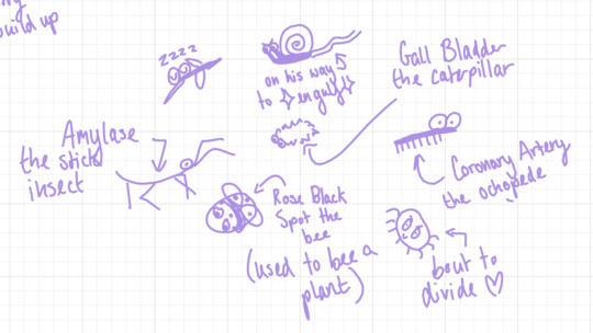


....no, i do not "use flashcards" to revise
#they're my little guys!#phagocytosis and pathogen and amylase and gall bladder and rose black spot and mitosis and coronary artery :D#the bugs ever#gcse biology#gcse studyblr#sorry aesthetic study moodboards#i'm coming to infest your tag with mitosis but spiders#gcses 2024#exam season#exams#good luck tomorrow everyone!!!
11 notes
·
View notes
Note
it's still a happy pride month so here's a joke i've been holding onto for the longest time but was afraid of actually making it its own post -- a cell eats out another cell, call that faggocytosis /j
ASDFGHJK 10/10. me in the pussy if im being fully honest
you should make this a post so i can reblog it
#red replies#Anonymous#no need to add the /j for the record - you already said it was a joke earlier#for those not getting it - phagocytosis (pronounced faggocytosis) is when a cell eats another cell
9 notes
·
View notes
Text
I might change my persona to a moth theme.... who knows
#im just bored with the fish#i might use both but ive got an oc i never use who is ncly already a self insert#so maybe she will become me through phagocytosis#ive already got stuff cooking with my friends that involves using a lil persona of me who is moth coded#and i REALLY like it#so i think yeah. moth and dirt and decay and a moth to the flame coded for me teehee :3#like i love the fish. dont get me wrong. but the moth???? 10/10#randy rambles
2 notes
·
View notes
Text
one of my microbiology teachers accidentally said "phaggotocytes" instead of phagocyte like THREE times before realizing and just went "uh im sorry" adjvgsdfdg im fucking lmfaooooooooooooo
#its especially funny bc the first thing i thought when i read phagocytosis was indeed phaggotcytosis#but she's an old southern white lady so.... idt she can reclaim that one#but maybe shes a phag you never really know!
3 notes
·
View notes
Text
constantly thinking about the word “fagocitosis”
8 notes
·
View notes
Text
guys guys stop this i don't care about any of this and all eight billion people in the world should be carefully curating their dashboards to align with my taste
6 notes
·
View notes
Text
Macrophage Phagocytosis Question and Answers
#DentalExpertise#DentalWellness#SmileBrighter#Bdsnotescom#Periodontics#Macrophage Phagocytosis Question and Answers
0 notes
Text

thank you discord
1 note
·
View note
Text
Japan really said "Sure, I'll integrate that into my system" about anything it was introduced to uh
0 notes
Last Seen Blogs

passionsalon-blog
Untitled

ankaratravestiester
Ankara Travesti Ester

meiqisu-blog
无标题

carindermalhisblog
Carinder

slavic-irishcryptid
Pluto