#Interhemispheric
Explore tagged Tumblr posts
Visit Tumblr Blog
Explore Tumblr blogs with no restrictions, modern design and the best experience.
Last Seen Tumblr Blogs


Fun Fact
Total funding amounts to $125.3M.
Text

New data on atmosphere from Earth to the edge of space
Two-decade-long dataset for entire atmosphere could benefit seasonal forecasts, climate models and space weather prediction
A team led by researchers at the University of Tokyo have created a dataset of the whole atmosphere, enabling new research to be conducted on previously difficult-to-study regions. Using a new data-assimilation system called JAGUAR-DAS, which combines numerical modeling with observational data, the team created a nearly 20-yearlong set of data spanning multiple levels of the atmosphere from ground level up to the lower edges of space. Being able to study the interactions of these layers vertically and around the globe could improve climate modeling and seasonal weather forecasting. There is also potential for interdisciplinary research between atmospheric scientists and space scientists, to investigate the interplay between space and our atmosphere and how it affects us on Earth.
Complaining about the weather, and about weather forecasters when they get things wrong, is a popular pastime for many. But a meteorologist’s job is not easy. Our atmosphere is multilayered, interconnected and complex, and global climate change is making it even harder to forecast both long-term and sudden, extreme weather events.
To help overcome these increasing challenges, researchers have created a dataset of the entire atmosphere. Ranging from September 2004 to December 2023, it spans multiple levels of the atmosphere from ground level up to the lower edge of space, about 110 kilometers above Earth’s surface. The region between about 50 km to 110 km (though exact ranges vary) is particularly of interest, as it is so notoriously difficult to study that it had previously been dubbed the “ignorosphere.” This region is too low for satellites and too high for weather balloons to observe, resulting in a shortage of data and consequently research. However, it is a fascinating area, characterized by vast global atmospheric tides and small-scale gravity waves which affect wind and temperature. It also plays an important role in the intensity of the impact of space weather events.
“The JAWARA (JAGUAR-DAS Whole neutral Atmosphere Reanalysis) dataset is a strong research tool which, for the first time, makes it possible to quantitatively understand atmospheric general circulation and the hierarchal structure of waves and vorticies in the mesospheric layer (which is above the stratosphere and about 50-90 km above Earth’s surface) and lower thermospheric layer (about 90-110 km above Earth’s surface) of the atmosphere, including the ignorosphere,” explained Professor Kaoru Sato from the University of Tokyo. “If we can better understand these layers, it would improve our ability to respond to climate change, extend the lead time of seasonal forecasts and advance our understanding of space weather phenomena.”
The team developed its new JAGUAR-DAS high-speed data assimilation system as part of an international project led by Sato. The system integrates observational data into a numerical model which can then produce data on atmospheric conditions. The resulting dataset, named JAWARA, makes it possible to perform detailed analysis of the general circulation of the atmosphere and its hierarchical structure.
“Atmospheric general circulation models which range up to the lower edge of space have only been developed by a limited number of research institutions around the world, including our own,” said Sato. “Recent studies indicate that extreme stratospheric phenomena can start at least in the upper mesosphere. Therefore, quantitative elucidation of phenomena in the mesosphere and lower thermosphere is extremely important for weather forecasting.”
The dataset is now openly available, and the team intends to use it to study the large-scale circulation and the hierarchical structure in the atmosphere, as well as vertical and interhemispheric (i.e., between the Northern and Southern hemisphere) couplings. They also hope to work in collaboration with space scientists to study the interactions between the atmosphere and space, particularly the mesosphere (where the highest clouds form) and ionosphere (located within the thermosphere and about 60-300 km above Earth’s surface, where many satellites are based).
IMAGE: This infographic shows the multiple layers of our atmosphere, extending from the ground up into space, and how the new dataset compares in coverage to those currently available. Credit D.Koshin, K. Sato, S. Watanabe and K. Miyazaki, 2025/ Progress in Earth and Planetary Science (PEPS)
3 notes
·
View notes
Text
What is the brain?
The brain is a complex organ that controls thought, memory, emotion, touch, motor skills, vision, breathing, temperature, hunger and every process that regulates our body. Together, the brain and spinal cord that extends from it make up the central nervous system, or CNS.
The brain sends and receives chemical and electrical signals throughout the body. Different signals control different processes, and your brain interprets each. Some make you feel tired, for example, while others make you feel pain.
Some messages are kept within the brain, while others are relayed through the spine and across the body’s vast network of nerves to distant extremities. To do this, the central nervous system relies on billions of neurons (nerve cells).
Main Parts of the Brain and Their Functions
At a high level, the brain can be divided into the cerebrum, brainstem and cerebellum.
Cerebrum
The cerebrum (front of brain) comprises gray matter (the cerebral cortex) and white matter at its center. The largest part of the brain, the cerebrum initiates and coordinates movement and regulates temperature. Other areas of the cerebrum enable speech, judgment, thinking and reasoning, problem-solving, emotions and learning. Other functions relate to vision, hearing, touch and other senses.
Cerebral Cortex
Cortex is Latin for “bark,” and describes the outer gray matter covering of the cerebrum. The cortex has a large surface area due to its folds, and comprises about half of the brain’s weight.
The cerebral cortex is divided into two halves, or hemispheres. It is covered with ridges (gyri) and folds (sulci). The two halves join at a large, deep sulcus (the interhemispheric fissure, AKA the medial longitudinal fissure) that runs from the front of the head to the back. The right hemisphere controls the left side of the body, and the left half controls the right side of the body. The two halves communicate with one another through a large, C-shaped structure of white matter and nerve pathways called the corpus callosum. The corpus callosum is in the center of the cerebrum.
Brainstem
The brainstem (middle of brain) connects the cerebrum with the spinal cord. The brainstem includes the midbrain, the pons and the medulla.
Midbrain. The midbrain (or mesencephalon) is a very complex structure with a range of different neuron clusters (nuclei and colliculi), neural pathways and other structures. These features facilitate various functions, from hearing and movement to calculating responses and environmental changes. The midbrain also contains the substantia nigra, an area affected by Parkinson’s disease that is rich in dopamine neurons and part of the basal ganglia, which enables movement and coordination.
Pons. The pons is the origin for four of the 12 cranial nerves, which enable a range of activities such as tear production, chewing, blinking, focusing vision, balance, hearing and facial expression. Named for the Latin word for “bridge,” the pons is the connection between the midbrain and the medulla.
Medulla. At the bottom of the brainstem, the medulla is where the brain meets the spinal cord. The medulla is essential to survival. Functions of the medulla regulate many bodily activities, including heart rhythm, breathing, blood flow, and oxygen and carbon dioxide levels. The medulla produces reflexive activities such as sneezing, vomiting, coughing and swallowing.
The spinal cord extends from the bottom of the medulla and through a large opening in the bottom of the skull. Supported by the vertebrae, the spinal cord carries messages to and from the brain and the rest of the body.

2 notes
·
View notes
Text
15th May 2024
Yale: Introduction to Radiology - Introduction to Brain Surface Anatomy
youtube
Introduction to Brain Surface Anatomy - Learning Objective:
Identify surface anatomy of the brain on CT and MRI Scans.
Surface of the brain is very convoluted and complex.
Brain Surface
Sulcus is a depression on the surface of the brain (valleys)
Gyrus is a ridge on the surface of the brain (hills)
Sulci and gyri are concepts
Variability
Superior Surface (Axial Image)
Midline Surface (Sagittal Image)
Lateral Surface (Sagittal Image)
Inferior Surface (Axial and Coronal Image)
Brain Surface - Superior Surface (Axial Image):
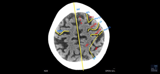
When looking at the axial images, look at it like a clock face.
12:00pm - 6:00pm - IHF (Interhemispheric Fissue)
Clockwise Rotation:
Next Gyrus - Superior Frontal Gyrus (SFG)
Next Sulcus - Superior Frontal Sulcus (SFS)
Next Gyrus - Middle Frontal Gyrus (MFG)
Next Sulcus - Pre Central Sulcus (Pre CS)
Notice how the Superior Frontal Sulcus joins the Pre Central Sulcus.
Next Gyrus - Pre Central Gyrus.
Next Sulcus - Central Sulcus.
Notice how the Central Suclus has a notch/knob that resembles the Greek alphabet Omega.
This is a very important concept.
This corresponds to the Hand Motor Area (HMA) on the Hohmann class.
Posterior to Central Sulcus, Post Central Gyrus (PCG).
Next Sulcus - Post Central Sulcus.
Anything anterior to the Central Sulcus is the Frontal Lobe.
Anything posterior to the Central Sulcus is the Parietal Lobe.
In the Parietal Lobe is a deep gash which runs from 3:00pm - 5:00pm position and from 9:00pm - 7:00pm position, Intraparietal Sulcus (IPS).
Superior to that is the Superior Parietal Lobule.
Inferior to that is Inferior Parietal Lobule.
Notice that the Intraparietal Sulcus laterally joins the Post Central Sulcus.
In order to proceed further, we need to look at the surface anatomy on a midline image.
Brain Surface - Midline Surface (Sagittal Image):
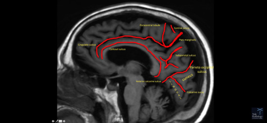
In the midline, anatomy is either co curvilinear(?) to the corpus callosum or perpendicular to it.
Paralleling the Corpus Callosum is the Colossal Sulcus.
Next Gyrus - Cingulate Gyrus
Next Sulcus - Cingulate Sulcus
Cingulate Sulcus is parallel to the Corpus Callosum 2/3 of its way, then suddenly makes a 90 degree swoop. This part of the Cingulate Sulcus which extends to the margain of the brain is Pars Marginalis.
Anterior to it is Central Sulcus.
(This is going to be the Pre Central Gyrus and Post Central Gyrus).
As we go down the Pars Marginalis, it makes a U-shaped curvature. The part of the brain which it encloses is Para Central Lobule.
Another deep gash on Midline Image is Parieto Occipital Sulcus.
It separates Parietal Lobe from Occipital Lobe.
Another deep gash is called Calcarine Sulcus.
The part of the Occipital Lobe between the Calcarine Sulcus and Parieto Occipital Sulcus, triangle part of the brain, is Cuneus.
The part of the Occipital Lobe inferior to Calcarine Sulcus, resembling a tongue sticking out, is Lingual Gyrus.
Between Pars Marginalis and Parietal Occipital Suclus, uppercase "H"-shaped fissure, is Subparietal Sulcus.
An important concept on the Midline Sagittal Image is when Parieto Occipital Sulcus and Calcarine Sulcus join, they extend down and form a lazy "Y" - fissure is known as Anterior Calcarine Sulcus.
It separates the Lingual Gyrus from the continuation of the Cingulate Sulcus.
Midline Sagittal Image - Axial Image:
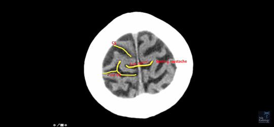
As we move from inferior to superior direction, the Pars Marginalis is seen as a deep cleft in the midline.
From inferiorly, it appears like a drooping mustache.
As we go a slice above (?), it appears like a straight mustache.
On most superior images, it appears like a smiling mustache.
Notice how it forms like a basket, Pars Basket.
As seen on the Midline Sagittal Image:
The Central Sulcus (CS) is anterior to the Pars Basket, and dips into the basket.
Post Central Sulcus (Post CS) is byford(?) and holds the basket.
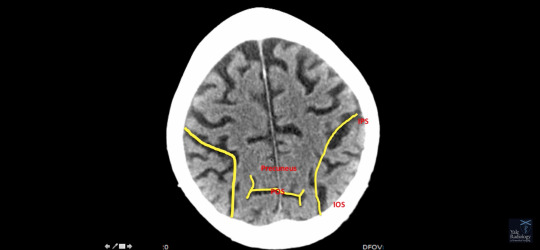
Intraparietal Sulcus (IPS) runs from 3:00pm - 5:00pm position and 9:00pm - 7:00pm position.
Part of the Intraparietal Sulcus, which is in the Occipital Lobe, is the Intra Occipital Sulcus (IOS).
Notice how the Intra Occipital Sulcus is parallel to the Inter Hemispheric Fissure (IHF).
Parieto Occipital Sulcus appears like a bifid/fish tail, along with the Intra Occipital Sulcus (IOS), it forms like a broken uppercase "m".
Anything anteriro to Parietal Occipital Sulcus is the pre-kunis/cuneus(?) of the Parietal Lobe.
Posterior to Parietal Occipital Sulcus is the Cuneus Occipital Lobe.
Brain Surface - Lateral Surface (Sagittal View)
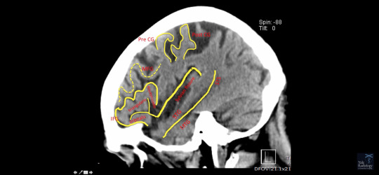
Deep fissure is Lateral Fissure/Sylvian Fissure.
Sylvian Fissure separates Frontal Lobe and Parietal Lobe from Temporal Lobe.
The most lateral gyrus in Frontal Lobe is Inferior Frontal Gyrus (IFG).
It appears like a molar tooth/triangle. A.k.a Triangle Gyrus.
It is divided by most anterior part of Sylvian Fissure into Orbitalis.
Between division of Sylvian Fissure, we have Triangularis then we have Opercularisis.
Above Inferior Frontal Gyrus is Middle Frontal Gyrus (MFG). MFG is zigzag, like Mississippi Riber, which joins the Precentral Gyrus (Pre CG).
Notice how Precentral Gyrus (Pre CG) forms like a hook, which is held by Post Central Gyrus (Post CG).
Important Concept - Hook Sign
It'll help to identify Pre CG and Post CG.
Just below Sylvian Fissure is Superior Temporal Sulcus (STS).
Above that is Superior Temporal Gyrus (STG).
Bellow is Middle Temporal Gyrus (MTG).
Inferior Surface (Coronal Image):
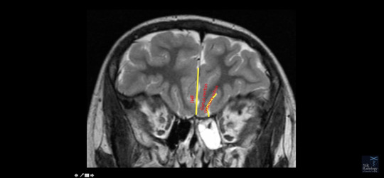
Interim Spirit Fissure(?)/Interhemispheric Fissure (IHF)
Olfactory Sulcus.
Between IHF and Olfactory Sulcus is Gryus Rectus/Straight Gyrus.

Lateral to Olfactory Sulcus is Orbitofrontal Gyrus, which is sitting on the orbit.
Brain Surface - Inferior Surface (Axial Image)

Divided into 4 parts by Orbitofrontal Sulcus, which represents the uppercase "H" into Anterior (A), Median (M), Posterior (P) and Lateral (L) Orbital Frontal Gyrus.
Brain Surface - Inferior Surface (Coronal Image)
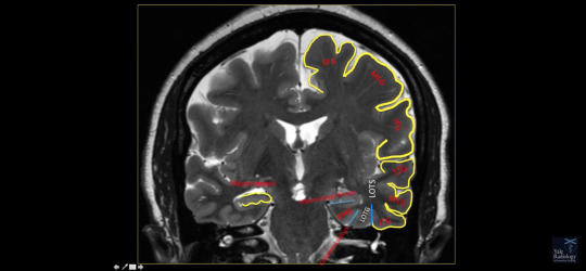
As we move posteriorly on the coronal image...
We can identify structures already seen.
Superior Frontal Gyrus (SFG)
Middle Frontal Gyrus (MFG)
Inferior Frontal Gyrus (IFG)
Superior Temporal Gyrus (STG)
Middle Temporal Gyrus (MTG)
Inferior Temporal Gyrus (ITG)
ITG is separated by Lateral Occipital Temporal Sulcus (LOTS) from Lateral Occipital Temporal Gyrus (LOTG).
This is separated by Collateral Sulcus from Parrahipocampal Gyrus (PHG).
PHG is separated by Hippocampal Fisher from Hippocampus.
Revision:
From 12:00pm - 6:00pm position is Interhemispheric Fissure (IHF).
Gyrus Order: Superior Frontal Gyrus (SFG), Superior Frontal Sulcus
Superior Frontal Sulcus joins Pre Central Sulcus
Just posterior to Pre Central Sulcus is Pre Central Gyrus.
Central Sulcus has notch that corresponds to Hand Motor Area, resembling Greek alphabet Omega.
Posterior is Post Central Gyrus
Post Central Sulcus, which is bifid.
Notice how Pre Central Gyrus is much thicker than Post Central Gyrus.
Pars. Basket.
Central Sulcus dips into Pars Basket.
Post Central Sulcus holds Pars Basket.
Intraparietal Intra Occipital Sulcus joins Post Central Sulcus.
We can identify Parietooccipital Sulcus by bifid fish tail like appearance.
0 notes
Text
Research Technician Washington University Join our team to study the development and function of interhemispheric connections in the mammalian brain. See the full job description on jobRxiv: https://jobrxiv.org/job/washington-university-27778-research-technician/?feed_id=58862 #ScienceJobs #hiring #research #neuroscience #neurotwitter St. Louis #UnitedStatesUS #ResearchTechnician
0 notes
Quote
The inferior frontal sulcus is conceptualized as the landmark delineating ventro-from dorsolateral prefrontal cortex. Functional imaging studies report activations within the sulcus during tasks addressing cognitive control and verbal working memory, while their microstructural correlates are not well defined. Existing microstructural maps, e.g., Brodmann's map, do not distinguish separate areas within the sulcus. We identified six new areas in the inferior frontal sulcus and its junction to the precentral sulcus, ifs1-4, ifj1-ifj2, by combined cytoarchitectonic analysis and receptor autoradiography. A hierarchical cluster analysis of receptor densities of these and neighbouring prefrontal areas revealed that they form a distinct cluster within the prefrontal cortex. Major interhemispheric differences were found in both cyto- and receptorarchitecture. The function of cytoarchitectonically identified areas was explored by comparing probabilistic maps of the areas in stereotaxic space with their functions and co-activation patterns as analysed by means of a coordinate-based meta-analysis. We found a bilateral involvement in working memory, as well as a lateralization of different language-related processes to the left hemisphere, and of music processing and attention to the right-hemispheric areas. Particularly ifj2 might act as a functional hub between the networks. The cytoarchitectonic maps and receptor densities provide a powerful tool to further elucidate the function of these areas. The maps are available through the Human Brain Atlas of the Human Brain Project and serve in combination with the information on the cyto- and receptor architecture of the areas as a resource for brain models and simulations.
The inferior frontal sulcus: Cortical segregation, molecular architecture and function - ScienceDirect
0 notes
Text
The ipsilateral interhemispheric transprecuneal approach: microsurgical anatomy, indications, and neurosurgical applications
The ipsilateral interhemispheric transprecuneal approach: microsurgical anatomy, indications, and neurosurgical applications
Neurosurgical Review (2021) 44:529–541 Surgical treatment of intraventricular lesions is challenging because of their deep location, vascularization, and their complex relationships with white matter fibers. The authors undertook this study to describe the microsurgical anatomy of the white matter fibers covering the lateral wall of the atrium and temporal horn and to demonstrate how the…

View On WordPress
#interhemispheric approach#intraventricular tumor#superior longitudinal fasciculus#Surgical management . Atrium
1 note
·
View note
Note
does eye movement have a part in programming? Like emdr type thing where they can influence your brain by giving you something g to focus on and moving it around or something like they do in emdr to reprocess. Thank you
Bilateral eye movements appear to enhance true memory and decrease the extent to which subjects rely or make use of *gist based false memory. Findings for recognition memory build on earlier work showing sideways eye movements improve the accuracy of recall.
There is a positive correlation between the number of eye movements that a participant made while viewing a picture and their subsequent memory for that picture. Researchers have linked eye movements to **visuo-spatial working memory and external projection of mental imagery.
Research suggests that bilateral eye movements can enhance interhemispheric interaction which in turn was theorised to have a role to play in episodic memory processes
You could also look into the Saccade-induced retrieval enhancement (SIRE) effect.
*Gist-based errors occur when people extract the gist, or general information about thematic content, but fail to encode or retrieve verbatim, item-specific distinguishing details (Brainerd & Reyna, 2002; Koutstaal & Schacter, 1997). **Visuospatial working memory is the ability to retain and process an object's identity and spatial location is essential for many daily tasks, often referred to as visual-spatial working memory.
Oz
10 notes
·
View notes
Text

Request or randomized kisses meme || No longer accepting
@technodromes sent: 'I want the K' (yeeting Bishop Rick's way. Confused croaking and burbling intensifies)

{ Randomly generated number: 6. Gentle Peck }
It isn't often that you can find Bishop outside his Norman-Body. While Krang and Subprime are more than content to use their walker or pod respectively, the other Utrom seems to favour the android, even when he is in the company of the Technodrome crew. Morty says that it's both out of habit and because it makes the alien feel like he fits more, while Rick simply calls it "bullshit behaviour", which is the lazy label he assigned to all behaviours he can't be bothered to decipher.
Today, however, is one of the occasions when the Kraang had decided to forgo his human-looking disguise and to move around in his pod. His teen friend probably had something to do with this choice, but Rick doesn't care enough to speculate. All that matters is that this is the perfect chance to literally get his hands on Bishop's real body and he isn't going to waste it.
So, he lurks around in the room where Morty is trying to explain to Bishop this or that human tradition, fixing up some device for Krang as he waits for the right moment to strike. It's a win-win, because the repairs he needs to do is simple enough not to require his full attention, which means that he can easily pretend to be busy while appeasing the aspiring overlord.
...He has no idea of what has gotten into the goddamn Utrom lately, but he's been busting his balls trying to get him to fix his shit, even more than he usually does.
Eventually, his effort to be patient is rewarded when his grandson stands up and leaves the room, most likely to retrieve something that can help him dispel Bishop's obvious confusion over whatever they have been discussing. Rick has to bite back a derisive scoff. Between the Utrom's cluelessness and Morty's verbal clumsiness is no surprise that their conversation hasn't gotten anywhere yet.
Under other circumstances, he might have throw out a mocking comment, but right now he needs the element of surprise.
So, as soon as the automatic doors have closed behind the teen, he makes his move, exploiting the fact that Bishop is still too busy squinting at his tablet to notice him approaching from behind.
Rick's hands close around the sides of the pod without a warning and he spins it around, so that he and the Kraang are face to face. His lips are curled in a predatory grin, and he's almost looking like he's getting read to feast on the poor alien.

"C-C'mere, bubblegum.~"
Once again without waiting for a reaction, he pries the Utrom away from the pod, bringing him closer to his own face. His smirk widens, showing too much teeth for everyone's comfort and strengthening the impression that he's about to do something unspeakable.
However, instead of a lick or a forceful kiss or, even worse, a bite as one would expect from such an act, what Bishop gets is a gentle, even if perhaps a little too slobbery, smooch between his eyes, right under the spot where his interhemispheric fissure begins.
When he breaks away, Rick's expression has turned from rapacious to smug, showing how pleased with himself he is with the prank he had just played. Snickering in delighted amusement, he sets the Kraang back in his pod, giving him a light tap on the top of his brain-like body, before shoving his hands hands in his pockets and heading out of the room, still laughing to himself.
#[ ic :: c137 Rick ]#&& Bishop#technodromes#[ relationship :: Rick & Bishop ]#[[ I was tempted to randomise it again when I got this ]]#[[ but then I realise that it was perfect xD ]]#[[ Rick basically looking like he's about to jump Bishop's spine ]]#[[ but instead he just *smooch* xD ]]#[[ and ofc the bastard is laughing his ass off -facepalm- ]]#;; queue#[ v. TransMortional Ricknections: Technodrome edition ; tmnt xover :: c137 Rick ]
3 notes
·
View notes
Text
Interesting Papers for Week 33, 2021
Interhemispheric transfer of working memories. Brincat, S. L., Donoghue, J. A., Mahnke, M. K., Kornblith, S., Lundqvist, M., & Miller, E. K. (2021). Neuron, 109(6), 1055-1066.e4.
Behavioral Time Scale Plasticity of Place Fields: Mathematical Analysis. Cone, I., & Shouval, H. Z. (2021). Frontiers in Computational Neuroscience, 15, 19.
Gating of hippocampal rhythms and memory by synaptic plasticity in inhibitory interneurons. He, X., Li, J., Zhou, G., Yang, J., McKenzie, S., Li, Y., … Ma, H. (2021). Neuron, 109(6), 1013-1028.e9.
Robust information routing by dorsal subiculum neurons. Kitanishi, T., Umaba, R., & Mizuseki, K. (2021). Science Advances, 7(11), eabf1913.
Frequency of theta rhythm is controlled by acceleration, but not speed, in running rats. Kropff, E., Carmichael, J. E., Moser, E. I., & Moser, M.-B. (2021). Neuron, 109(6), 1029-1039.e8.
Preexisting hippocampal network dynamics constrain optogenetically induced place fields. McKenzie, S., Huszár, R., English, D. F., Kim, K., Christensen, F., Yoon, E., & Buzsáki, G. (2021). Neuron, 109(6), 1040-1054.e7.
Does Attention Increase the Value of Choice Alternatives? Mormann, M., & Russo, J. E. (2021). Trends in Cognitive Sciences, 25(4), 305–315.
Stress undermines reward-guided cognitive performance through synaptic depression in the lateral habenula. Nuno-Perez, A., Trusel, M., Lalive, A. L., Congiu, M., Gastaldo, D., Tchenio, A., … Mameli, M. (2021). Neuron, 109(6), 947-956.e5.
A neural correlate of visual feature binding in primate lateral prefrontal cortex. Parto Dezfouli, M., Schwedhelm, P., Wibral, M., Treue, S., Daliri, M. R., & Esghaei, M. (2021). NeuroImage, 229, 117757.
Striatal activity topographically reflects cortical activity. Peters, A. J., Fabre, J. M. J., Steinmetz, N. A., Harris, K. D., & Carandini, M. (2021). Nature, 591(7850), 420–425.
The structure dilemma in biological and artificial neural networks. Pircher, T., Pircher, B., Schlücker, E., & Feigenspan, A. (2021). Scientific Reports, 11(1), 5621.
A predictive account of how novelty influences declarative memory. Quent, J. A., Henson, R. N., & Greve, A. (2021). Neurobiology of Learning and Memory, 179, 107382.
Cochlear neural degeneration disrupts hearing in background noise by increasing auditory cortex internal noise. Resnik, J., & Polley, D. B. (2021). Neuron, 109(6), 984-996.e4.
Dopamine-based mechanism for transient forgetting. Sabandal, J. M., Berry, J. A., & Davis, R. L. (2021). Nature, 591(7850), 426–430.
An autonomous debating system. Slonim, N., Bilu, Y., Alzate, C., Bar-Haim, R., Bogin, B., Bonin, F., … Aharonov, R. (2021). Nature, 591(7850), 379–384.
Looking for Image Statistics: Active Vision With Avatars in a Naturalistic Virtual Environment. Straub, D., & Rothkopf, C. A. (2021). Frontiers in Psychology, 12, 431.
Tonic firing mode of midbrain dopamine neurons continuously tracks reward values changing moment-by-moment. Wang, Y., Toyoshima, O., Kunimatsu, J., Yamada, H., & Matsumoto, M. (2021). eLife, 10, e63166.
Noninvasive neuromagnetic single-trial analysis of human neocortical population spikes. Waterstraat, G., Körber, R., Storm, J.-H., & Curio, G. (2021). Proceedings of the National Academy of Sciences of the United States of America, 118(11).
Adaptation of spontaneous activity in the developing visual cortex. Wosniack, M. E., Kirchner, J. H., Chao, L.-Y., Zabouri, N., Lohmann, C., & Gjorgjieva, J. (2021). eLife, 10, e61619.
Individual differences in experienced and observational decision-making illuminate interactions between reinforcement learning and declarative memory. Yifrah, B., Ramaty, A., Morris, G., & Mendelsohn, A. (2021). Scientific Reports, 11(1), 5899.
#science#Neuroscience#computational neuroscience#Brain science#research#cognition#cognitive science#neurobiology#neurons#psychophysics#neural networks#neural computation#scientific publications
16 notes
·
View notes
Text
Anterior interhemispheric transsplenial approach to pineal region tumors
J Neurosurg 128:182–192, 2018
Pineal region tumors are challenging to access because they are centrally located within the calvaria and surrounded by critical neurovascular structures.
The goal of this work is to describe a new surgical trajectory, the anterior interhemispheric transsplenial approach, to the pineal region and falcotentorial junction area. To demonstrate this approach, the…
View On WordPress
#anatomy#Interhemispheric#Microsurgical anatomy#pineal#surgical approach#Surgical technique#transsplenial
0 notes
Text

Name: Victor John Adley
Age: 26
Birthday: October 31st
Zodiac: Scorpio
Scorpio-born are passionate and assertive people. They are determined and decisive, and will research until they find out the truth. Scorpio is a great leader, always aware of the situation and also features prominently in resourcefulness. Scorpio is a Water sign and lives to experience and express emotions. Although emotions are very important for Scorpio, they manifest them differently than other water signs. In any case, you can be sure that the Scorpio will keep your secrets, whatever they may be.
Birth Place: London England
Gender: Male
Sexuality: Asexual, Demiromantic
Alignment: Neutral
Blood type: O
Personality construct/disorder: Alexithymia
Alexithymia is essentially a dysfunction in the normal emotional awareness processes that makes it tough for people to put a label on their feelings, Richey explains. In research, it has been described as a “personality construct characterized by altered emotional awareness” and something that “negatively impacts empathic processing.” In practice, alexithymia makes it difficult to recognize when you're feeling something and even more difficult to assign a name to it.
Early studies showed evidence that there may be an interhemispheric transfer deficit among people with alexithymia; that is, the emotional information from the right hemisphere of the brain is not being properly transferred to the language regions in the left hemisphere, as can be caused by a decreased corpus callosum, often present in psychiatric patients who have suffered severe childhood abuse. This would tie in with Victors childhood, as the experiments done on him did mess with his brain quite a bit and could have changed quite a few things around.
Voice Claim?: https://m.youtube.com/watch?autoplay=1&v=3egIslyHnLE
Quote(s)-
“Why fight...when I’ve already won...?”
Physical Appearance
Hair Type: short
Hair Color: Burgundy-Brown
Eye Color: emerald green
Height: 5’10
Weight: 155
Strength: 7/10
Intelligence: 10/10
Charisma: 6/10
Loyalty: 3/10
Hacking skills: 10/10
BioEngineering skills: 8/10
Interrogation skills: 8/10
Cruelty level: 9/10
Outfit: he wears a grey shirt with a black trench coat with lights that changes colors depending on his emotions (there’s something implanted in his head that’s connected to his nervous system to help this work, this jacket was his first invention as an older teen/young adult), he also wears black pants and black shoes
What do the lights on his coat mean?:
• Green-Calm/neutral
• Powder Blue-Sadness
• Red-Anger
• Orange-Embarrassment
• Yellow-Happiness
• Purple-N/A
• Pink-Love
• White-confusion
• Black-Fear/afraid
• Blood red- rage
• Dark Blue- Depression
• Gold yellow-excitement/overjoyed/amusement
• Gray-pain (because why not)
• Brown- nervous/anxious
Personal Life
Hobbies: N/A
Likes: calm tranquil areas, piece and quiet, interrogation {easy interrogation}
Dislikes: the organization (obviously), being touched, incompetence.
Fears: Electrocution
Status: Hacker, Inventor, scientist, Bioengineer.
Old Alliance: an old evil organization, a long time ago
New Alliance: He works for Himself
Personality: he isn’t the nicest person, he can be rude, grumpy, stubborn and cruel. He likes to mess with his patients/‘victims’ (kidnapped agents) from time to time depending on how well they behave for him. He takes his job very seriously and is extremely intelligent with a lack of empathy for others well being, though at times he can be kind depending on if you get on his good side or not. When he is polite it unfortunately could only be a farce, Ruthless and willing to do what it takes to get what he wants from a situation, it is notable that he is more than a bit of a psychopath.
Back Story
Backstory- Victor was a former agent of The organization, though not much is known about him or his backstory. However the obvious scaring on his neck to the back of his head show an obvious sign of experimentations done on him, electricity scars to be more precise about what type they are
Childhood/early teens: At a young age he went through what is called supercharge sessions, which enhances one’s cognitive abilities through stimulation done by performing direct magnetic and electrical pulses to specific regions of the brain. This can be used to either quiet some mental processes and to activate or enhance others. direct stimulation to the brain through magnetic or electrical pulses can improve memory, pattern recognition, the ability to pay attention, mathematical skills, among many others including athletic skills. Side effects of these methods can include the alteration of brain regions they didn’t intend to touch, boosting one ability that comes with an unexpected trade-off where one mental process is sacrificed. It also leaves mental/psychological scars, especially since the ones who did this to him were his own family. He was basically their intelligence experiment, he ran away after the experiment was successful.
• His past is very unknown but it is known that he started inventing at a very young age.
• He was one of Novitatis’ favorite agents for obvious reasons when he was apart of the organization.
• He has a daughter, Celena. However she stays with the mother at the agency. He does actually care for her though, even if he won’t admit to it.
• He tends to keep a monotone voice and blank/neutral expression if not at all times, unless you actually get to him
• He doesn’t like being touched, at all.
• He isn’t easy to interrogate, very stubborn.
• He has hypermobility in his arms/shoulders so keep an eye on him when he’s handcuffed. It’s better to tie him to a chair than handcuff his hands behind his back.
• He’s better at shooting a gun from a far distance rather than fighting hand to hand combat, get close up to him and you can take him down with ease.
• He’s made a serum that causes the bones in ones body to break easily, as easy as cracking a peanut.
• he has also made a serum for paralyzing his victims and of course to help himself fall asleep at night, knocks him out cold and anyone else injected with it
• Victors favorite invention is his force field belt, it looks just like any other regular belt so its easily disguised. The name itself is explanatory enough, it envelopes the body in a blue protective force field once turned on.
1 note
·
View note
Quote
Thus, while language emerged in the left hemisphere at the cost of pre-existing perceptual systems, the critical features of the bilaterally present perceptual system were spared in the opposite half-brain. By having the callosum serve as the great communication link between redundant systems, a pre-existing system could be jettisoned as new functions developed in one hemisphere, while the other hemisphere could continue to perform the previous functions for both half-brains. Split-brain studies have also revealed the complex mosaic of mental processes that participate in human cognition. And yet, even though each cerebral hemisphere has its own set of capacities, with the left hemisphere specialized for language and speech and major problem-solving capacities and the right hemisphere specialized for tasks such as facial recognition and attentional monitoring, we all have the subjective experience of feeling totally integrated. Indeed, even though many of these functions have an automatic quality to them and are carried out by the brain prior to our conscious awareness of them, our subjective belief and feeling is that we are in charge of our actions. These phenomena appear to be related to our left hemisphere's interpreter, a device that allows us to construct theories about the relationship between perceived events, actions and feelings.
Cerebral specialization and interhemispheric communication | Brain | Oxford Academic
1 note
·
View note
Text
Contralateral anterior interhemispheric transcallosal- transrostral approach to the subcallosal region
Contralateral anterior interhemispheric transcallosal- transrostral approach to the subcallosal region

J Neurosurg 129:508–514, 2018
The authors report a novel surgical route from a superior anatomical aspect—the contralateral anterior interhemispheric-transcallosal-transrostral approach—to a lesion located in the subcallosal region. The neurosurgical approach to the subcallosal region is challenging due to its deep location and close relationship with important vascular structures. Anterior and…
View On WordPress
#anatomy#cavernous malformation#corpus callosum#interhemispheric transcallosal#subcallosal region#surgical approach#Surgical technique
1 note
·
View note
Text
3D-printed 'protective cap' helps repair missing skull
3D-printed 'protective cap' helps repair missing skull Recently, the neurosurgery craniocerebral trauma team of the First Affiliated Hospital of Anhui Medical University, in conjunction with the Clinical Digital Medicine Transformation Center, successfully customized a 3D printed "protective cap" for a patient with a skull defect. The patient survives the skull defect safely and smoothly The patient Mr. Dou, due to subdural effusion after cerebral hemorrhage, went to the Department of Neurosurgery of the High-tech Hospital District of the First Affiliated Hospital of Anhui Medical University. "The skull defect was more than 20 days after the operation, and the head CT showed the right subdural effusion. Considering a customized skull defect protection device." According to Mao Xiang, the deputy chief physician of the neurosurgery of the hospital, the neurosurgery communicated with the Clinical Digital Medicine Transformation Center. , On the basis of the patient's 3D CT data, through personalized design and 3D printing technology, a "protective cap" for the skull defect was tailored for him. After wearing it for a week, Mr. Dou found that the interhemispheric effusion was less than before, and the intracranial structure was basically restored to physiological and anatomical reduction. According to Elimold, the "protective cap" is a three-dimensional reconstruction map of the patient's skull defect by performing continuous thin-slice CT scans of the patient's head, and realizing a personalized design that matches the reconstructed model 100% with the real head. , adopts the polymer material PLA polylactic acid with suitable hardness and toughness that can effectively withstand and disperse the impact force and has a good anti-collision effect. The cap body is light (less than 100g) and has a good comfort; Timeliness of clinical application. According to Mao Xiang, for patients with skull defects caused by surgery, it generally requires a recovery period of about three months before cranial repair. "For the patient during the three-month skull defect period, because the brain tissue in the defect area has lost the protection of the skull, strict attention should be paid to avoiding bumps in daily life. In addition, subdural accumulation may also be caused by unbalanced pressure of cerebral effusion. fluid and hydrocephalus.” The "protective cap" tailored for each patient's 3D printing can better fit the skull defect, protect brain tissue, prevent hydrocephalus and subdural effusion, promote brain tissue repair, and help patients Returning to normal life as soon as possible provides a strong guarantee for them to safely and smoothly pass through the skull defect period.
0 notes
Text
Interesting Papers for Week 29, 2021
Entorhinal and ventromedial prefrontal cortices abstract and generalize the structure of reinforcement learning problems. Baram, A. B., Muller, T. H., Nili, H., Garvert, M. M., & Behrens, T. E. J. (2021). Neuron, 109(4), 713-723.e7.
Midbrain activity shapes high-level visual properties in the primate temporal cortex. Bogadhi, A. R., Katz, L. N., Bollimunta, A., Leopold, D. A., & Krauzlis, R. J. (2021). Neuron, 109(4), 690-699.e5.
The sensory representation of causally controlled objects. Clancy, K. B., & Mrsic-Flogel, T. D. (2021). Neuron, 109(4), 677-689.e4.
Using deep reinforcement learning to reveal how the brain encodes abstract state-space representations in high-dimensional environments. Cross, L., Cockburn, J., Yue, Y., & O’Doherty, J. P. (2021). Neuron, 109(4), 724-738.e7.
Crossed-uncrossed projections from primate retina are adapted to disparities of natural scenes. Gibaldi, A., Benson, N. C., & Banks, M. S. (2021). Proceedings of the National Academy of Sciences of the United States of America, 118(7).
The misrepresentation of spatial uncertainty in visual search: Single- versus joint-distribution probability cues. Gibson, B. S., Pauszek, J. R., Trost, J. M., & Wenger, M. J. (2021). Attention, Perception, & Psychophysics, 83(2), 603–623.
Learning hierarchical sequence representations across human cortex and hippocampus. Henin, S., Turk-Browne, N. B., Friedman, D., Liu, A., Dugan, P., Flinker, A., … Melloni, L. (2021). Science Advances, 7(8), eabc4530.
Opposing force fields induce direction-specific sensorimotor adaptation but a non-specific perceptual shift consistent with a contraction of peripersonal space representation. Leclere, N. X., Sarlegna, F. R., Coello, Y., & Bourdin, C. (2021). Experimental Brain Research, 239(1), 31–46.
Hippocampal spatial memory representations in mice are heterogeneously stable. Levy, S. J., Kinsky, N. R., Mau, W., Sullivan, D. W., & Hasselmo, M. E. (2021). Hippocampus, 31(3), 244–260.
Subthalamic stimulation impairs stopping of ongoing movements. Lofredi, R., Auernig, G. C., Irmen, F., Nieweler, J., Neumann, W.-J., Horn, A., … Kühn, A. A. (2021). Brain, 144(1), 44–52.
Spontaneous brain activity underlying auditory hallucinations in the hearing-impaired. Marschall, T. M., Ćurčić-Blake, B., Brederoo, S. G., Renken, R. J., Linszen, M. M. J., Koops, S., & Sommer, I. E. C. (2021). Cortex, 136, 1–13.
Memory Load Alters Perception-Related Neural Oscillations during Multisensory Integration. Michail, G., Senkowski, D., Niedeggen, M., & Keil, J. (2021). Journal of Neuroscience, 41(7), 1505–1515.
A distinct population of heterogeneously color-tuned neurons in macaque visual cortex. Nigam, S., Pojoga, S., & Dragoi, V. (2021). Science Advances, 7(8), eabc5837.
Selective modulation of interhemispheric connectivity by transcranial alternating current stimulation influences binaural integration. Preisig, B. C., Riecke, L., Sjerps, M. J., Kösem, A., Kop, B. R., Bramson, B., … Hervais-Adelman, A. (2021). Proceedings of the National Academy of Sciences of the United States of America, 118(7).
Extracting the dynamics of behavior in sensory decision-making experiments. Roy, N. A., Bak, J. H., Akrami, A., Brody, C. D., & Pillow, J. W. (2021). Neuron, 109(4), 597-610.e6.
Functional Differentiation of Mouse Visual Cortical Areas Depends upon Early Binocular Experience. Salinas, K. J., Huh, C. Y. L., Zeitoun, J. H., & Gandhi, S. P. (2021). Journal of Neuroscience, 41(7), 1470–1488.
Temporal binding and agency under startle. Scott, M., & Chiu, C. (2021). Experimental Brain Research, 239(1), 289–300.
Dissociation between asymmetric value updating and perseverance in human reinforcement learning. Sugawara, M., & Katahira, K. (2021). Scientific Reports, 11(1), 3574.
Learning to Synchronize: Midfrontal Theta Dynamics during Rule Switching. Verbeke, P., Ergo, K., De Loof, E., & Verguts, T. (2021). Journal of Neuroscience, 41(7), 1516–1528.
Prolactin enhances hippocampal synaptic plasticity in female mice of reproductive age. Zamora‐Moratalla, A., & Martín, E. D. (2021). Hippocampus, 31(3), 281–293.
#science#Neuroscience#computational neuroscience#Brain science#research#neurobiology#mirror neurons#cognition#cognitive science#psychophysics#scientific publications
12 notes
·
View notes
Photo

The female mind is wired for leadership, creating complicated decisions, empathy, and then effort.
Nature's default is female - we begin with an X chromosome.
If perhaps you're the fortunate recipient of other X chromosome (thus causing you to female), you are going to continue to produce neurons together with the Jedi like power to read language, communication, and faces.
The female mind, as you are going to see from several key gender dimorphisms I talk about below, is created to have positions of power.
Naturally, this's Not saying males shouldn't stay in positions of power.
But if you glance at the gorgeous nuances within the female mind, and also just how she's uniquely qualified to direct with compassion, elegance, along with arguably a greater executive choice developer, it begs the question
It will be useless to try to fit females right into a masculine design of attitudes, abilities and skills and disastrous to push them to control their specifically female qualities and capabilities by staying in touch the pretense that you'll find no variations between the sexes.? Arianna Huffington
The prefrontal cortex will be the queen mother and it is liable for preparing the long term, expression, personality, decision making and moderating interpersonal behavior.
It's the executive function, the SheEO, nay, the CEO, of the human brain.
The prefrontal cortex is much larger and matures faster in females than in males.
Developmentally, the prefrontal cortex is much larger and grows more quickly in females.
Exactly why is the?
Among the prevailing theories will be the effect of estrogen, the main hormone within the female mind, which strongly influences the quicker growth and upkeep of the female prefrontal cortex.
One more reason to explain this behavioural impact (early keener vs previous minute) which could co-exist with a bigger prefrontal cortex may have a thing to do with the variations inside our serotonin receptors.
I've written earlier on the reduced serotonin receptors in females as compared to the male counterparts of theirs.
I talked about males being more dopaminergic than females.
Meaning, they're less dependent on the environment of theirs, and may remain motivated and interested in a task more to complete it to completion.
Obtaining the job done in advance alleviates some stress response the female may have around the strain on the deadline.
We see the contrary behaviour in males. Guys will frequently wait until the last second so they use neurotransmitters as norepinepherine and dopamine to push them to complete.
Sound familiar?
Do not ever make choices based on fear. Make choices based on possibility and hope. Make choices based on what must occur, not what mustn't.? Michelle Obama
One of the primary functions of the bigger mind centers just like the frontal lobe is inhibiting lower regions of the human brain, particularly, the temporal lobe, the place that the amygdala lives.
This's known as our primitive, instinctual brain.
This's exactly where the female mind is superbly nuanced for leadership.
Having the ability to keep the cool of her, and also carry on and issue solvethat's the making of any world leader.
Older employees are much less impulsive, more creative, less reactive, and also more centered? Deepak Chopra
Neuroscientists have extended claimed the improved prevalence of ADHD, impulsivity, violence as well as aggression occurring a lot more often in guys than women.
This may be within portion on the anatomically lesser anterior cingulate cortex in guys, versus females.
Once again, in the context of leadership, this's an advantage of female mind.
In high stress situations, appropriating impulsive views and feelings, while still being in a position to consider top solutions is important.
What this usually means in an adult females is traditional over risky behaviour.
4. How We Connect :The Corpus Callosum
This's what neuroscientists phone an intrahemispheric mind.
He has a tendency to remain more in the left brain of his.
Guys are systematizers.
The female brain has much less neurons in general than the male counterparts of her BUT has far more connections between them.
She has a tendency to remain more in her right mind, having the ability to empathize and join with other people and create community.
Women are empathizers.
Women have what neuroscientists phone an interhemispheric mind. Significance, the female is much more effective with her neuronal contacts, and also employs more aspects of the brain of her across the cortices.
This enables her, with accuracy and speed, to engage a lot more areas of the brain of her.
In order to call female the weakened sex is a libel; it's male's injustice to female. If by strength is designed brute strength, indeed, then, is female much less brute compared to male. If by strength is designed moral energy, subsequently female is immeasurably male's superior. Who is able to make a far more effective appeal to the center than woman?? Mahatma Gandhi
Each time you see someone, or maybe in order to make use of the expression? choose your gut? it's the insula in which the signals from gut feelings, or the microbiota, are processed.
The insula is much larger and much more productive in females.
Once again, this's mainly because the female mind is under the good impact of estrogen.
An excellent mind is among the most valuable assets of the religious living? Max Anders
That is since the hippocampus of her, the part of the mind where memories are created, is much larger and much more active than within the man.
What's awesome about the hippocampus is it's estrogen very sensitive (specifically estradiol), and of course, has regulatory influences on her memory and learning.
Oh, I should also note the auditory cortex, in which learning, hearing and words facilities are located, are eleven % larger in women, too.
And so Why Is The Future Female?
Empathy
Collaboration
Intuition
Person Control
Suitable Worry
We make this happen through the special ways we, as females, are different.
Through the prefrontal cortex of ours, we effortlessly and easily plan ahead, really inhibit aggression and anger, learn information that is new, and also build executive communication styles.
Ladies will in addition work with their good language abilities to cultivate collaboration and consensus among peers (particularly various other females) much more effectively compared to males, and the language of ours will give to being ready to navigate through vulnerable negotiations.
We've even more communication and interconnectedness across the Corpus Callosum, which means we're far more effective and also utilize additional aspects of the brain of ours for tasks.
We've exceptional impulse control, and also much better inhibition of aggression facilities in the brain, while nevertheless able to resolve challenging problems.
We've a much better sense of we and some think, through our bigger and much more energetic insula, and that is the spot the gut feelings of ours are processed.
Today everything to say, this's NOT to say males aren't qualified to have positions in leadership.
Obviously they're.
It's very different statement to state males are not competent compared to women are uniquely qualified.
The female mind, is exclusively gifted, provided with her gender dimorphisms, to direct with empathy, instilling collaboration, intuition, and self management.
It's a neurological explanation for exactly why we need to have more females, with their distinctive neurological disparities in positions of energy.
4 notes
·
View notes