#Molecular gastronomy
Text
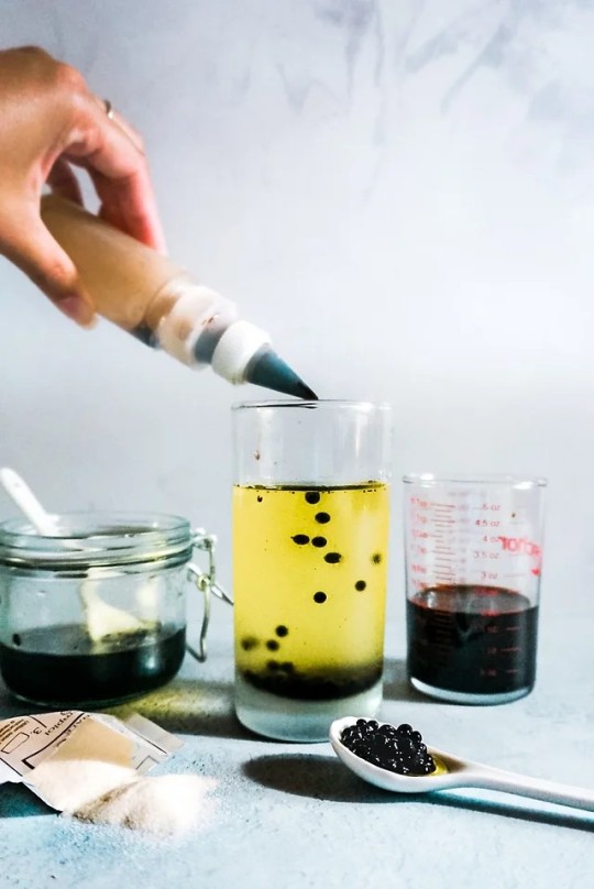
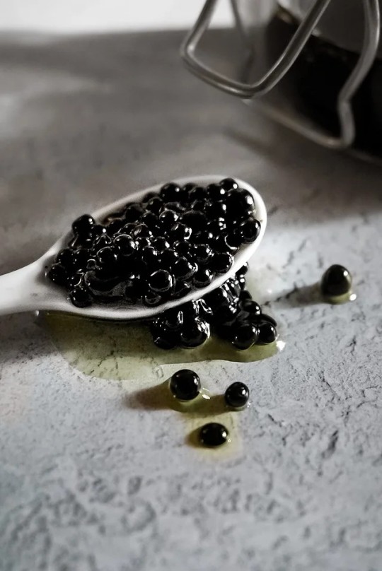
Balsamic ‘Caviar’ Pearls (Vegan)
#vegan#condiments#veganized#caviar#diy#molecular gastronomy#agar agar#balsamic vinegar#maple syrup#olive oil#sea salt
23 notes
·
View notes
Photo

Arttrober Day16: Alice Nakiri (Food Wars)
Molecular gastronomy is pretty cool.
Time-Lapse: https://youtu.be/2G-PPP0sx-g
Here's a link to the Arttrober2022 prompt for those interested:
https://twitter.com/Artgerm/status/1564274285858140160?s=20
#Arttrober#Arttrober2022#Alice Nakiri#Food Wars#Shokugeki no Soma#Shokugeki#Molecular Gastronomy#White Viper#digitalart#anime#manga
35 notes
·
View notes
Video
youtube
Unique 25 COURSE Las Vegas Tasting Menu at é by José Andrés!
#youtube#michelin chef#travelling foodie#foodie#travel#las vegas#las vegas food#restaurants#tasting menu#molecular gastronomy#spanish food
8 notes
·
View notes
Text

Agatha Agar
An Agar agar Candy Cutie! Agatha is a brilliant scientist and inventor. She's very soft spoken but her newfound friends at college are helping her grow.
Day 30 of #cutietober
Available on my RBand TeePubl!c.
#art#freelance#commissions are open#cute#commissions#pinup#candy#halloween#candy cuties#cutietober#science#nerd#scifi#agar agar#chemistry#molecular gastronomy#shy#geek#adorable
6 notes
·
View notes
Text


Could be fried egg and toast…could also be coconut jelly with a mango spherification and gingerbread toast!
7 notes
·
View notes
Text
Ham chemical breakdown through ripening and hydrolysis

ID: a flow chart. Dry cured ham, triglycerides, phospholipids, muscle & adipose tissues lipases, muscle phospholipases: free fatty acids. Short chain, (oxidation,) carbonyl compounds, volatile compounds, aroma. Proteins, muscle calpains, & cathepsins, peptides, muscle exopeptidases, free amino acids, strecker/maillard reaction, taste & aroma.
#ham#chemical breakdown#molecular gastronomy#ripening#hydrolysis#ham ripening#dry cured ham#cured ham#fermented whole muscle#cured whole muscle#whole muscle cure#Dry cured ham#triglycerides#phospholipids#muscle & adipose tissues lipases#muscle phospholipases#free fatty acids#Short chain#oxidation#carbonyl compounds#volatile compounds#aroma. Proteins#muscle calpains#& cathepsins#peptides#muscle exopeptidases#free amino acids#strecker/maillard reaction#taste & aroma
0 notes
Text
Theories on the Philosophy of Food
The philosophy of food is a multidisciplinary field that explores the various philosophical dimensions related to food, eating, and culinary practices. It delves into questions about the ethical, cultural, environmental, and existential aspects of food production, consumption, and distribution. Philosophers examine topics such as the ethics of food choices, the cultural significance of cuisine, the environmental impact of food systems, the aesthetics of food preparation, and the relationship between food and identity. Additionally, the philosophy of food considers broader issues such as hunger, food justice, food sovereignty, and the role of food in shaping human societies and relationships.
The philosophy of food encompasses various theories and perspectives that explore the ethical, cultural, and existential dimensions of food. Some of the prominent theories in this field include:
Gastronomic Essentialism: This theory posits that certain foods or culinary traditions possess inherent qualities or essences that make them valuable or meaningful. It emphasizes the authenticity and uniqueness of different cuisines and ingredients, highlighting the importance of preserving culinary traditions.
Food Ethics: Food ethics examines the moral principles and values that guide our food-related decisions and behaviors. It addresses questions about the ethical treatment of animals, environmental sustainability, food justice, and fair trade practices. Food ethics theories aim to promote ethical consumption practices and address ethical dilemmas in food production and distribution.
Slow Food Movement: The Slow Food movement advocates for a more mindful and sustainable approach to food consumption. It emphasizes the importance of local and traditional food cultures, promotes biodiversity in agriculture, and encourages people to savor and appreciate the pleasures of eating. Slow Food philosophy emphasizes the connection between food, culture, and the environment.
Veganism and Vegetarianism: Veganism and vegetarianism are dietary practices based on ethical, environmental, or health-related principles. Veganism advocates for the complete avoidance of animal products, while vegetarianism typically involves abstaining from meat but may include other animal products such as dairy and eggs. These dietary philosophies often stem from concerns about animal welfare, environmental sustainability, and personal health.
Commensality: Commensality refers to the social and cultural significance of sharing meals together. This theory explores the rituals, symbols, and meanings associated with communal eating experiences. Commensality emphasizes the role of food in fostering social bonds, strengthening community ties, and promoting cultural identity.
Molecular Gastronomy: Molecular gastronomy is a scientific approach to cooking that explores the physical and chemical processes behind food preparation. This theory investigates the interplay between taste, texture, and aroma, using techniques such as sous vide, foams, and spherification to create innovative culinary experiences. Molecular gastronomy challenges traditional notions of cooking and gastronomic aesthetics, pushing the boundaries of culinary artistry.
Food as Art: The concept of food as art explores the aesthetic dimensions of culinary creation and consumption. This theory considers food preparation and presentation as artistic expressions, akin to painting, sculpture, or music. It highlights the creativity, craftsmanship, and sensory pleasure involved in culinary endeavors, viewing food as a medium for artistic expression and cultural communication.
These are just a few examples of the diverse theories within the philosophy of food, each offering unique perspectives on the cultural, ethical, and existential significance of food in human life.
#philosophy#epistemology#knowledge#learning#chatgpt#education#ethics#psychology#metaphysics#aesthetics#Gastronomic essentialism#Food ethics#Slow Food movement#Veganism#Vegetarianism#Commensality#Molecular gastronomy#Food as art#Culinary traditions#Environmental sustainability#Philosophy of food#Culinary philosophy#Food culture#Gastronomy#Food justice#Environmental ethics#Sustainable food systems#Food sovereignty#Food aesthetics#Food identity
1 note
·
View note
Photo

✿ 分子ガストロノミー | Molecular Gastronomy
・1992年、当時84歳だったニコラス・クルティは「Molecular and Physical Gastronomy」(分子・物理美食学)の国際ワークショップを開催し、料理人や大学の基礎研究者、食品工業の専門家などを集めて、料理の発展について論じました。この会議は現在も方向性を変えつつ受け継がれ、料理の楽しみを広げています。この流れのなかで生まれたのが「Molecular Gastronomy」(分子ガストロノミー)という言葉です。
・ニコラス・クルティと、フランスの物理化学者エルヴェ・ティスによって創設されたもので、科学者とシェフが協力し、料理のなかで起こるプロセスを分析することで、従来伝わってきた調理法などを見直すといったもの。ニコラス・クルティが亡くなったあと、この動きはエルヴェ・ティスに引き継がれます。
・分子ガストロノミーの技術には、テクスチャーの操作からフレーバーの変更まで、あらゆるものが含まれます。シェフはこれらのテクニックを使用して、伝統的な料理の境界を打ち破る、見た目に美しい料理を作成します。球状化、ゲル化、乳化、泡、燻製、凍結療法などさまざまなテクニックがあります。
#molecular gastronomy#分子ガストロノミー#style:サイエンス#food:化学#style:化学#food:サイエンス#tip#tip:サイエンス#tip:化学#tip:科学#科学#tip:分子ガストロノミー#分子#ガストロノミー#ニコラス・クルティ#1992#food:分子ガストロノミー
1 note
·
View note
Text
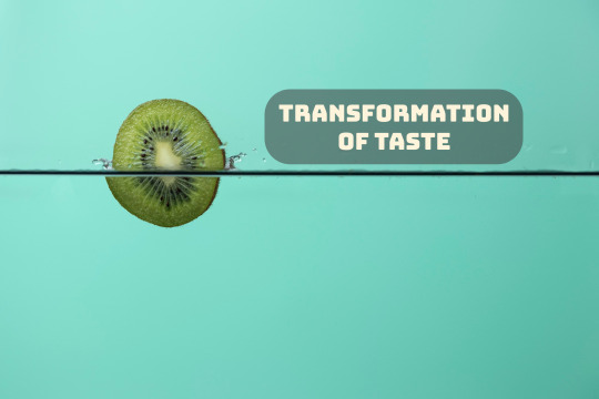
Flavors Unveiled
A Sarcastic Journey into the Transformation of Taste
Image by Freepik
Dear reader, do you ever find yourself pondering the peculiar evolution of our palates? From savoring the simplest ingredients to experimenting with avant-garde dishes, it seems our taste buds have embarked on a relentless journey through time. The culinary landscape has undergone a metamorphosis, leaving us to question the…
View On WordPress
#Culinary evolution#Culinary rebellion#Food for thought#Fusion cuisine#Historical flavors#Molecular gastronomy#Palate evolution#Taste and memory#Tradition vs. progression#Transformation of taste
0 notes
Text
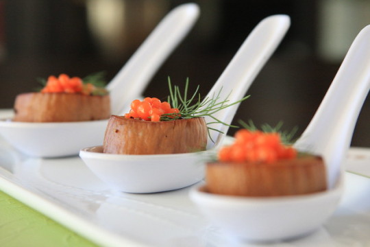
Molecular Vegan: Scallops with Carrot-Ginger Caviar
#vegan#appetizer#veganized#scallops#caviar#carrots#ginger#mushrooms#vegan butter#sodium alginate#calcium chloride#fennel#molecular gastronomy
25 notes
·
View notes
Text
Antiproliferative Effect of Green Papaya on Lymphocytic Leukemic Cells
Abstract
Numerous edible plants have been reported to interfere with the carcinogenic process, and therefore, the regular consumption of these plant products may reduce the risk of developing cancer. We investigated the effect of papaya fruit and leaves on the cell proliferation of Jurkat T-lymphocytic and Daudi B-lymphocytic leukemia cells. Cells were treated with aqueous or methanolic extracts from leaves, skin, pulp, and seeds from green papaya. The papaya fractions were tested for total phenolic content, total flavonoids content, and anti-oxidation activity using chemical assays. Cell proliferation was measured using a WST-1 assay. Our data indicate that methanol and water extracts of seeds and leaves contained higher concentrations of total phenolic compounds and higher anti-oxidation activity than that of extracts from skin and pulp.
Both methanol and water extracts from leaves and skin potently inhibited the proliferation of leukemic Jurkat T-cells and Daudi B-cells. However, the effect was more potent on Jurkat T-cells, and the leaf extracts were more effective than that of skin extracts. None of the pulp or seed extracts showed inhibitory activity on leukemic cell proliferation. Although papaya leaves are not consumed as a food, leaf extracts have been used for the treatment of various conditions, including dengue and malaria fevers, gastric ulcers, low platelet counts, and cancers of the breast, lung, and cervix. Our data suggest that the consumption of papaya leaf extracts may also be beneficial in preventing and/or treating lymphocytic cancer. Isolation of active compounds from papaya leaves will also help in developing new drugs for cancer treatment.
Keywords: Polyphenols; Antioxidants; Anticancer; Jurkat cells; Daudi cells; Leukemia
Introduction
Leukemia is a cancer of the blood or bone marrow, as described by an anomalous multiplication of immature clonal hematopoietic cells. Leukemia is one of the cancers that can affect all races in the United States. The American Cancer Society estimates that there will be around 60,530 new cases of leukemia in the United States in 2020, resulting in 23,100 deaths [1]. Leukemia is classified based on its onset (acute or chronic), the affected blood cell type (lymphoblastic/lymphocytic or myeloid/myelogenous), the maturity stage of the blood cell, and phenotypic expression of the disease [2]. Leukemia, like other forms of cancer, can be treated by using a number of methods, including chemotherapy, radiation therapy, targeted therapy, or stem cell therapy, though it is easier to treat acute leukemia than chronic leukemia [3]. Although leukemia remains a common medical issue, there has been a decrease in incidence due to preventive measures, and death rates have also declined since 1991 due to recent advances in treatment strategies [4]. The 5-year survival rate for children with acute lymphocytic leukemia has greatly increased over time to about 90% [5]. Although there are improved treatment strategies and survival rates, the cost of such therapies remains an issue that hinders access for all patients [6]. There is a need for less expensive alternative strategies to increase treatment access and survival outcomes for all patients.
Recent advances in alternative and complementary medicine have shown that various herbs, fruits, and vegetables have compounds that could act as inhibitors of cancer formation, blockers of carcinogen interaction, and suppressors of tumor progression. For example, Korean red ginseng extract treatment inhibited the growth of U937 leukemia cells by downregulating the expression of human telomerase reverse transcriptase [7]. Another study confirmed that Kenaf seed oil causes the death of human leukemia cells HL60 and K562 by induction of apoptosis [8]. Studies demonstrated that a hot water extract of Euphorbia formosana (EFW) restrained the growth of THP-1 human leukemic cells by inducing apoptosis through caspase-dependent cell death via Fas and mitochondrial pathways [9]. Water, ethanol, and methanol extracts of garlic have exhibited lethal cytotoxic effects of 50-75% on cells harvested from 3 patients with acute lymphoblastic leukemia (ALL) and three patients with acute myeloid leukemia (AML) [10]. Typhonium flagelliforme (TF), a herbal plant in Malaysia, exhibited cytotoxic effects in P388 [11] and WEHI-3 [12] murine leukemia cells. Further, in vivo studies showed a significant decline in the cell count of immature monocytes and immature granulocytes, when the TF extract was orally administered for 28 days in a BALB/c leukemic mouse model [12]. Studies with grape seed extracts reported induction of apoptosis in Jurkat leukemic cells through a process that involves sustained JNK activation and Cip1/p21 up-regulation, culminating in caspase activation [13].
Papaya fruit has also been studied for its anticancer activities for colorectal [14], prostate [15,16], cervical [17], breast [18- 20], and skin cancers [21]. Papaya also effectively inhibits cancers of hematopoietic cell lines, including T cell lymphoma (Jurkat), plasma cell leukemia (ARH77), Burkitt’s lymphoma (Raji), anaplastic large cell lymphoma (Karpas-299), and human promyelocytic leukemia (HL-60) [22-23]. A study demonstrated that Benzyl isothiocyanate (BITC), a promising chemo preventive agent found in papaya fruits, when given followed by cisplatin (BITC/cisplatin) caused significant death in HL-60 cells [24]. Furthermore, a recent study reported that papaya leaf extract improved bone marrow blast counts and stabilized the hematological parameters of a 76-year-old male with untreated chronic myelomonocytic leukemia [25].
It is well Known that fruits, vegetables, and other medicinal plants contain polyphenols and flavonoids that exhibit various biological and pharmacological properties [26], which are often mediated by their anti-oxidation activity [27]. Based on these earlier studies, we evaluated water and methanol extracts from leaves, skin, pulp, and seeds of green papaya on T-lymphocytic leukemia and B-cell lymphocytic leukemia using Jurakt and Daudi cell lines, respectively, and hypothesized that the effect of papaya fractions on cell proliferation of leukemic cells is correlated to their phenolic contents and/or anti-oxidation activity.
Materials and Methods
Materials
The Jurkat T-cell line and Daudi B-cell line were purchased from ATCC (Manassas, VA). RPMI 1640 medium was purchased from Gibco (Grand Island, NY). Fetal Bovine Serum (FBS) was purchased from RAMBIO (Missoula, MT). Penicillin and streptomycin antibiotics (BP2959) and Phosphate Buffered Saline (PBS; BP399-550) were purchased from Fisher Scientific (Fair Ln, NJ). Folin Ciocalteu, sodium carbonate, aluminum chloride, Diphenyl-1-picrylhydrazyl (DPPH), quercetin, gallic acid, and trolox were purchased from Sigma Chemical Co. (St Louis, MO). WST-1 (MK400) was purchased from Takara (Kusatsu, Shiga, Japan). Green papaya was obtained from Randolph Farm at Virginia State University, Petersburg, VA.
Isolation of papaya leaves, skin, pulp, and seeds
The papaya samples were isolated as described previously [28]. Briefly, the papaya was washed with distilled water and blotted dry with a paper towel. The skin was peeled off using a kitchen peeler. An unskinned papaya was cut into half to remove seeds, and then the pulp was cut into small pieces. The leaves and seeds were washed with distilled water. All fractions (leaves, skin, pulp, and seeds) were freeze-dried. The dried leaves, skin, pulp, and seeds were ground to a fine powder using a mortar and pestle with liquid nitrogen added to keep the powder frozen. The dried powder was flash-frozen with liquid nitrogen and stored at -80 ℃ until used.
Preparation of extracts from papaya leaves, skin, pulp and seeds
The methanol and water extracts of papaya fractions were prepared as described previously [28]. Briefly, a known quantity (5g) of different fractions of freeze-dried papaya powder was homogenized with 200ml of 80% methanol or distilled water. The mixtures were placed on a shaker at room temperature overnight. The next day, the mixture was centrifuged at 2000 x g for 20 minutes using a Thermo Scientific centrifuge (Waltham, MA). The supernatants were collected, and the residues were washed two times by resuspending in the respective solutions, mixing, and placing on a shaker overnight. The collected supernatants were pooled, and the insoluble residues were discarded. The methanol extracts were dried in a nitrogen evaporator (Organomation Associates, Inc, Berlin, MA), whereas the water extract was freezedried. The dried extracts were stored in a -20℃ freezer. The dried extract was suspended into DMSO at a concentration of 250mg/ ml and was further diluted into media for cellular studies.
Characterization of anti-oxidation activity of papaya extracts
The oxidative properties of papaya were determined by assaying their total polyphenolic content (TPC), total flavonoid content (TFC), and anti-oxidation capacity (DPPH) as previously described [29].
Cell culture
Jurkat T-cells and Daudi B-cells were maintained in RPMI 1640 media supplemented with penicillin (100 units/ml), streptomycin (100μg/ml) and 10% FBS. All cell cultures were incubated in a humidified incubator at 37 °C and 5% CO2. Media was changed every three days, and cells were subcultured when they became confluent.
Cell proliferation assay
The effects of papaya leaf, skin, pulp, and seed extracts on cell proliferation were determined using a WST-1 assay as per manufacturer instructions and described previously [28]. Briefly, cells were isolated from a confluent flask, and about 10,000 cells were placed in each well of a 96 well plate and incubated in a CO2 incubator for 24 hours. The next day, media was removed, and cells were incubated in serum-free media containing varying concentrations of papaya leaf, skin, pulp, or seed extract for 24 hours. After treatment with papaya extract, 10 μl of WST-1 was added in each well, and the plate was read at 420 nm in a 96 well plate reader for the background reading. The plate was then incubated for 3 hours at 37 ℃ in a CO2 incubator, and the reduction of WST-1 dye due to mitochondrial oxidation was recorded at 420 nm. The change in absorbance was calculated by subtracting the initial reading from the final reading. The results are expressed as % change from control (untreated cells).
Data analysis
The data are expressed as mean ± SD for at least three replicates. All comparisons were made by one-way ANOVA with Tukey’s -HSD-post-hoc test using SPSS Statistics 20 software (IBM, Armonk, NY). All significant differences are reported as P < 0.05 and indicated by “*” whereas non-significant differences are not marked.
Results
Characterization of papaya extract for anti-oxidation activity
The anti-oxidation potentials of papaya extracts were determined by assessing their total polyphenol content, total flavonoid content, and by assaying their anti-oxidation capacity. The data showing the total phenolic contents are presented in Table 1 and reported previously [28]. The highest amounts of polyphenols were found in the seed extracts that ranged 14 -16 mg/g dry weight. There was no significant difference in total phenolic contents between water and methanol extracts from papaya seeds. The leaves were second highest in total phenolic content but had a significantly lower amount of total phenolic content than that of seeds (P < 0.05). The leaves contained 1- 4 mg/g dry weight total phenolic content. Water extracts of leaves contained less total phenolic content (~ 1 mg/g dry weight) compared to that of methanol extracts. The skin extracts contained total phenolic content ~ 0.5 - 1 mg/g dry weight, whereas the pulp extracts have very small amounts of total phenolic content (0.05 - 0.5 mg/g dry weight).
The data for the total flavonoid contents in various papaya fractions are presented in (Table 2) and reported previously [28]. The water extract of seeds contained the highest amounts of total flavonoid content (5 mg/g dry weight), whereas the methanol extracts contained lower amounts (2-2.5 mg/g dry weight, P < 0.05). In leaves, the methanol extract contained more total flavonoid content (~ 5mg/g dry weight) than that of water (~2 mg/g dry weight). The total flavonoid contents in pulp and skin were less than those of seeds and leaves. In the skin, higher total flavonoid contents were present in the methanol extracts (~ 3.2 - 3.5 mg/g dry weight) than in water extracts (1 mg/g dry weight). The pulp contained the lowest total flavonoid contents compared to other fractions. The total flavonoid content in pulp ranged from 1 - 1.5 mg/g dry weight.
The anti-oxidation capacities of papaya fractions were measured by assaying the inhibition of DPPH oxidation and are shown in (Table 3) and reported previously [28]. The seeds and leaves contained significantly more (P < 0.05) anti-oxidation capacity than that of the skin and pulp. The methanol fractions of seeds and leaves contained more anti-oxidation activity than water extracts. The methanol fractions of seeds and leaves inhibited DPPH oxidation by 75 - 85%, whereas the water extracts of these fractions inhibited DPPH oxidation by 50 - 70%. The skin and pulp inhibited DPPH oxidation from 25 - 35%. There were no significant differences between water or methanol extracts of skin and pulp.
Effect of papaya water extracts on T- cell leukemia
The effects of papaya water extracts from different fractions are presented in Figure 1. The treatment with leaf extract caused a dose-dependent decrease in cell viability. A concentration of the extract as low as 50 μg/ml caused significant cell death by 30% (P < 0.05). Upon increasing the extract concentration more cells died, reaching a plateau of almost 70% cell death (P < 0.05) at 150 μg/ml. Additional increases to 250 μg/ml concentration resulted in a very small change in cell viability (Figure 1).
The treatment with water extract from papaya seeds caused a dose-dependent decrease in cell viability. Extract concentrations as low as 150 μg/ml caused a significant 20% increase in cell death (P < 0.05). On further increasing the extract concentration, no further decrease in cell viability occurred, which reached a plateau at 250 μg/ml with 25% cell death (P < 0.05) (Figure 1)
The water extract of papaya skin started to significantly (P < 0.05) inhibit leukemic cell proliferation at 25 μg/ml, causing about 30% cell death. However, the leukemic cells progressively died in a dose-dependent manner reaching to about 80% cell death at 250 μg/ml of the dried papaya skin extract.
The water extract from papaya pulp was unable to cause significant cell death of Jurkat leukemic T-cells.
Effect of papaya methanol extracts on T- cell leukemia
The effects of papaya methanol extracts from different fractions are presented in Figure 2. The methanolic extract of papaya leaves appeared to be less effective than water extract. At 25 μg/ml a significant (P < 0.05) 30% decrease in cell viability resulted; however, the cell viability was decreased by only 60% (P < 0.05) on further increasing the methanolic extract concentration to 250 μg/ml. Similarly, the methanolic extract of papaya seeds appeared to be less effective than water extract. At 200 μg/ml, a significant P < 0.05) decrease in cell viability resulted (10%); however, the further decrease in cell viability was not significant at 250 μg/ml. The methanolic extract of skin also caused significant cell death by 25% at 25 μg/ml (P < 0.05). Further increase of the extract concentration resulted in a dose-dependent response reaching 80% cell death at 250 μg/ml .
The methanol extract from papaya pulp caused a modest significant effect by decreasing cell viability by 20% at 50 μg/ml ; however, on increasing the concentration of pulp extract to 250 μg/ml , there was no further increase in cell death.
Effect of papaya water extracts on B- cell leukemia
The effects of water extract from different papaya fractions on Daudi leukemia B-cells are presented in Figure 3. Treatment with leaf extract caused a dose-dependent decrease in cell viability. Concentrations as low as 25 μg/ml caused a significant 20% cell death (P < 0.05). On further increasing the extract concentration, significantly more cells (P < 0.05) died in a dosedependent manner, with approximately 50% cells dead at the highest concentration of 250 μg/ml. The water extracts from seeds and pulp have no significant effect on cell viability at any concentration. The extract of papaya skin started to significantly (P < 0.05) inhibit leukemic cell proliferation at 25 μg/ml causing about 25% cell death. However, on further increasing the extract concentration, no change in cell death was observed
Effect of papaya methanol extracts on B- cell leukemia
The effects of methanol extracts from different papaya fractions on Daudi leukemia B-cells are presented in Figure 4. The methanolic extract of papaya leaves appeared to affect Daudi B-cells, similar to water extracts. At 50 μg/ml, a significant 30% (P < 0.05) of cells died and the cell death was further increased in a dose-dependent manner to 80% (P < 0.05) at 250 μg/ml. Consistent with the water extract, the methanol extract of papaya pulp has no effect on Daudi B-cells. The methanolic extract of skin had a similar effect on Daudi B-cells as the water extract. It caused significant cell death by 25% at 25 μg/ml (P < 0.05). Increasing the concentration of extract resulted in a dose-dependent effect, reaching 50% cell death at 250 μg/ml.
Discussion
The present study was carried out to investigate the effect of water and methanol extracts of papaya leaves, skin, pulp, and seeds on Jurkat leukemia T- cells and Daudi leukemia B-cells. Water and methanol were used because phenolic compounds have different chemical characteristics and polarities, and because of these properties, compounds in different fractions of papaya may or may not be soluble [30]. We initially tested these fractions for their phenolic contents and their anti-oxidation activity. Most of the anti-oxidation activity in fruits and vegetables is due to their total polyphenolic contents [31]. Our data indicate that papaya seeds have higher amounts of polyphenols than that of other fractions (Table 1). Papaya leaves also contained substantial amounts of total phenolic contents but were 1/3 that of seeds. Other fractions have very little total phenolic content. In seeds, the amount of total phenolic content was similar in both extracts, but the leaf water extract has less total phenolic content than that of methanol. However, the water extract of skin contained almost twice the polyphenols than that of the methanol extract. Organic solvents are generally used to extract bioactive agents from fruits and vegetables. The most suitable solvents for the extraction of polyphenols are aqueous mixtures containing ethanol, methanol, acetone, and ethyl acetate. Ethanol has been known as a good solvent for polyphenol extraction and is safe for human consumption. Methanol has been generally found to be more efficient in the extraction of lower molecular weight polyphenols, whereas aqueous acetone is good for the extraction of higher molecular weight flavonoids [32]. Currently, over 8000 phenolic structures have been identified in fruits and vegetables [33], and flavonoids are one of the major phenolic classes comprising almost 4000 compounds present in different edible plants [33]. We also determined the total flavonoids in the papaya extracts. Our data show that water extracts of seeds and methanol extracts of leaves contained the highest amount of total flavonoid content, which represented about 30- 40% of total phenolic compounds in seeds and leaves (Table 2). The data suggest that most of the polyphenols in seeds are water-soluble flavonoids, whereas a higher proportion of polyphenols in leaves are less water-soluble flavonoids.
Next, the anti-oxidation activities were determined in these fractions. The leaves and seeds possess a higher anti-oxidation activity compared to that of other fractions (Table 3). Although leaves and seeds have different phenolic and flavonoid contents, they have similar profiles for their anti-oxidation activity. It was interesting to note that methanol extracts exhibited higher antioxidation activity than the water extracts. This difference could be due to the different natures of polyphenols.
Next, we analyzed the anticancer properties of papaya fractions on Jurkat cells and Daudi cells. The Jurkat cells are an immortalized line of human T lymphocyte cells that are often used to study acute T cell leukemia. The Jurkat cell line was established in the late 1970s from the peripheral blood of a 14-year-old boy with T cell leukemia [34] whereas the Daudi cell line is a wellcharacterized B lymphoblast cell line which was derived from a 16-year-old black male with Burkitt’s lymphoma [35].
A large number of phytochemicals from vegetables, fruits, and grains have been reported to possess anticancer properties and are promoted for cancer prevention and treatment [36-40]. Recently, multiple scientific reports have emerged about the medicinal benefits of papaya [16,41-43].
Our data indicated that none of the extracts from seeds or pulp possess significant inhibitory activity for cell proliferation in Jurkat leukemic T-cells or Daudi leukemic B-cells. The extracts from leaves and skin, however, showed significant inhibition of both Jurkat leukemic T-cells and Daudi B-cell growth. This effect was stronger on Jurkat T-cells and modest on Daudi B-cells. During the present study, we found that papaya seeds contained the highest amounts of phenolic compounds; however, none of the extracts from papaya seeds have any inhibitory activity for Jurkat leukemic cells. Our data clearly indicated that the inhibitory activity of these extracts was neither dependent on their phenolic contents, nor was it dependent on their anti-oxidation activity. It appears from this observation that the inhibitory compounds in papaya leaves and skin extracts acted on cellular mechanisms linked to cell growth. For example, phytochemicals have been shown to interfere with the stabilization of the microtubule structure, thereby inhibiting mitosis and cancer cell propagation [44]. Vincristine and vinblastine, isolated from the leaves of Madagascar periwinkle, were the first phytochemicals to be used clinically in combination with other anticancer agents in lymphomas, leukemias, and breast, lung, and ovarian cancers [45,46].
Epigallocatechin-3-gallate (EGCG), a polyphenol from the leaves of Camellia sinensis (processed to green tea), possesses anti-oxidation activity but it limited cancer cell proliferation by reducing DNA methylation through the inhibition of DNA methyltransferase together with reactivation of the silenced tumor suppressor genes [47]. Curcumin (diferuloylmethane), a polyphenol isolated from the rhizome of the turmeric plant, has also shown therapeutic efficacy on numerous disorders, including cancer [48]. Curcumin is reported to inhibit NF-κB signaling that triggers the intracellular inflammatory response as well as cellcycle- associated genes. By arresting the cell cycle and inducing apoptosis through the relaying pathways, curcumin interferes with angiogenesis and reduces tumor invasion.
An additional group of anticancer phytochemicals functions as inhibitors of topoisomerase I or II, which are the nuclear enzymes that control DNA supercoiling, eliminate tangles in the chromatin structure, and allow DNA to be replicated and transcribed. Thus, topoisomerase inhibitors can act as anticancer agents by inducing a delay of the cell cycle, followed by cell death [36]. β-Lapachone from the bark of the Lapacho (Handroanthus impetiginosus) plant, camptothecin from the bark/stem of Camptotheca acuminata (the Chinese happy tree), and podophyllotoxin from the root of the Mayapple (Podophyllum peltatum) plant are examples of phytochemicals inhibiting topoisomerases in cancer cells [49- 51]. We have not yet characterized the chemical composition of aqueous and methanolic extracts of leaves and skin or explored the molecular mechanisms that may be involved in inhibition of Jurkat leukemic cell growth. Clearly, additional experiments are required for isolation and identification of active compounds in papaya leaves and skin to determine the cellular mechanisms of their anticancer activity.
Our data suggest that consumption of papaya leaves or skin may be beneficial for preventing and/or treating leukemia, particularly T-cell leukemia. Historically, papaya leaves have been used in different cultures around the world, which suggests that the consumption of leaves extract is not toxic [52]. It is possible that anticancer compounds from leaves can be extracted by boiling them in water. However, the skin is not typically consumed because of its bitter taste. The skin can be dried and made into a powder that can be consumed in the form of pills. Alternatively, the active compounds from leaves and skin can be extracted and isolated in pure form, which can be the basis for new therapeutic leukemia drugs. In conclusion, this study suggests that leaves and skin of papaya contained anticancer activity for leukemic cells.
To Know More About Nutrition and Food Science International Journal
Please click on: https://juniperpublishers.com/nfsij/index.php
For more Open Access Journals in Juniper Publishers
please click on: https://juniperpublishers.com/index.php
#molecular gastronomy#food physics#food packaging#food physical chemistry#Juniper Publishers#juniper publishers open access
0 notes
Text
Molecular Gastronomy: A Scientific Approach to Cooking
The world of cooking has always been an interesting field, where experimentation and creativity have been the driving forces behind every masterpiece. Chefs are constantly pushing the boundaries of traditional cooking methods, finding new ways to tantalize the taste buds of food enthusiasts all over the world. One of the most recent and exciting developments in this field is the emergence of…

View On WordPress
1 note
·
View note
Text
2nd International Conference on Gastroenterology and Hepatology
I would like to take the privilege to invite you to be a Distinguished Speaker and deliver a presentation of the same at our 2nd International Conference on Gastroenterology and Hepatology held during October 16-17, 2023 at Online Event. Gastro Hepato 2023 is a conference aimed at providing a platform for gastroenterologists, internal medicine specialists, and hepatologists to exchange knowledge, discuss innovative techniques, and review new advances in the field of gastroenterology. The program appears to cover a wide range of topics such as new medications, innovative surgical procedures, the latest research and technological advancements, and other fundamental aspects of gastroenterology research. It is also designed to provide participants with up-to-date education on the latest systems, procedures, and current updates in the field of gastroenterology, hepatology, endoscopy, and unites leading gastroenterologists, specialists, physicians, researchers, students, and mechanics and pharmacy experts to share their perspectives on fundamental parts of gastroenterology research.
Why to attend?
Attending Gastro Hepato 2023 conference will be an excellent opportunity for networking with other professionals in the field. Here are some tips for networking at gastroenterology conferences:
Building global network of contacts: Networking at international Gastro Hepato 2023 conference provides an opportunity to meet and connect with professionals from all over the world, building lasting relationships that can lead to collaboration on research projects, referrals, and future business opportunities.
Sharing knowledge: Networking at International Gastro Hepato 2023 conferences allows attendees to share their knowledge, expertise, and experiences, providing a unique opportunity to learn from others and gain valuable insights into new developments in the field.
Career development: Attending International Gastro Hepato 2023 conferences and networking with other professionals can be an excellent way to enhance one's professional reputation and build a global network of contacts, which can lead to new job opportunities, speaking engagements, or other career advancement opportunities.
Staying updated: Attending International Gastroenterology and Hepatology 2023 conferences provides an opportunity to stay updated on the latest research, technology, and trends in the field, ensuring that attendees remain at the forefront of the latest developments.
Collaboration opportunities: Networking at International Gastro Hepato 2023 conferences can lead to collaboration opportunities on research projects or other initiatives, providing an opportunity to work with professionals from all over the world and enhance one's knowledge and expertise in the field.
Target Audience::
• Gastroenterologists
• Hepatologists
• Surgeons
• Internal Medicine Physicians
• Oncologists
• Scientists
• Researchers and Scholars
• Physician's Assistant and Nurses
• Academicians
• Delegates
• Business visionaries
• Pharmaceutical organizations
• Companies producing clinical gadgets
• Healthcare Institutions • Research and Medical Laboratories
0 notes
Text
Examples of Gastronomy.
Gastronomy is the culture of food production, preparation, selection, service and appreciation. This includes the End-To-End Culture of Food from cultivation of ingredients to the consumption of a social meal.
#Ingredients#Gatronomy#Cultivation#selection#sustainability#Nutrition#diets#Molecular Gastronomy#Technical gastronomy#Meal Presentation#foodpairing#tablescape
0 notes
Last Seen Blogs

mooncakehk-blog
月餅 - 至尊月餅 | 五仁 | 低糖 | 傳統 | 奶黃 |

emilysfnafchronicles
GGY Enthusiast

mercyref-blog
Untitled

broadwaybabymomma
Broadway Baby Momma

ja3yun
enha