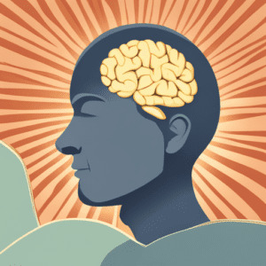
#basal forebrain
Explore tagged Tumblr posts
Visit Tumblr Blog
Explore Tumblr blogs with no restrictions, modern design and the best experience.
Last Seen Tumblr Blogs


Fun Fact
If you dial 1-866-584-6757, you can leave an audio post for your followers.
Text
we've updated the terminology of the triune brain model to match our newest evidence for the development of the relevant vertebrate forebrain structures:
reptilian complex (basal ganglia) -> vertebrate complex paleomammalian complex (limbic system) -> amniotic complex neomammalian (neocortex) -> mammalian complex
this has not affected the falsity of its science. thank you
30 notes
·
View notes
Text
Interesting Papers for Week 44, 2023
Rhythmic temporal coordination of neural activity prevents representational conflict during working memory. Abdalaziz, M., Redding, Z. V., & Fiebelkorn, I. C. (2023). Current Biology, 33(9), 1855-1863.e3.
Which processes dominate visual search: Bottom-up feature contrast, top-down tuning or trial history? Becker, S. I., Grubert, A., Horstmann, G., & Ansorge, U. (2023). Cognition, 236, 105420.
Neural dynamics underlying associative learning in the dorsal and ventral hippocampus. Biane, J. S., Ladow, M. A., Stefanini, F., Boddu, S. P., Fan, A., Hassan, S., … Kheirbek, M. A. (2023). Nature Neuroscience, 26(5), 798–809.
A reservoir of foraging decision variables in the mouse brain. Cazettes, F., Mazzucato, L., Murakami, M., Morais, J. P., Augusto, E., Renart, A., & Mainen, Z. F. (2023). Nature Neuroscience, 26(5), 840–849.
Spike-phase coupling patterns reveal laminar identity in primate cortex. Davis, Z. W., Dotson, N. M., Franken, T. P., Muller, L., & Reynolds, J. H. (2023). eLife, 12, e84512.
Is all mental effort equal? The role of cognitive demand-type on effort avoidance. Embrey, J. R., Donkin, C., & Newell, B. R. (2023). Cognition, 236, 105440.
Ventral striatum dopamine release encodes unique properties of visual stimuli in mice. Gonzalez, L. S., Fisher, A. A., D’Souza, S. P., Cotella, E. M., Lang, R. A., & Robinson, J. E. (2023). eLife, 12, e85064.
Computational complexity drives sustained deliberation. Hong, T., & Stauffer, W. R. (2023). Nature Neuroscience, 26(5), 850–857.
Mathematical Model of Synaptic Long-Term Potentiation as a Bistability in a Chain of Biochemical Reactions with a Positive Feedback. Katauskis, P., Ivanauskas, F., & Alaburda, A. (2023). Acta Biotheoretica, 71(3), 16.
Geometric determinants of the postrhinal egocentric spatial map. LaChance, P. A., & Taube, J. S. (2023). Current Biology, 33(9), 1728-1743.e7.
Learning at your brain’s rhythm: individualized entrainment boosts learning for perceptual decisions. Michael, E., Covarrubias, L. S., Leong, V., & Kourtzi, Z. (2023). Cerebral Cortex, 33(9), 5382–5394.
Retinal motion statistics during natural locomotion. Muller, K. S., Matthis, J., Bonnen, K., Cormack, L. K., Huk, A. C., & Hayhoe, M. (2023). eLife, 12, e82410.
Neural dynamics and architecture of the heading direction circuit in zebrafish. Petrucco, L., Lavian, H., Wu, Y. K., Svara, F., Štih, V., & Portugues, R. (2023). Nature Neuroscience, 26(5), 765–773.
Elucidating a locus coeruleus-dentate gyrus dopamine pathway for operant reinforcement. Petter, E. A., Fallon, I. P., Hughes, R. N., Watson, G. D., Meck, W. H., Ulloa Severino, F. P., & Yin, H. H. (2023). eLife, 12, e83600.
Principles for coding associative memories in a compact neural network. Pritz, C., Itskovits, E., Bokman, E., Ruach, R., Gritsenko, V., Nelken, T., … Zaslaver, A. (2023). eLife, 12, e74434.
Critical Drift in a Neuro-Inspired Adaptive Network. Sormunen, S., Gross, T., & Saramäki, J. (2023). Physical Review Letters, 130(18), 188401.
Dopaminergic prediction errors in the ventral tegmental area reflect a multithreaded predictive model. Takahashi, Y. K., Stalnaker, T. A., Mueller, L. E., Harootonian, S. K., Langdon, A. J., & Schoenbaum, G. (2023). Nature Neuroscience, 26(5), 830–839.
Inhibitory control of sharp-wave ripple duration during learning in hippocampal recurrent networks. Vancura, B., Geiller, T., Grosmark, A., Zhao, V., & Losonczy, A. (2023). Nature Neuroscience, 26(5), 788–797.
Optogenetics reveals paradoxical network stabilizations in hippocampal CA1 and CA3. Watkins de Jong, L., Nejad, M. M., Yoon, E., Cheng, S., & Diba, K. (2023). Current Biology, 33(9), 1689-1703.e5.
The cholinergic basal forebrain provides a parallel channel for state-dependent sensory signaling to auditory cortex. Zhu, F., Elnozahy, S., Lawlor, J., & Kuchibhotla, K. V. (2023). Nature Neuroscience, 26(5), 810–819.
#neuroscience#science#research#brain science#scientific publications#cognitive science#neurobiology#cognition#psychophysics#neurons#neural computation#neural networks#computational neuroscience
18 notes
·
View notes
Text
Basal Nuclei
-- initiates and terminates body movement
-- at forebrain base and midbrain top
-- interconnected with cerebral cortex, thalamus, and brainstem
#studyblr#notes#my notes#biology#bio#human biology#human bio#anatomy#human anatomy#anatomical structures#anatomical vocabulary#anatomical variation#medblr#medical notes#med notes#human anatomical structures#anatomy and physiology#anatomy & physiology#life science#biological science#note cards#flashcards#flash cards
4 notes
·
View notes
Text
The Habit Loop Explained: Why Your Brain Loves Repetition

As creatures of habit, our daily actions are largely driven by the habit cycle, which accounts for 40 to 50% of what we do each day instead of conscious decision making. The failure rate of New Year’s resolutions proves this point — only 35% of people kept their 2020 resolutions, and a mere 19% stuck with them beyond two years, highlighting the challenges of changing habits.
The habit cycle’s three components are the cue, routine, and reward. This cycle shapes how our brain’s habits form and creates automatic behaviors that become part of our daily lives. The repeated combination of these elements develops a craving that powers the whole process, making it a habit-forming mechanism.
The science behind habits shows why creating new behaviors isn’t simple. Research indicates people need between 18 and 254 days to establish a new habit. This significant variation demonstrates the complexity of habit formation and explains why quick fixes rarely work when it comes to changing a habit. We’ll explore the habit cycle’s mechanics, the neuroscience of habit formation, and practical ways to make use of this knowledge to build better routines and change your habits.
What is the habit cycle and why it matters
A powerful neurological pattern shapes our daily actions beneath every automatic behavior. The habit cycle functions as a sophisticated feedback system. This mechanism allows our brains to transform repeated behaviors into automatic routines and frees up mental energy for complex tasks, illustrating how habits work in our daily lives.
Four distinct elements work together in the habit cycle to create automatic behaviors. The cue acts as a trigger that tells your brain to start a behavior. Your brain might respond to location-based cues, time-specific triggers, emotional states, people around you, or actions that came before. The cue then creates a craving that drives every habit. People don’t actually crave the habit — they want the change in their internal state it brings.
The response comes next as the actual habit you perform through thoughts or actions. Your motivation levels and the required effort determine whether a response happens. The reward finishes the loop. This satisfies your craving and teaches your brain which actions deserve remembering, playing a crucial role in habit-forming processes.
These four steps create an endless neurological feedback loop in our brains. The process splits into two phases. The problem phase (cue and craving) shows us something needs to change. The solution phase (response and reward) pushes us to take action.
This system holds great value for our brains because it optimizes our functioning. The brain creates hardwired information about our responses through repeated actions. This allows us to complete tasks without conscious awareness. Our daily actions reflect this efficiency — 40–50% happen through habits rather than conscious decisions.
The brain’s reward system plays a vital role here. Dopamine, a key neurotransmitter in our neurotransmitter systems, doesn’t spike during the reward. Instead, it increases in anticipation — right when cravings become active. This biological mechanism explains why breaking habits proves so challenging and why immediate rewards are so powerful in reinforcing behavioral patterns.
The habit cycle developed as a way to conserve energy. The basal ganglia in the forebrain takes control once habits form and lets actions happen automatically. The prefrontal cortex manages which habits activate moment by moment, demonstrating the interplay between different brain regions in habit formation.
Understanding what a habit is and how the habit cycle works provides the foundations for changing unwanted behaviors and building beneficial ones. We’ll explore these concepts more deeply in the sections that follow, including how to change a habit effectively.
How habits are formed in the brain
Our brains physically change as habits take root. A remarkable transition happens inside our skulls. The brain moves from conscious decisions to automatic behaviors, and a group of subcortical nuclei called the basal ganglia coordinates this change, showcasing the intricate process of developing a habit.
The prefrontal cortex actively works when we learn new behaviors. This demands our full attention and conscious effort. This brain region handles complex decision making while the dorsomedial striatum (DMS) manages goal-directed actions that need awareness of desired outcomes. Neurons in the striatum fire continuously as we perform these tasks.
Something fascinating happens with consistent practice. The dorsolateral striatum (DLS) starts to take control and lets habitual behavior run without conscious oversight. Scientists have noticed a dramatic change in neuron firing patterns as animals master habitual tasks. These patterns cluster at the start and end of routines instead of firing throughout. The brain uses this “bracketing pattern” to package behaviors it sees as valuable.
The striatum’s inhibitory neurons follow a matching pattern. They activate during learned sequences and might prevent interruptions until completion. This handoff from prefrontal cortex to striatum helps explain why 10-year old habits persist even when scientists experimentally deactivate the cortex. The involvement of the sensorimotor cortex in this process further illustrates the complexity of habit formation in the brain.
Dopamine works as the key reinforcement mechanism here. Each time we successfully perform a behavior, dopamine releases and deepens the thalamostriatal synapses. These neural pathways then become quicker until they work automatically without cortical input. This process involves complex corticostriatal circuits that are crucial for habit formation and maintenance.
This brain transformation shows neuroplasticity at work — knowing how to reorganize and strengthen connections between neurons. Studies reveal that repeated behaviors create new neural pathways while old habit connections become weaker. Neural networks grow stronger and more efficient as we repeat actions more often. They need smaller habit cues to activate the same network of brain cells.
The process of habit formation in the brain can be seen as a form of mental programming, where repeated behaviors become ingrained in our neural circuitry. This understanding of how habits are formed at a neurological level provides valuable insights into why changing habits can be challenging and how we can leverage this knowledge to create more effective behavioral strategies.
Using habit science to build better routines
Understanding how the habit cycle works gives you a powerful edge in creating new routines. You can use habit science to make positive behaviors automatic instead of depending on willpower alone, whether you’re trying to improve study habits or develop organizational habits.
Consistent tiny changes create most important results. Research shows that your brain adapts better to small habits compared to overwhelming ambitious goals. To name just one example, start with five-minute exercise sessions rather than committing to long workouts when you want to create a habit of regular physical activity.
Science proves that habit stacking helps embed new behaviors effectively. This technique uses existing neural pathways by connecting new habits to 10-year old ones. The simple formula works like this: “After [CURRENT HABIT], I will [NEW HABIT]”. Your morning coffee ritual can naturally trigger one minute of meditation, demonstrating how you can change your habits by building on existing ones.
The space around you shapes your habits powerfully. Studies show that visible good habits and hidden bad ones work better than motivation. Keep your workout clothes visible, place fruits where you can see them, and remove distracting apps from your phone’s home screen to make better choices easier. This approach to environmental design is one of the most effective behavioral strategies for habit change.
Identity-based habits are maybe even the deepest path to lasting change. Your brain processes habits differently when you focus on becoming someone who exercises daily rather than just wanting to lose weight. This shift in perspective can significantly impact the success of your habit-forming efforts.
Visual feedback drives your brain’s progress. People who track their journey toward goals like weight loss succeed twice as often as others. Remember that staying consistent matters more than being perfect. The “never miss twice” rule helps you bounce back from occasional slip-ups, which is crucial when changing a habit.
These proven strategies help you turn habit science into daily routines that last. By understanding concepts like reward devaluation and contingency degradation, you can design more effective habit-change interventions. For instance, recognizing how immediate rewards influence behavior can help you create more compelling incentives for sticking to new habits.
Conclusion
The habit cycle shapes nearly half of what we do each day — driven not by conscious decision-making, but by automatic routines. This guide unpacked how habits form through a cycle of cue, craving, response, and reward, and how consistent repetition rewires the brain for long-term change.
That’s exactly why Mevolve’s Habit Tracker is built to support this process — not fight it. Whether you’re forming a new fitness habit, breaking a screen-time loop, or adding mindful routines, Mevolve helps by:
Tracking your progress with visual feedback (streaks and charts)
Supporting multiple habit types (Yes/No, Timed, Number-based, and more)
Letting you start small, stack habits, and grow over time
Because behavior change isn’t about willpower — it’s about designing systems that match how your brain actually works. With Mevolve, you’re not just setting habits. You’re rewiring your life, one loop at a time.
Lasting behavior change needs patience and understanding. Many people want overnight changes, but science shows habit formation gradually reshapes our neural structure. All the same, this slower approach ended up creating more lasting results as new behaviors become automatic and need less conscious effort.
The habit cycle presents both a challenge and a chance. It explains why changing old habits is tough but also provides a blueprint to design better routines. So, people who become skilled at using this basic brain mechanism gain remarkable influence over their daily actions and future outcomes. By understanding and leveraging the principles of habit science, we can create more effective strategies for personal growth and development, transforming our behavioral patterns one habit at a time.
0 notes
Text
The Habit Loop Explained: Why Your Brain Loves Repetition
As creatures of habit, our daily actions are largely driven by the habit cycle, which accounts for 40 to 50% of what we do each day instead of conscious decision making. The failure rate of New Year’s resolutions proves this point — only 35% of people kept their 2020 resolutions, and a mere 19% stuck with them beyond two years, highlighting the challenges of changing habits.
The habit cycle’s three components are the cue, routine, and reward. This cycle shapes how our brain’s habits form and creates automatic behaviors that become part of our daily lives. The repeated combination of these elements develops a craving that powers the whole process, making it a habit-forming mechanism.
The science behind habits shows why creating new behaviors isn’t simple. Research indicates people need between 18 and 254 days to establish a new habit. This significant variation demonstrates the complexity of habit formation and explains why quick fixes rarely work when it comes to changing a habit. We’ll explore the habit cycle’s mechanics, the neuroscience of habit formation, and practical ways to make use of this knowledge to build better routines and change your habits.
What is the habit cycle and why it matters
A powerful neurological pattern shapes our daily actions beneath every automatic behavior. The habit cycle functions as a sophisticated feedback system. This mechanism allows our brains to transform repeated behaviors into automatic routines and frees up mental energy for complex tasks, illustrating how habits work in our daily lives.
Four distinct elements work together in the habit cycle to create automatic behaviors. The cue acts as a trigger that tells your brain to start a behavior. Your brain might respond to location-based cues, time-specific triggers, emotional states, people around you, or actions that came before. The cue then creates a craving that drives every habit. People don’t actually crave the habit — they want the change in their internal state it brings.
The response comes next as the actual habit you perform through thoughts or actions. Your motivation levels and the required effort determine whether a response happens. The reward finishes the loop. This satisfies your craving and teaches your brain which actions deserve remembering, playing a crucial role in habit-forming processes.
These four steps create an endless neurological feedback loop in our brains. The process splits into two phases. The problem phase (cue and craving) shows us something needs to change. The solution phase (response and reward) pushes us to take action.
This system holds great value for our brains because it optimizes our functioning. The brain creates hardwired information about our responses through repeated actions. This allows us to complete tasks without conscious awareness. Our daily actions reflect this efficiency — 40–50% happen through habits rather than conscious decisions.
The brain’s reward system plays a vital role here. Dopamine, a key neurotransmitter in our neurotransmitter systems, doesn’t spike during the reward. Instead, it increases in anticipation — right when cravings become active. This biological mechanism explains why breaking habits proves so challenging and why immediate rewards are so powerful in reinforcing behavioral patterns.
The habit cycle developed as a way to conserve energy. The basal ganglia in the forebrain takes control once habits form and lets actions happen automatically. The prefrontal cortex manages which habits activate moment by moment, demonstrating the interplay between different brain regions in habit formation.
Understanding what a habit is and how the habit cycle works provides the foundations for changing unwanted behaviors and building beneficial ones. We’ll explore these concepts more deeply in the sections that follow, including how to change a habit effectively.
How habits are formed in the brain
Our brains physically change as habits take root. A remarkable transition happens inside our skulls. The brain moves from conscious decisions to automatic behaviors, and a group of subcortical nuclei called the basal ganglia coordinates this change, showcasing the intricate process of developing a habit.
The prefrontal cortex actively works when we learn new behaviors. This demands our full attention and conscious effort. This brain region handles complex decision making while the dorsomedial striatum (DMS) manages goal-directed actions that need awareness of desired outcomes. Neurons in the striatum fire continuously as we perform these tasks.
Something fascinating happens with consistent practice. The dorsolateral striatum (DLS) starts to take control and lets habitual behavior run without conscious oversight. Scientists have noticed a dramatic change in neuron firing patterns as animals master habitual tasks. These patterns cluster at the start and end of routines instead of firing throughout. The brain uses this “bracketing pattern” to package behaviors it sees as valuable.
The striatum’s inhibitory neurons follow a matching pattern. They activate during learned sequences and might prevent interruptions until completion. This handoff from prefrontal cortex to striatum helps explain why 10-year old habits persist even when scientists experimentally deactivate the cortex. The involvement of the sensorimotor cortex in this process further illustrates the complexity of habit formation in the brain.
Dopamine works as the key reinforcement mechanism here. Each time we successfully perform a behavior, dopamine releases and deepens the thalamostriatal synapses. These neural pathways then become quicker until they work automatically without cortical input. This process involves complex corticostriatal circuits that are crucial for habit formation and maintenance.
This brain transformation shows neuroplasticity at work — knowing how to reorganize and strengthen connections between neurons. Studies reveal that repeated behaviors create new neural pathways while old habit connections become weaker. Neural networks grow stronger and more efficient as we repeat actions more often. They need smaller habit cues to activate the same network of brain cells.
The process of habit formation in the brain can be seen as a form of mental programming, where repeated behaviors become ingrained in our neural circuitry. This understanding of how habits are formed at a neurological level provides valuable insights into why changing habits can be challenging and how we can leverage this knowledge to create more effective behavioral strategies.
Using habit science to build better routines
Understanding how the habit cycle works gives you a powerful edge in creating new routines. You can use habit science to make positive behaviors automatic instead of depending on willpower alone, whether you’re trying to improve study habits or develop organizational habits.
Consistent tiny changes create most important results. Research shows that your brain adapts better to small habits compared to overwhelming ambitious goals. To name just one example, start with five-minute exercise sessions rather than committing to long workouts when you want to create a habit of regular physical activity.
Science proves that habit stacking helps embed new behaviors effectively. This technique uses existing neural pathways by connecting new habits to 10-year old ones. The simple formula works like this: “After [CURRENT HABIT], I will [NEW HABIT]”. Your morning coffee ritual can naturally trigger one minute of meditation, demonstrating how you can change your habits by building on existing ones.
The space around you shapes your habits powerfully. Studies show that visible good habits and hidden bad ones work better than motivation. Keep your workout clothes visible, place fruits where you can see them, and remove distracting apps from your phone’s home screen to make better choices easier. This approach to environmental design is one of the most effective behavioral strategies for habit change.
Identity-based habits are maybe even the deepest path to lasting change. Your brain processes habits differently when you focus on becoming someone who exercises daily rather than just wanting to lose weight. This shift in perspective can significantly impact the success of your habit-forming efforts.
Visual feedback drives your brain’s progress. People who track their journey toward goals like weight loss succeed twice as often as others. Remember that staying consistent matters more than being perfect. The “never miss twice” rule helps you bounce back from occasional slip-ups, which is crucial when changing a habit.
These proven strategies help you turn habit science into daily routines that last. By understanding concepts like reward devaluation and contingency degradation, you can design more effective habit-change interventions. For instance, recognizing how immediate rewards influence behavior can help you create more compelling incentives for sticking to new habits.
Conclusion
The habit cycle shapes nearly half of what we do each day — driven not by conscious decision-making, but by automatic routines. This guide unpacked how habits form through a cycle of cue, craving, response, and reward, and how consistent repetition rewires the brain for long-term change.
That’s exactly why Mevolve’s Habit Tracker is built to support this process — not fight it. Whether you’re forming a new fitness habit, breaking a screen-time loop, or adding mindful routines, Mevolve helps by:
Tracking your progress with visual feedback (streaks and charts)
Supporting multiple habit types (Yes/No, Timed, Number-based, and more)
Letting you start small, stack habits, and grow over time
Because behavior change isn’t about willpower — it’s about designing systems that match how your brain actually works. With Mevolve, you’re not just setting habits. You’re rewiring your life, one loop at a time.
Lasting behavior change needs patience and understanding. Many people want overnight changes, but science shows habit formation gradually reshapes our neural structure. All the same, this slower approach ended up creating more lasting results as new behaviors become automatic and need less conscious effort.
The habit cycle presents both a challenge and a chance. It explains why changing old habits is tough but also provides a blueprint to design better routines. So, people who become skilled at using this basic brain mechanism gain remarkable influence over their daily actions and future outcomes. By understanding and leveraging the principles of habit science, we can create more effective strategies for personal growth and development, transforming our behavioral patterns one habit at a time.
Talk to Us : https://go.mevolve.app/AE
Visit Website : https://go.mevolve.app/AF
0 notes
Text
Tangled in Tingles: The effect of ASMR on Mental Health
If you’re reading this, you have likely heard of something called the Autonomous Sensory Meridian Response (ASMR). Colloquially known as ‘Brain Massage’ or ‘Tingles’ ; ASMR refers to the sensation that originates in the scalp, neck, and shoulders in response to visual or auditory stimuli. Many people experience the ‘classic’ tingles in the form of a pleasant prickling sensation down the head, neck, and shoulders. Others feel more of a ‘fog’ sensation that rolls down their entire bodies. While still others feel nothing at all! Crazy cool how people can experience the same phenomenon in different ways right?
So what makes this somatic experience so unique to everyone? How does it aid in anxiety relief?
Well first- let’s discuss what ‘tingles’ actually are.
ASMR is a fairly new and abstract phenomenon that is being studied. It is currently being related to the phenomenon of frisson, or musical chills, since both are characteristically described as a ‘tingling’ sensation that is induced through stimuli deemed pleasant by the perceiver. MRI research has shown that multiple brain areas are activated during frisson like nucleus accumbens, the basal forebrain, and insular cortices. That’s some big vocabulary there! What is important to take away here is that ASMR utilizes all of these areas to provide a dopamine pathway to the limbic system that induces relaxation which can aid in sleep. What makes ASMR interesting is the sheer variety in how people experience it. Some experience a dynamic that rapidly spreads through the whole body and originates in various parts of the body, while others experience sensations that start in the top of the scalp and radiate downward. The perceived differences in experiences may possibly be explained by medial prefrontal cortex activity, which is heightened for some during frisson. Meaning how your nervous system reacts to stimuli is conditioned by your environment.
TLDR: ASMR is the nervous system directly responding to visual and auditory stimuli, but the experiences are different for everyone based on their own life experiences to various stimuli.
Can we skip to the good part? The effect of ASMR on Mental Health!
ASMR can be defined as a mindfulness tool, which in therapy is able to help empower and provide feelings of action for clients towards uncertainties such as anxiety and depression. ASMR utilization allows for a sensory experience that disrupts the nervous system and provides feelings of primal comfort.
From a wider perspective actions that are portrayed in ASMR videos are often considered primal emotional expressions that stimulate feelings of belonging within a ‘pack’. Things like grooming, whispering, tapping, and watching another person complete something carefully are social or intimate activities that promote bonding and feelings of safety.
So, How does one do ASMR?
Very simply, you just do it! There are millions of stimuli and applications for comfort or the pursuit of these tingles are really endless, find what works for you! There are many different apps available for mobile and pc devices, but the most accessible is definitely Youtube. Look for something that seems interesting to you and try it out. I personally enjoy videos that read and follow the words across the page, or videos ofintricate miniature builds that people perform. I don’t experience the tingle sensation, but I do get intense relaxation and feelings of calm.
Please get in touch. We will be happy to discuss how I may be able to help.
0 notes
Text
Alzheimer’s disease (AD) is a neurodegenerative disease significantly impacting cognitive function; the pathogenesis of Alzheimer’s is complex and is due to the interplay of genetic, biochemical, and inflammatory factors. Afflicted individuals typically exhibit severe memory loss, confusion, difficulty with problem-solving and language, and disorientation. The excessive accumulation of β-amyloid proteins is caused by mutations in genes such as amyloid precursor protein (APP), presenilin 1 (PS-1), and presenilin 2 (PS-2)– which cause glial damage and amyloid plaques. Plaques are significant for being pathological markers of disease,
Amyloid plaques, neurofibrillary tangles, and significant neuronal loss are the three features that signify Alzheimer’s presence. The tau protein, known for forming a structure of the microtubule, maintains the health of neurons; in Alzheimer’s, tau is phosphorylated abnormally, causing microtubule structures to collapse. Without any structural integrity, no neurotransmitter pathway can work properly. This degeneration of neural systems affects the cholinergic system, which is linked to memory and learning systems– more specifically, the basal forebrain which is a contributor to the cognitive critical area.

Cholinergic systems are significant for modulating the brain’s processing of information, acetylcholine facilitates synaptic plasticity, which measures the ability for the brain to adapt to new information. In Alzheimer’s specifically, there is a deficiency of cholinergic neurons, resulting in the deterioration of cognitive function.
The monoaminergic system, consisting of monoamines such as serotonin, dopamine, and norepinephrine, plays an important role in neurological behavior, and the degradation of any of these, contribute to the significant degradation of the disease. To begin with, the serotonergic system, involved in mood regulation, cognitive function, and sleep, interact with amyloidogenic processes, which play a major role in behavior, along with depression symptoms. Serotonergic neurons are typically found in the raphe nuclei in the brainstem. Next, dopaminergic neurons, located mostly in the substantia nigra and ventral tegmental area, mostly affect motor function and motivation– affecting one’s cognitive control, degenerated dopaminergic neurons will affect one’s physical function and influence the spread of neurofibrillary tangles (tau protein instability.) Lastly, norepinephrine is critical for stress responses and arousal. Located primarily in the locus coeruleus, noradrenergic neurons can affect energy levels, and normal anti-inflammatory properties; if this system is damaged, the disease process may be accelerated.
The recognition of the cholinergic, serotonergic, dopaminergic, and noradrenergic neurons in Alzheimer’s pathophysiology can lead to drugs to help treat the degenerative disease. To begin with, using cholinesterase inhibitors can inhibit the enzyme that breaks down acetylcholine, in order to protect its degradation and increase its availability in the brain. Increasing cholinergic function can slow down the progression of cognitive decline and foster temporary mitigation of symptoms. Next, focusing on the monoaminergic systems, one can improve quality of life through the focus of mood regulation and stability. Currently, serotonin and norepinephrine reuptake inhibitors (SSRIs and SNRIs) are used to assist with mood and depressive symptoms. SSRIs work through the natural releasing of serotonin into the presynaptic neuron into the synaptic cleft. Once neurotransmission occurs, serotonin binds to specific receptors that allow for cellular responses to influence mood, SSRIs block the natural reuptake of serotonin by blocking the serotonin transporters; by preventing the reuptake of serotonin, more serotonin is left in the synaptic cleft, causing an enhanced and prolonged effect of improved mood. SNRIs are similar, in that they influence both serotonin and norepinephrine. Enhanced levels of neurotransmission help alleviate symptoms of depression, which is incredibly important to manage the symptoms of Alzheimer’s and slow down degeneration. Lastly, using dopaminergic agonists and antagonists, drugs can mimic dopamine by binding to receptors (agonists), or they can block dopamine receptors (antagonists) in order to treat comorbid conditions.
The cholinergic and monoaminergic systems both play key roles in the degradation of cognitive function seen in Alzheimer’s. Alzheimer’s disease affects multiple neural pathways and it is important to understand the interplay between systems and markers of disease.
0 notes
Quote
Molecular diversity of microglia, the resident immune cells in the CNS, is reported. Whether microglial subsets characterized by the expression of specific proteins constitute subtypes with distinct functions has not been fully elucidated. Here we describe a microglial subtype expressing the enzyme arginase-1 (ARG1; that is, ARG1+ microglia) that is found predominantly in the basal forebrain and ventral striatum during early postnatal mouse development. ARG1+ microglia are enriched in phagocytic inclusions and exhibit a distinct molecular signature, including upregulation of genes such as Apoe, Clec7a, Igf1, Lgals3 and Mgl2, compared to ARG1– microglia. Microglial-specific knockdown of Arg1 results in deficient cholinergic innervation and impaired dendritic spine maturation in the hippocampus where cholinergic neurons project, which in turn results in impaired long-term potentiation and cognitive behavioral deficiencies in female mice. Our results expand on microglia diversity and provide insights into microglia subtype-specific functions.
ARG1-expressing microglia show a distinct molecular signature and modulate postnatal development and function of the mouse brain | Nature Neuroscience
0 notes
Text
Caffeine Effects On The Brain
Have you ever thought about what caffeine does to your body? Well, I have and I did some research on it. Some might think it is just a cup of coffee or a can of energy drink but there is so much more to it than that. You might feel good at first but eventually, you might feel a lang in energy. That is from the caffeine's blockade of your adenosine receptors and increase of the autonomic nervous system (Gonzaga). Adenosine is said to act as a homeostatic regulator of sleep and helps regulate when a person is awake or asleep (National Institute of Health). Adenosine is found in ATP. ATP is the source of energy for use and storage at a cellular level (Biology). The structure of ATP is a nucleotide triphosphate, consisting of a nitrogen base (adenine), ribose sugar, and three serially bonded phosphate groups (Biology). An increase in adenosine increases a person's need for sleep. There are many parts of the brain that are involved for sleep such as the hypothalamus, brainstem, thalamus, cerebral cortex, pineal gland, basal forebrain, midbrain, and amygdala (National Institute of Health). Some signs or symptoms of too much caffeine include a rise in body temperature, rapid heartbeat, and frequent urination which leads to dehydration, restlessness, anxiety, or shaky hands (Mayo Clinic). If caffeine intake is done in moderation then it can have good health effects. Such good health effects like one could live longer, your body may process glucose better, and a lesser chance of developing Parkinson's disease, it might also help keep the chances of colon cancer down (John Hopkins). Caffeine also can affect multiple systems of the body. The renal system, cardiovascular system, limbic system, and gastrointestinal system just to name a few (Leonard). Caffeine is a central nervous system stimulant. Other central system stimulants are used to treat ADD, narcolepsy, and excessive sleepiness. Some central nervous system stimulants are illegal due to people misusing them such as cocaine and ecstasy (Livertox) Located in the limbic system is the hippocampus. The hippocampus manages the function of feeling and reacting. Other known as the "fight or flight" response. The hippocampus also helps process and retrieve declarative memories and spatial relationships. Short-term memories are converted into long-term memories in the hippocampus. (MedicalNews Today)Caffeine affects everyone differently. The amount of caffeine also contributes to its lasting effects.
Gonzaga, L.A., Vanderlei, L.C.M., Gomes, R.L. et al. Caffeine affects autonomic control of heart rate and blood pressure recovery after aerobic exercise in young adults: a crossover study. Sci Rep 7, 14091 (2017). https://doi.org/10.1038/s41598-017-14540-4
Johns Hopkins Medicine. https://www.hopkinsmedicine.org/health/wellness-and-prevention/9-reasons-why-the-right-amount-of-coffee-is-good-for-you
Leonard TK, Watson RR, Mohs ME. The effects of caffeine on various body systems: a review. J Am Diet Assoc. 1987 Aug;87(8):1048-53. PMID: 3301987.
LiverTox: Clinical and Research Information on Drug-Induced Liver Injury [Internet]. Bethesda (MD): National Institute of Diabetes and Digestive and Kidney Diseases; 2012-. Central Nervous System (CNS) Stimulants. [Updated 2021 Aug 12]. Available from: https://www.ncbi.nlm.nih.gov/books/NBK548702/
Mayo Clinic Staff. https://www.mayoclinic.org/healthy-lifestyle/nutrition-and-healthy-eating/in-depth/caffeine/art-20045678 March 19, 2022.
MedicalNewsToday. What is the Hippocampus. Medically reviewed by Nancy Hammond, M.D. — By Rachael Ajmera, MS, RD — Updated on February 17, 2023 https://www.medicalnewstoday.com/articles/313295
National Institute of Neurological Disorders and Stroke. Brain Basics: Understanding Sleep. https://www.ninds.nih.gov/health-information/public-education/brain-basics/brain-basics-understanding-sleep#:~:text=The%20brain%20stem%2C%20at%20the,%2C%20medulla%2C%20and%20midbrain.)
1 note
·
View note
Text
I’d learned by this point that comparing brains is a difficult business in general. In explaining how clever humans are, we often point out the extraordinarily large size of our thinking organs. Their bulk is the bane of childbirth and consumes 90 percent of the glucose in our blood. But size itself is not a clear guide for comparing animal intelligences, as some bigger animals with larger brains seem to lack the cognitive abilities of smaller ones. Size, as the saying goes, isn’t everything. Relative brain-to-body size, how wrinkled and complex brains are, the thickness of their layers, the structures within them, and the types of neurons these are made of are all helpful—though our human brains are, naturally, the yardstick that other brains are measured against. And yet it is impossible to look at a whale brain and not be surprised by its size. When Hof first saw one, despite knowing they were big, its mass still shocked him. The human brain is about 1,350 grams, three times larger than our big-brained relative, the chimpanzee. A sperm whale or killer whale brain can be 10 kilograms. These are the biggest brains on Earth and possibly the biggest brains ever, anywhere. It’s perhaps not a fair comparison: in relation to the size of our bodies, our brains are bigger than those of whales. Ours are similar in proportion to our body mass, as are the brains of some rodents; mice and men both invest a lot of themselves in their thinking organs. But we both lag far behind small birds and ants, which have much bigger brains compared to their body size than any big animals.
The outer layer of a mammal’s brain is called the cerebral cortex. In cross section, it looks a little like a wraparound bicycle helmet sitting on top of the other parts of the brain. This is the most recently evolved part of our brains, and it was by using their own cerebral cortexes that brain scientists have learned that this area is responsible for rational, conscious thought.
It handles tasks like perceiving senses, thinking, movement, figuring out how you relate to the space around you, and language. You are using yours now to read and think about this sentence. Many biologists define “intelligence” as something along the lines of the mental and behavioral flexibility of an organism to solve problems and come up with novel solutions. In humans, the cerebral cortex, acting with other bits of the brain (the basal ganglia, basal forebrain, and dorsal thalamus), appears to be the seat of this form of “intelligence.” The more cortex you have and the more wrinkled it is, the more surface area available for making connections—and voila! More thinking.
Humans have a really large neocortex surface area, but it’s still just over half that of a common dolphin, and miles behind the sperm whale. Even if you divide the cortex area by the total weight of the brain to remove the cetacean size advantage, humans still lag behind dolphins and killer whales. But there are other measurements in the cortex that seem to be associated with intelligence, and here, dolphins and whales lag behind humans.
The more neurons are packed in, how closely and effectively they are wired, and how fast they transmit impulses are also extremely important in brain function. Just as the composition and layout of the chipset in your tiny, cheap cellphone allows it to pack more computing power than a five-tonne room-sized 1970s supercomputer. Both cetaceans and elephants, the biggest mammals on sea and land, seem to have large distances between their neurons and slower conduction speeds. In raw numbers of neurons, humans here, too, have the edge, with a human cortex containing an estimated 15 billion neurons. Given the larger size of cetacean brains, you’d think they’d have more, but in fact their cerebral cortex is thinner, and the neurons are fatter, taking up more room.
Nevertheless, some cetaceans such as the false killer whale are close behind human levels with 10.5 billion cerebral neurons, about the same as an elephant. Chimps have 6.2 billion and gorillas 4.3 billion. Further complicating comparisons, whales have huge numbers of other kinds of cells, called glia, packing their cortexes. Until recently, we believed these glial cells to be an unthinking filler, but we’ve now discovered that they actually seem important for cognition, too. I don’t know about you, but all this cortex measurement and comparison makes my own feeble organ hurt.
— In the Mind of a Whale
#tom mustill#in the mind of a whale#science#zoology#biology#human biology#marine biology#neuroscience#brain#cerebral cortex#basal ganglia#basal forebrain#thalamus#neocortex#neurons#glia#patrick hof#whales#intelligence
1 note
·
View note
Text
Interesting Papers for Week 25, 2024
Silencing CA1 pyramidal cells output reveals the role of feedback inhibition in hippocampal oscillations. Adaikkan, C., Joseph, J., Foustoukos, G., Wang, J., Polygalov, D., Boehringer, R., … McHugh, T. J. (2024). Nature Communications, 15, 2190.
A multi-demand operating system underlying diverse cognitive tasks. Cai, W., Taghia, J., & Menon, V. (2024). Nature Communications, 15, 2185.
A view-based decision mechanism for rewards in the primate amygdala. Grabenhorst, F., Ponce-Alvarez, A., Battaglia-Mayer, A., Deco, G., & Schultz, W. (2023). Neuron, 111(23), 3871-3884.e14.
Local and global predictors of synapse elimination during motor learning. Hedrick, N. G., Wright, W. J., & Komiyama, T. (2024). Science Advances, 10(11).
Laminar evoked responses in mouse somatosensory cortex suggest a special role for deep layers in cortical complexity. Hönigsperger, C., Storm, J. F., & Arena, A. (2024). European Journal of Neuroscience, 59(5), 752–770.
Synaptic wiring motifs in posterior parietal cortex support decision-making. Kuan, A. T., Bondanelli, G., Driscoll, L. N., Han, J., Kim, M., Hildebrand, D. G. C., … Lee, W.-C. A. (2024). Nature, 627(8003), 367–373.
Organization of reward and movement signals in the basal ganglia and cerebellum. Larry, N., Zur, G., & Joshua, M. (2024). Nature Communications, 15, 2119.
Autokinesis Reveals a Threshold for Perception of Visual Motion. Liu, Y., Tian, J., Martin-Gomez, A., Arshad, Q., Armand, M., & Kheradmand, A. (2024). Neuroscience, 543, 101–107.
Temporally organized representations of reward and risk in the human brain. Man, V., Cockburn, J., Flouty, O., Gander, P. E., Sawada, M., Kovach, C. K., … O’Doherty, J. P. (2024). Nature Communications, 15, 2162.
Neural timescales reflect behavioral demands in freely moving rhesus macaques. Manea, A. M. G., Maisson, D. J.-N., Voloh, B., Zilverstand, A., Hayden, B., & Zimmermann, J. (2024). Nature Communications, 15, 2151.
Changes in spatial self-consciousness elicit grid cell–like representation in the entorhinal cortex. Moon, H.-J., Albert, L., De Falco, E., Tasu, C., Gauthier, B., Park, H.-D., & Blanke, O. (2024). Proceedings of the National Academy of Sciences, 121(12), e2315758121.
Goal-seeking compresses neural codes for space in the human hippocampus and orbitofrontal cortex. Muhle-Karbe, P. S., Sheahan, H., Pezzulo, G., Spiers, H. J., Chien, S., Schuck, N. W., & Summerfield, C. (2023). Neuron, 111(23), 3885-3899.e6.
A persistent prefrontal reference frame across time and task rules. Muysers, H., Chen, H.-L., Hahn, J., Folschweiller, S., Sigurdsson, T., Sauer, J.-F., & Bartos, M. (2024). Nature Communications, 15, 2115.
Interactions between circuit architecture and plasticity in a closed-loop cerebellar system. Payne, H. L., Raymond, J. L., & Goldman, M. S. (2024). eLife, 13, e84770.
Functionally refined encoding of threat memory by distinct populations of basal forebrain cholinergic projection neurons. Rajebhosale, P., Ananth, M. R., Kim, R., Crouse, R., Jiang, L., López-Hernández, G., … Talmage, D. A. (2024). eLife, 13, e86581.
Functional architecture of dopamine neurons driving fear extinction learning. Salinas-Hernández, X. I., Zafiri, D., Sigurdsson, T., & Duvarci, S. (2023). Neuron, 111(23), 3854-3870.e5.
Neural attentional filters and behavioural outcome follow independent individual trajectories over the adult lifespan. Tune, S., & Obleser, J. (2024). eLife, 12, e92079.3.
Coordinated head direction representations in mouse anterodorsal thalamic nucleus and retrosplenial cortex. van der Goes, M.-S. H., Voigts, J., Newman, J. P., Toloza, E. H., Brown, N. J., Murugan, P., & Harnett, M. T. (2024). eLife, 13, e82952.
Specific rules for time and space of multisensory plasticity in the superior colliculus. Wang, L., Xin, H., Buren, Q., Zhang, Y., Han, Y., Ouyang, B., … Dong, C. (2024). Brain Research, 1828, 148774.
Structural constraints on the emergence of oscillations in multi-population neural networks. Zang, J., Liu, S., Helson, P., & Kumar, A. (2024). eLife, 12, e88777.3.
#neuroscience#science#research#brain science#scientific publications#cognitive science#neurobiology#cognition#psychophysics#neurons#neural computation#neural networks#computational neuroscience
6 notes
·
View notes
Text
Reticular Activating System and the Law of Attraction
Our brains are extremely intricate. At any given time, we may sift through billions of pieces of data, specifically 400000 million bits per second in our subconscious mind, compared to only 200 million bits per second in our conscious mind. And we have to organize the information in some way so that we don't short circuit. That's where the Reticular Activating System comes in.
The Reticular Activating System (RAS) is a network of nerves in our brainstem that filters away irrelevant information so that the crucial information can get through.
The RAS is what causes you to learn a new term and then hear it all over the place. It's why you can tune out a crowded room full of people chatting but jump to attention instantly when someone mentions your name or something that sounds like it. Your RAS develops a filter for whatever you're focusing on. It then sorts through the information and displays only the parts that are relevant to you.
RAS looks for evidence that supports your beliefs. It filters the environment through the parameters you set for it, and those parameters are shaped by your ideas. You will most likely be awful at giving speeches if you believe you are. You most certainly function efficiently if you believe you do. The RAS influences your actions by assisting you in seeing what you want to see.
Some people believe that you can train your RAS by connecting your subconscious thoughts to your conscious thoughts. It's known as "intent setting." This means that if you concentrate hard on your objectives, your RAS will expose the people, information, and chances that will assist you in achieving them.
If you care about something, such as optimism, you will become more conscious of it and seek it out. If you're serious about acquiring a pet turtle and have set your mind to it, you'll seek out the correct information to assist you.
Awake, asleep (resting or slow-wave sleep), and asleep and dreaming are the three sleep and arousal stages that humans have (REM rapid eye movement sleep). The reticular activating system in the mesopons interacts with the descending reticulospinal and ascending hypothalamic, basal forebrain, and thalamocortical systems to modulate these states.
The RAS is a network of neurons in the brain stem that project anteriorly to the hypothalamus to influence behavior, as well as posteriorly to the thalamus and directly to the cortex to activate awake, desynchronized cortical EEG patterns. Visceral, somatic, and sensory systems all provide input to the RAS. Acetylcholine, serotonin, noradrenalin, dopamine, histamine, and hypocretin are among the neurotransmitters used in this system (orexin).
How does this relate to the Law of Attraction, given that we now know that the brain actively scans massive volumes of data every second and that the RAS selects crucial information for you? Consider something you've wanted for a long time, such as a car...as soon as the concept of wanting this specific kind of automobile enters your mind, what do you see all of the time? Almost every time you're on the road, you'll see this particular car drive by (again, the RAS is pointing out this car for you). As a result, because the RAS is constantly pointing out this car to you, it's more probable that you'll be open to learning more about it.

E. Garcia, Rill, 2009. https://doi.org/10.1016/B978-008045046-9.01767-8
Suzanne Stevens, Wayne A. Hening,2007. https://doi.org/10.1016/B978-141603618-0.10002-5
68 notes
·
View notes
Text
The Mysteries of Motivation: Deep Dive into the Brain's Role
Our ideas, feelings, and, most crucially, our motivations all originate in the human brain, a natural miracle. But what is it in the human brain that causes us to take action, strive, and succeed? This article explores the exciting issue of Motivation by delving into the intricate neurological systems that support it. We'll learn about the brain regions that play a part in Motivation, the chemicals that play a part in that process, and the effects that neurological illnesses may have on our drive. We'll also discuss where this field of Study is going and how we can tap into the brain's latent motivational powers.
Which Part of Your Brain is Involved in Your Motivation?
Which Part Of Your Brain Is Involved In Your Motivation? The Limbic System, the Source of All Drive The limbic system, or "emotional brain," is fundamental regarding our drive and Motivation. The hippocampus and the amygdala are the only parts of this intricate network that govern our reward and punishment systems. The limbic system is pivotal in memory formation and regulates our emotional lives. The emotional component of memory causes us to anticipate or fear certain occurrences in the future. The Function of the Basal Forebrain in Memory and Learning The basal forebrain is engaged in learning and memory and is a crucial component of Motivation. It aids in shaping future behavior by associating it with positive or negative outcomes. This brain region is also essential for maintaining focus, regulating wakefulness, and falling asleep. It's vital for sensory perception and free-willed movement because it supplies the cerebral cortex with acetylcholine.
How Neurotransmitters Like Dopamine Drive Behavior
Dopamine, the Heroin of the Reward System Dopamine is a neurotransmitter often known as the "feel-good chemical of the brain." When we experience pleasure, our brains produce dopamine, which reinforces and motivates us to pursue similar experiences in the future. Dopamine regulates the secretion of other hormones and is also involved in motor control. Regarding the motivational aspect of reward-motivated behavior, these dopamine pathways are crucial. The Nucleus Accumbens and the Ventral Tegmental Area Work Together to Process Rewards The nucleus accumbens and ventral tegmental area (VTA) are inseparable when processing rewards and inspiring action. The anticipation of a reward triggers the release of dopamine in these regions. The nucleus accumbens, among other brain regions, receives dopamine from the ventral tegmental area (VTA), which ultimately causes a sensation of reward or pleasure. This drives us to keep doing things that are rewarding in the first place. Dopamine's Effects on Mood and Reinforcement Learning Dopamine is also crucial in the formation of emotional responses and long-term memory. It reinforces existing synapses by reinforcing positive connections in the hippocampus (the brain's learning and memory hub). This step is essential for reward learning to take place, in which we learn to repeat actions that resulted in satisfying results in the past.
Which region of the brain is responsible for our desires and emotions?
Physiology of the Amygdala The Brain's Emotional Control Center Emotional processing relies heavily on the amygdala, a little almond-shaped structure in the brain. It influences our motives and emotions and plays a pivotal role in the fear and pleasure responses. The amygdala also helps choose which memories to keep and where in the brain to keep them. This metric considers the intensity of the felt impact of an inevitable occurrence. For example, if a person goes through an emotionally taxing experience, the amygdala directs them to file away their thoughts and feelings about that time. The Motivating Power of the Prefrontal Cortex The frontal lobe of the brain, known as the prefrontal cortex, is responsible for executive functions such as planning, decision-making, and social conduct. It's essential to our Motivation since it aids in weighing potential gains against costs and deciding how best to proceed. The prefrontal cortex also controls executive tasks, including time management, focus switching, detail recall, self-control, and incorporating previous experiences into current actions. One of the Brain's Most Important Reward and Motivation Centers Another important brain region involved in Motivation is the striatum in the basal ganglia. It's active when we're feeling driven to do something since it plays a role in the brain's reward processing system. In addition to its function in motor and movement planning, the striatum is also involved in decision-making, Motivation, reinforcement, and the perception of rewards.
Where Do You Find Your Drive? Frontal Lobe?
Motivation and Self-Control Rely on the Lateral Prefrontal Cortex. The frontal lobe's lateral prefrontal cortex has a role in self-control. A key component of Motivation, willpower allows us to rein in urges and make choices that support our long-term objectives. Working memory, cognitive flexibility, planning, inhibition, and abstract thinking depend on activity in this part of the brain. Implications of Frontal Lobe Development on Risk-Taking and Social Acceptance During this time, a lot of development occurs in the brain's frontal lobe. Teenagers are often highly driven by peer approval and may participate in dangerous activities to get it, which might impact Motivation throughout this development stage. The frontal lobe is also responsible for movement, problem-solving, spontaneity, memory, language, beginning new tasks, judging, resisting temptation, and engaging in social and sexual conduct.
Imbalances in the Brain's Reward System and Psychiatric Illness
Brain's Reward System The Lateral Habenula Is an Important Part of the Brain's Reward System The small brain region known as the lateral habenula is pivotal in the brain's reward system. Dysfunction in this region has been associated with mental diseases characterized by excessive aggressiveness, which is thought to encode punishment by suppressing dopamine release. The lateral habenula is engaged in more than only pain processing; it also plays a role in reproduction, eating, sleeping, stress management, and immune function. Damage to the Brain's Reward System as a Cause of Aggression Misdirected activation of the brain's reward system in response to aggressive social cues is one possible cause of aggression. When stimulated, some parts of the amygdala may cause anger and aggressiveness, while other portions, when removed, can make laboratory animals more submissive. This points to the amygdala as a critical player in aggressive and violent behavior.
New Frontiers and Unanswered Questions in the Study of Motivation
Basal forebrain involvement in Motivation: a promising area for Study The basal forebrain is a potential topic for future Study because of its role in learning and memory. Studying this factor's influence on Motivation might light up fundamental questions about cognition and behavior. The basal forebrain plays a role in sleep, waking, attention, and different states of consciousness. A Look into the Future of Brain Stimulation and Its Effect on Motivation The potential of brain stimulation methods like transcranial magnetic stimulation to increase Motivation is being investigated. Preliminary studies indicate that activating specific brain regions might boost Motivation and performance. A lack of Motivation typically characterizes depression and attention deficit hyperactivity disorder (ADHD). Therefore, this might lead to novel therapies for both disorders.
Using Your Brain to Its Full Potential Through Inspiration
Using Your Brain To Its Full Potential Through Inspiration Cognitive Improvement: Fueling Your Brain with Inspiration Increase your brain's Motivation by engaging in cognitive improvement practices like brain training activities and mindfulness meditation. Motivated individuals may benefit from these methods because they enhance the brain circuits responsible for Motivation. For instance, brain training activities help you improve your memory and problem-solving abilities, simplifying planning and accomplishing your objectives. In contrast, mindfulness meditation can help you focus on your goals by lowering stress and worry. Motivation Improvement Through Mental Exercise

Motivation Improvement Through Mental Exercise Puzzles, memory games, and other forms of mental exercise have been shown to boost Motivation and cognitive performance. These mental workouts help strengthen brain areas responsible for processing Motivation and reward. Doing these things will improve your brain's Motivation and help you reach your objectives.
An Intricate Tango Between the Brain and Motivation
Summary: The Brain's Hidden Role in Optimal Motivation
The brain's part in Motivation involves complex structures, chemicals, and neuronal connections. Each region of the brain, from the primitive brain to the more evolved prefrontal cortex, is essential in determining our choices and actions. Learning about these processes may help you get insight into your motivations and use your brain more effectively to accomplish your objectives.
Exploring the Path Forward in Our Knowledge of What Drives People
We set out on a path of research and invention as we strive to decipher the secrets of the brain and Motivation. Discoveries, enhanced therapies, and a better knowledge of the human brain are all in store for motivational research in the years to come. The Study of Motivation is an academic exercise and a personal search for insight. Read the full article
#BasalForebrain#Brain#CognitiveScience#Dopamine#EmotionalBrain#LearningandMemory#LimbicSystem#Motivation#Neuroscience#PrefrontalCortex#Psychology#RewardSystem#whichpartofyourbrainisinvolvedinyourmotivation?
2 notes
·
View notes
Text
Drugs & Behavior Week 1
Intro
drugs: chemical compounds administered to produce a desired change in the body.
pharmacology:
(a) study of drug action on the body AKA pharmacodynamics
(b) study of the fate of drugs in the body AKA pharmacokinetics
psychoactive drugs: affect behavior by altering the functions of the nervous system.
psychopharmacology: study of how drugs affect the nervous system and behavior.
What You Will Need to Know for the Exam
Difference between the Central Nervous System (CNS) and Peripheral Nervous System (PNS), components of each system
Difference between somatic and autonomic nervous systems
Difference between sensory and motor function, definitions of each
Definition of plasticity
Explain the three different cuts to view brain structures: horizontal, sagittal, coronal
Three brain divisions based on surface features: cerebrum, cerebellum, brain stem
Three brain divisions based on brain development: forebrain, midbrain, hindbrain
Components of forebrain:
A. End brain
1. Cortex
2. Corpus callosum
3. Limbic system
4. Basal ganglia
5. Olfactory bulb
B. Between brain
1. Thalamus
2. Hypothalamus
Components of midbrain:
A. Tectum
B. Tegmentum
Components of hindbrain:
A. Pons
B. Cerebellum
C. Medulla
Divisions of the cortex:
1. Frontal lobe
2. Temporal lobe
3. Parietal lobe
4. Occipital lobe
Difference between sulci and gyri (i.e. which is a bump and which is a groove?)
Three areas of cortex:
1. primary motor cortex
2. primary sensory cortex
3. association cortex
Which brain structure can be severed as a last-resort treatment for severe epilepsy?
Structures of the limbic system:
1. Amygdala
2. Hippocampus
3. Cingulate cortex
Structures of basal ganglia:
1. Striatum
2. Globus pallidus
3. Nucleus accumbens
Difference between thalamus and hypothalamus, including locations and their major functions
What is the function of the pineal gland?
Structures of midbrain:
1. Substantia nigra
2. Ventral tegmental area (VTA)
3. Periaqueductal gray (PAG)
What happens when there is cerebellar dysfunction? Is there complete paralysis?
What is the medulla’s role in respiration?
What drugs suppress respiratory control?
How does the medulla act as a vomiting center?
Definition of a ventricle; know the four ventricle locations
Why is CSF so important?
What causes hydrocephalus?
Difference between cranial and spinal nerves
How many segments does the spinal cord consist of?
Major functions of the spinal cord
How is either the gray or white matter organized in the spinal cord?
What are the functions of the Autonomic Nervous System (ANS)?
Difference between the Sympathetic and Parasympathetic divisions of ANS:
(A) Which is rest & digest vs. fight or flight?
(B) Know general body controls with each division (i.e. heart rate, digestion, etc.)
What do neurotransmitters are released by the ANS?
10 notes
·
View notes
Link
Highlights
Dysfunction of anterior insula during anesthesia disables brain network transitions
Prestimulus activity of anterior insula predicts conscious access of visual stimuli
Anterior insula might be a gate for conscious access of sensory information
This cortical gate occupies an intermediate position along a neurocognitive hierarchy
Summary
Conscious access to sensory information is likely gated at an intermediate site between primary sensory and transmodal association cortices, but the structure responsible remains unknown. We perform functional neuroimaging to determine the neural correlates of conscious access using a volitional mental imagery task, a report paradigm not confounded by motor behavior. Titrating propofol to loss of behavioral responsiveness in healthy volunteers creates dysfunction of the anterior insular cortex (AIC) in association with an impairment of dynamic transitions of default-mode and dorsal attention networks. Candidate subcortical regions mediating sensory gating or arousal (thalamus, basal forebrain) fail to show this association. The gating role of the AIC is consistent with findings in awake participants, whose conscious access is predicted by pre-stimulus AIC activity near perceptual threshold. These data support the hypothesis that AIC, situated at an intermediate position of the cortical hierarchy, regulates brain network transitions that gate conscious access.
#anterior insular cortex#consciousness#quantum consciousness#Neuroscience#neurology#fav#print this off later
1 note
·
View note
Text
Is Lion’s Mane Mushroom effective for memory function?
Neurotrophic components are important in advertising and marketing the growth and differentiation of neurons. Nerve growth issue (NGF) is essential for the maintenance with the basal forebrain cholinergic process. Hericenones and erinacines isolated in the medicinal mushroom Hericium erinaceus can induce NGF synthesis in nerve cells. In this particular analyze, we evaluated the synergistic interaction among H. erinaceus aqueous extract and exogenous NGF to the neurite outgrowth stimulation of neuroblastoma-glioma mobile NG108-fifteen. The neuroprotective result from the mushroom extract toward oxidative tension was also examined. Aqueous extract of H. erinaceus was revealed to be non-cytotoxic to human lung fibroblast MRC-five and NG108-fifteen cells. The mix of ten ng/mL NGF with 1 μg/mL mushroom extract yielded the highest share improve of 60.six% neurite outgrowth. The extract contained neuroactive compounds that induced the secretion of extracellular NGF in NG108-15 cells, thereby promoting neurite outgrowth action. Nevertheless, the H. erinaceus extract unsuccessful to protect NG108-15 cells subjected to oxidative stress when applied in pre-remedy and co-remedy modes. In conclusion, the aqueous extract of H. erinaceus contained neuroactive compounds which induced NGF-synthesis and promoted neurite outgrowth in NG108-fifteen cells. The extract also Increased the neurite outgrowth stimulation activity of NGF when utilized together. The aqueous planning of H. erinaceus had neurotrophic although not neuroprotective activities.
References
Johnson B. Mind Lab Pro Review: The complete package nootropic?. Safety. 2019 May 22;9:10.
Johnson B. Alpha Brain Review: Onnit’s OLD nootropic looks STALE. Safety. 2019 Apr 20;9:10.
1 note
·
View note