#digital microscope handheld
Explore tagged Tumblr posts
Visit Tumblr Blog
Explore Tumblr blogs with no restrictions, modern design and the best experience.
Last Seen Tumblr Blogs


Fun Fact
Tumblr has a low social media market share in South America.
Text
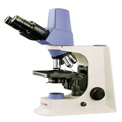
Digital Microscope - Labnic
Digital microscope is an advanced digital microscope with built-in 5.0 MP camera and USB 2.0 output. Equipped with an infinity semi-plan optical system and a standard magnification of 40X to 1000X. Its ergonomic design enables reliable operation for long hours.
#digital microscope#digital optical microscope#digital microscope online#digital microscope uses#digital microscope handheld#digital microscope for laboratory
0 notes
Text

Portable digital microscope
Portable digital microscope is an independent and easy to operate handheld unit. It provides powers from 20X and 300X with 5.0MP Image sensor. LED illumination with LED lamps for enhancing details in microscopic images.Compatible for PC usage with USB connection
#portable digital microscope#handheld microscope#best handheld microscope#tinyscope#digital usb microscope#portable microscope 1000x
0 notes
Text
Comprehensive Industrial Solutions by AxisValence: Advancing Productivity, Safety, and Efficiency
In today’s fast-paced manufacturing world, industrial productivity is driven by precision, consistency, safety, and compliance. Whether it’s printing, packaging, converting, textiles, plastics, or pharmaceuticals—modern production lines demand advanced electro-mechanical systems that minimize waste, ensure operational safety, and improve overall efficiency.
AxisValence, a business unit of A.T.E. India, addresses this demand with a complete range of industrial automation and enhancement products. From static elimination to print quality assurance, ink management, and solvent recovery, AxisValence solutions are engineered to optimize each critical point in the production cycle.
This article provides an overview of the key technologies and systems offered by AxisValence across its diverse portfolio:
Electrostatics: Managing Static for Quality and Safety
Electrostatics can compromise product quality, disrupt operations, and pose serious safety hazards, especially in high-speed processes involving films, paper, textiles, or volatile solvents. AxisValence offers a complete suite of static control solutions:
ATEX AC Static Eliminators: Certified for use in explosive or solvent-heavy environments such as rotogravure or flexo printing lines.
AC and DC Static Eliminators: Designed for long-range or close-range static charge neutralization across a range of substrates.
Passive Static Dischargers: Cost-effective, maintenance-free brushes for light-duty static elimination where power isn't available.
Air-based Static Eliminators / Ionisers: Use ionized air streams for dust blow-off and static removal, ideal for hard-to-reach areas.
Static Measurement & Online Monitoring: Includes handheld meters and IoT-enabled monitoring systems for real-time control and diagnostics.
Electrostatic Charging Systems: Generate controlled static charges for bonding or pinning applications in laminating or packaging lines.
Electrostatic Print Assist (ESA): Enhances ink transfer in rotogravure printing by improving ink pickup and registration.
Camera-Based Web Videos for Print Viewing: Real-Time Visual Inspection
High-speed printing applications require instant visibility into print quality. AxisValence’s ViewAXIS systems are high-performance, camera-based web viewing solutions:
ViewAXIS Mega: Entry-level system with high-resolution imaging for real-time visual inspection.
ViewAXIS Giga: Equipped with 14x optical zoom and X-ray vision for deeper inspection of layered prints.
ViewAXIS Tera: Full repeat system with a 55” display, allowing operators to monitor and inspect the complete print layout in real-time.
Camera-Based Web Videos for Print Viewing systems help identify print errors like registration issues, smudging, or color inconsistencies early in the production run—minimizing rework and improving efficiency.
100% Inspection Systems: Intelligent Defect Detection
Modern converters and packaging companies require automated systems that can identify microscopic flaws at high speeds. AxisValence’s DetectAXIS systems use AI-based image processing and line scan cameras for 100% inspection:

DetectAXIS Print: Identifies printing defects such as streaks, misregistration, color deviation, and missing text at speeds up to 750 m/min.
DetectAXIS Surface: Designed for detecting surface anomalies—scratches, gels, holes, fish-eyes—on films, textiles, and nonwovens.
Real-time alerts, digital roll-maps, and adaptive detection improve quality control while reducing material waste and production downtime.
Ink Handling Systems: Consistent Ink Quality and Reduced Waste
Stable ink flow and temperature directly impact print quality and solvent consumption. AxisValence’s Valflow range ensures optimal ink conditioning through:
Ink Filters: Eliminate contaminants like metallic particles, fibers, and dried pigments that can damage cylinders or cause print defects.
Ink Pumps & Tanks: Efficient centrifugal pumps and round stainless-steel tanks designed for continuous ink circulation and minimal ink residue.
Ink Temperature Stabilisers (ITS): Automatically control ink temperature to prevent viscosity drift and reduce solvent evaporation—delivering consistent print shade and odor-free operation.
Valflow Ink handling solutions are ideal for gravure and flexographic printing applications.
Print Register Control Systems: Precision Alignment in Every Print
Maintaining precise print registration control systemis critical in multi-color printing processes. AxisValence offers two specialized systems:
AlygnAXIS: For rotogravure presses, using fiber optic sensors and adaptive algorithms to deliver real-time register accuracy.
UniAXIS: A versatile controller for print-to-mark, coat-to-mark, and cut-to-mark applications—both inline and offline.
These controllers reduce waste, enhance print alignment, and speed up setup during job changes.
Safety and Heat Recovery Systems: Energy Efficiency and Explosion Prevention
Solvent-based processes require strict monitoring of air quality and heat management to meet compliance and reduce operational costs. AxisValence provides:
NIRA Residual Solvent Analyser: Lab-based gas chromatography system for quick analysis of residual solvents in films.
Air-to-Air Heat Exchangers (Lamiflow): Recover and reuse waste heat from drying processes—improving energy efficiency.
LEL Monitoring and Recirculation Systems: Ensure solvent vapor concentrations stay within safe limits in enclosed dryers using flame ionization or infrared detection.
Together, safety and heat recovery systems ensure both environmental safety and process optimization.
Surface Cleaning Systems: Contaminant-Free Production Lines
Particulate contamination can ruin coating, lamination, and printing jobs. AxisValence offers contactless surface cleaning systems that combine airflow and static control:
Non-Contact Web Cleaners: Use air curtains and vacuum to remove dust from moving substrates without physical contact.
Ionising Air Knives: High-velocity ionized air streams neutralize static and clean surfaces entering finishing zones.
Ionising Air Blowers: Cover larger surfaces with ionized air to eliminate static and debris.
Ionising Nozzles & Guns: Handheld or fixed, these tools offer targeted static and dust elimination at workstations.
Waste Solvent Recovery: Sustainable Ink and Solvent Reuse
Reducing solvent consumption and improving environmental compliance is critical for modern converters. AxisValence partners with IRAC (Italy) to offer:
Solvent Distillation Systems: Recover usable solvents from spent ink mixtures, reducing hazardous waste and cutting costs.
Parts Washers: Clean anilox rolls, gravure cylinders, and components through high-pressure, ultrasonic, or brush-based systems.
Waste solvent recovery systems offer a quick ROI and support zero-waste manufacturing goals.
Why Choose AxisValence?
AxisValence combines decades of industrial expertise with innovative product design to deliver reliable, safe, and efficient solutions for manufacturing processes. With a product portfolio spanning:
Electrostatics & Static Control Systems
Web Viewing & Print Inspection Solutions
Ink Handling and Conditioning Equipment
Register Control and Print Automation
Heat Recovery and Air Quality Monitoring
Surface Cleaning Technologies
Waste Solvent Management

…AxisValence serves diverse industries including printing, packaging, plastic and rubber, textile, pharma, and automotive.
From single-device retrofits to complete system integration, AxisValence enables manufacturers to improve output quality, reduce waste, meet safety norms, and gain a competitive edge.
Explore our full product range at www.axisvalence.com or contact our sales network for a customized consultation tailored to your industrial needs.
youtube
2 notes
·
View notes
Note
Hi, I enjoy your art a lot.
I used to make art but I've not been able to recently.
Is there anything that inspires you a lot? Or makes you feel the I want to draw/make this feeling?
Thank you in advance 🩷
hi! I was thinking over your message this week because this is something i struggle with a lot, so various media/things have worked at various times, but here are some things that have given me this feeling in the past: movies/tv: - kingdom of dreams and madness, song of the sea, from up on poppy hill, grand budapest hotel, phantom thread, the night is short walk on girl, urasawa naoki's manben books: invisible cities (italo calvino), piranesi (susanna clarke) underland (robert macfarlane). - skip & loafer, blue period, witch hat atelier - also various folklore and mythology collections other: visiting libraries or museums/digital collections online, keeping an eye out for unusual events/shows in your area, lectures/videos & interesting concepts (animals/architecture/culture, whatever), going on walks, & making cursed little crafts. - recently I got a little handheld microscope and while idk if it really counts as "inspo" it's been really special to be able to look at things up close, and makes you look at your everyday surroundings differently afterwards I just want to add that it's easy to be stuck in a cycle feeling so unhappy with yourself for being unable to make things like you used to. try not to worry too much, and pursue your interests without pressure of finding inspiration, the drive to create will follow. I hope you will encounter surprising and fun moments that fill you with joy!
#asks#anon#thank you for your message!! hope one of these can be a little helpful for you :^)#SORRY keep trotting out the same 4-5 movies sldkfjsldkfjslf#i just love media that can show you how strange and wonderful different interpretations of this world can be#if anyone has recommendations please feel free to suggest also!!! would love to find new things.......inspo hivemind
27 notes
·
View notes
Text
Can people with near sightedness see something far away if they use a close mirror?
People who can only see with glasses need them because their vision has degraded. And the structure of glasses is to enhance the image by placing a focusing apparatus a certain distance from their eye to get sharpest quality of what they're looking at. --Similar to how we use magnifying glasses and microscopes to see something outside of our regular range of vision-- right? At least that's the premise.
Cameras taking pictures, specifically old cameras, and not digital cameras, but also digital cameras. Expose themselves to light to capture the detail, which is engrained into some film or sensor. I remember hearing that the negatives of film cameras have all the detail up to extremely fine detail, but the method we had to expose them made the quality get loaded. Similar to how with increased pixels, we can sharpen the image of what is on a screen. Or polygons in 3D design in games.
Mirrors and other reflective surfaces reflect whatever light photons/waves hit it. Now the amount of light does decrease depending on how much light is absorbed, because mirrors are not perfect reflective surfaces.
So my question is this: would a near sighted person, one who cannot see far away from them, be able to see detail or far away objects, which they could not normally see, with clarity as if it was that close to them by using a handheld?
I will clarify that the person can read letters on a page very crisply at 10-12 size font at an arms length. However, no magnification of the object occurs. It is that simply what they are seeing is now "closer to them" because it is on the mirrored surface
My thought is that yes they could. Because it would be as if they had a picture or video of the thing they are looking at with great detail.
I want to know what anyone else thinks? I’ll do a poll for simplicity
#random polls#opinions#stem academia#stemblr#actually adhd#science hyperfixations#mirror#glasses#nearsightedness#optometry#focal point#eyes#I can’t see and I was staring at a tree that was blurry#then I flipped my mirror sunglasses and I thought I could see better#but I was like maybe I’m high#dude I’m so curious
5 notes
·
View notes
Text
Types of Digital Microscope
In today's world of advanced technology, digital microscopes have revolutionized the field of microscopy, offering enhanced imaging, precision, and ease of use. Unlike traditional optical microscopes, digital microscopes use a digital camera and a computer screen to display magnified images, making it easier to analyze and document samples. They have become essential tools in various industries, including healthcare, electronics, material science, and education. In this article, we will explore the different types of digital microscopes available, their unique features, and their applications.
1. USB Digital Microscopes
USB digital microscopes are among the most popular and widely used types of digital microscopes. They are compact, easy to use, and connect directly to a computer or mobile device via a USB cable. These microscopes are ideal for hobbyists, students, and professionals who need a portable and affordable solution for magnification and image capturing.
Key Features:
Direct connection to a computer or smartphone via USB.
High-resolution imaging (ranging from 2 MP to 14 MP).
Built-in LED lighting for enhanced visibility.
Easy to use with plug-and-play functionality.
Applications:
Educational purposes (biology, physics, and chemistry labs).
Jewelry inspection.
Coin and stamp collection analysis.
PCB and circuit board examination.
2. HDMI Digital Microscopes
HDMI digital microscopes are designed to provide high-quality, real-time imaging directly to an HDMI-compatible display. These microscopes offer high-resolution images and low latency, making them suitable for applications requiring real-time monitoring and precision.
Key Features:
Direct connection to a monitor or TV via HDMI.
High frame rates for smooth, real-time viewing.
High-definition resolution (up to 1080p or 4K).
Adjustable LED lighting for better illumination.
Applications:
Quality control in manufacturing.
Electronics repair and assembly.
Forensic examination.
Educational demonstrations.
3. Wireless Digital Microscopes
Wireless digital microscopes provide the convenience of wireless connectivity, allowing users to view and capture images on a smartphone, tablet, or computer without the hassle of cables. These microscopes are battery-operated and offer excellent portability.
Key Features:
Wi-Fi or Bluetooth connectivity.
High-resolution cameras (up to 5 MP).
Portable and lightweight design.
Long battery life for extended use.
Applications:
Outdoor biological research.
Mobile inspection of industrial equipment.
Educational fieldwork.
Dermatology and skin analysis.
4. Desktop Digital Microscopes
Desktop digital microscopes are larger, more powerful, and designed for high-precision tasks. They offer greater magnification levels, advanced image processing features, and a stable platform for accurate analysis.
Key Features:
Magnification power up to 1000x or more.
Adjustable stand for precise positioning.
Built-in measurement tools.
High-definition imaging and video recording.
Applications:
Material analysis in metallurgy.
Semiconductor and PCB inspection.
Medical research and pathology.
Biological sample examination.
5. Portable Handheld Digital Microscopes
Handheld digital microscopes are compact and designed for use on the go. They are battery-powered and lightweight, making them ideal for quick inspections and fieldwork.
Key Features:
Lightweight and easy to carry.
Battery-operated for enhanced portability.
High-resolution imaging with adjustable focus.
LED lighting for low-light conditions.
Applications:
Industrial inspections.
Environmental research.
Geological surveys.
Mobile forensic analysis.
6. Inverted Digital Microscopes
Inverted digital microscopes are designed for observing samples from below. This type of microscope is particularly useful for biological and medical research where samples are in petri dishes or liquid-filled containers.
Key Features:
High-quality optics for clear imaging.
Capability to observe live cell cultures.
Adjustable lighting and focus.
Compatibility with various imaging software.
Applications:
Cell culture and tissue analysis.
IVF and embryology research.
Microbiology and bacteriology.
Pharmaceutical development.
7. 3D Digital Microscopes
3D digital microscopes allow users to view samples in three dimensions, providing a more detailed and accurate analysis of the sample's surface structure. These microscopes use advanced imaging software to construct a 3D model of the sample.
Key Features:
High-resolution 3D imaging.
Advanced measurement and analysis tools.
Multiple lighting options for enhanced contrast.
User-friendly interface for data manipulation.
Applications:
Material surface analysis.
Forensic science.
Biological research.
Industrial quality control.
8. Confocal Digital Microscopes
Confocal digital microscopes use laser technology to capture high-resolution images at different depths. This type of microscope is ideal for analyzing complex structures and creating detailed 3D images.
Key Features:
Laser-based imaging system.
High-resolution, high-contrast imaging.
Ability to create 3D reconstructions.
Software integration for advanced analysis.
Applications:
Biological and medical research.
Material science and engineering.
Nanotechnology analysis.
Pharmaceutical research.
9. Polarized Light Digital Microscopes
Polarized light digital microscopes use polarized light to enhance contrast and detail in birefringent materials. They are widely used in material science and geology.
Key Features:
Polarized light system for enhanced detail.
High-resolution imaging.
Advanced contrast adjustment.
Compatible with various imaging software.
Applications:
Mineral and crystal analysis.
Textile and fiber inspection.
Pharmaceutical quality control.
Metallurgical studies.
10. Fluorescence Digital Microscopes
Fluorescence digital microscopes use specific wavelengths of light to excite fluorophores in the sample, producing highly detailed images with excellent contrast.
Key Features:
High sensitivity and resolution.
Multi-channel imaging capability.
Software-controlled image enhancement.
Compatibility with live cell imaging.
Applications:
Cellular and molecular biology.
Immunology research.
Cancer research.
Drug discovery and development.
Choosing the Right Digital Microscope
When selecting a digital microscope, it is essential to consider factors such as magnification power, resolution, lighting options, and software compatibility. The intended application, budget, and portability requirements should also guide your decision. For example, a USB or wireless digital microscope may be ideal for hobbyists and educators, while a 3D or confocal microscope would be more suitable for industrial and medical research.
Conclusion
Digital microscopes have transformed the way we examine and analyze samples. With various types available, including USB, HDMI, wireless, desktop, 3D, and confocal models, there is a suitable microscope for every application. Understanding the unique features and applications of each type will help you make an informed decision and enhance your research or inspection capabilities.
0 notes
Text
Microscope Manufacturers In India
Microscope manufacturers in India are revolutionizing the fields of science, healthcare, education, and industrial research. With growing investments in research and development (R&D) and cutting-edge technology, the industry is witnessing rapid expansion. Indian manufacturers are now offering high-quality microscopes that meet global standards, catering to both domestic and international markets.
This article explores the growth, key players, latest trends, and future prospects of the microscope industry in India.
Growth of the Microscope Industry in India
India’s booming scientific research sector and increasing demand for diagnostic and analytical tools have significantly contributed to the microscope industry's growth. Factors driving this expansion include:
✅ Government support through policies promoting R&D and infrastructure development ✅ Increasing investments in healthcare and educational institutions ✅ Technological advancements in digital and electronic microscopes ✅ Rising exports of high-precision microscopes to international markets
Leading Microscope Manufacturers in India
Several top microscope manufacturers in India are known for their innovation, quality, and reliability. These companies cater to diverse applications, including biological, industrial, metallurgical, and digital microscopy. Some key players include:
Olympus India – A global leader in advanced microscope technology
Radical Scientific Equipments – Specializing in educational and research microscopes
Optics India – Offering precision optical instruments
Quasmo Microscopes – Known for digital and industrial microscopes
Magnus Opto Systems – Providing high-end medical and laboratory microscopes
Emerging Trends in the Indian Microscope Market
The microscope manufacturing industry in India is experiencing several new trends, including:
🔹 Digital and Smart Microscopes – Advanced imaging solutions for real-time analysis 🔹 AI-Integrated Microscopes – Enhancing diagnostics with artificial intelligence 🔹 Portable and Handheld Microscopes – Making research and diagnostics more accessible 🔹 Increased Customization – Tailored solutions for specific industry needs
Future Outlook of Microscope Manufacturing in India
With the rising demand for high-tech microscopes, the future looks promising for Indian microscope manufacturers. The industry is expected to grow further with:
📈 Expanding exports to global markets 📈 Continuous R&D investments for next-generation microscopy 📈 Strong government initiatives to support scientific innovation
Conclusion
Microscope manufacturers in India are at the forefront of technological advancements, providing state-of-the-art solutions for various industries. As the sector continues to evolve, India is poised to become a global hub for microscope production.
For businesses and researchers seeking high-quality microscopes, India offers a wide range of innovative and cost-effective options.
0 notes
Text
Reliable Stability on the Go: OIS USB Cameras for Versatile Imaging Needs

What Keeps Your Images from Being Perfect?
Have you ever captured an important moment, only to find the image plagued by blur or distortion? Whether you’re a professional in medical imaging, a researcher in life sciences, or someone relying on vision systems in industrial automation, this frustration is universal. Motion blur and image instability can render even the most advanced cameras ineffective in dynamic environments.
Enter OIS USB cameras—a technological game-changer designed to combat these exact challenges. Combining portability, versatility, and stability, OIS USB cameras provide a reliable solution for professionals who demand precision.
Addressing Motion and Vibration Challenges
One of the primary customer pain points in imaging applications is the inability to achieve sharp visuals in challenging conditions. Movement, vibration, or unsteady surfaces often compromise the quality of conventional cameras, especially in:
Healthcare and Telemedicine: Blurred visuals during remote diagnostics can lead to misinterpretations.
Industrial Automation: Vibrations from machinery impact the accuracy of inspection systems.
Life Sciences Research: Microscopic imaging requires extreme precision, often disrupted by the slightest motion.
Surveillance: Dynamic monitoring environments call for consistently clear visuals to capture critical details.
Traditional cameras rely on software-based solutions like digital stabilization, which often degrade image quality. OIS USB cameras, however, provide a hardware-based stabilization approach, directly addressing the root cause of image instability without sacrificing clarity.
What Sets OIS USB Cameras Apart?
The Optical Image Stabilization (OIS) mechanism is the cornerstone of these cameras. Here’s how it works and why it’s so effective:
Mechanical Lens Adjustment: OIS uses micro-motors to adjust the lens or sensor in real-time, countering movements and vibrations.
High Precision: The stabilization occurs at a hardware level, maintaining the integrity of the image without relying on post-processing.
Adaptability: Whether you’re on a moving platform, dealing with hand tremors, or working in a high-vibration environment, OIS USB cameras excel in providing stable outputs.
This advanced stabilization is complemented by the USB connectivity, which ensures compatibility with a wide range of devices and systems.
Key Applications of OIS USB Cameras
OIS USB cameras aren’t just about stability; they’re about versatility. Here’s how they address specific challenges across industries:
Healthcare Diagnostics:
Problem: Motion blur in handheld diagnostic devices leads to inaccuracies.
Solution: OIS USB cameras stabilize imaging tools used for ultrasound, dermatology, or ophthalmology, ensuring precise diagnostics even in unsteady hands.
Microscopic Imaging in Life Sciences:
Problem: Environmental vibrations affect the clarity of microscopic images.
Solution: OIS-enabled cameras eliminate disruptions, allowing researchers to focus on critical observations.
Industrial Quality Control:
Problem: Vibrations from production lines compromise the accuracy of defect detection.
Solution: OIS technology ensures stable and clear visuals, improving operational efficiency.
Surveillance and Security:
Problem: Moving vehicles or unstable mounts distort surveillance footage.
Solution: OIS USB cameras provide steady visuals, even in dynamic environments, aiding law enforcement and security personnel.
Solving Real Problems with Real Solutions
Customers often face three key challenges when selecting imaging solutions:
Portability vs. Stability: Compact cameras are often less stable. OIS USB cameras bridge this gap by providing portability without sacrificing performance.
Compatibility Issues: Many advanced imaging devices require proprietary setups, creating integration challenges. USB-based connectivity ensures broad compatibility.
Cost-Effectiveness: Investing in high-end stabilization systems can be prohibitively expensive. OIS USB cameras offer an affordable alternative without compromising on quality.
Maximizing the Value of OIS USB Cameras
To truly harness the potential of OIS USB cameras, consider the following tips:
Evaluate Your Environment: Understand the specific challenges of your application. Are you dealing with vibrations, motion, or dynamic conditions? Choose an OIS USB camera model tailored to your needs.
Ensure System Compatibility: Check the USB standards and compatibility with your existing devices to avoid integration hurdles.
Focus on Image Quality: Look for cameras that balance stabilization with high resolution, ensuring you don’t trade one for the other.
Leverage Flexibility: Use the camera’s versatility to explore new applications—from drones to handheld imaging tools.
Redefining Imaging Reliability
OIS USB cameras represent a breakthrough for professionals who refuse to compromise on image quality. By addressing the pain points of motion and vibration, they unlock new possibilities for precision imaging across healthcare, research, industry, and security.
Say goodbye to motion blur and instability. With OIS USB cameras, you can achieve reliable, stable, and clear visuals—wherever your work takes you.
0 notes
Text
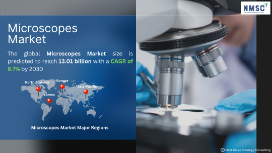
𝑴𝒊𝒄𝒓𝒐𝒔𝒄𝒐𝒑𝒆𝒔 𝑴𝒂𝒓𝒌𝒆𝒕: 𝑨 𝑪𝒍𝒐𝒔𝒆𝒓 𝑳𝒐𝒐𝒌 𝒂𝒕 𝒕𝒉𝒆 𝑭𝒖𝒕𝒖𝒓𝒆
𝑫𝒐𝒘𝒏𝒍𝒐𝒂𝒅 𝒂 𝑭𝑹𝑬𝑬 𝑺𝒂𝒎𝒑𝒍𝒆: https://www.nextmsc.com/microscopes-market/request-sample
The microscopes market is witnessing a transformative era driven by technological advancements, increasing research activities, and growing healthcare demands.
𝑴𝒂𝒓𝒌𝒆𝒕 𝑮𝒓𝒐𝒘𝒕𝒉:
The Microscopes Market size is predicted to reach 13.01 billion with a CAGR of 8.7% by 2030.
𝑲𝒆𝒚 𝑻𝒓𝒆𝒏𝒅𝒔:
𝑫𝒊𝒈𝒊𝒕𝒂𝒍 𝑴𝒊𝒄𝒓𝒐𝒔𝒄𝒐𝒑𝒚: The integration of AI and machine learning with digital microscopy is enhancing image analysis, making diagnostics faster and more accurate.
𝑷𝒐𝒓𝒕𝒂𝒃𝒍𝒆 𝑴𝒊𝒄𝒓𝒐𝒔𝒄𝒐𝒑𝒆𝒔: The demand for portable and handheld microscopes is rising, especially in field research and point-of-care diagnostics.
𝑺𝒖𝒑𝒆𝒓-𝑹𝒆𝒔𝒐𝒍𝒖𝒕𝒊𝒐𝒏 𝑴𝒊𝒄𝒓𝒐𝒔𝒄𝒐𝒑𝒚: Advances in super-resolution techniques are enabling scientists to observe biological processes at the molecular level, opening new frontiers in biomedical research.
𝑯𝒆𝒂𝒍𝒕𝒉𝒄𝒂𝒓𝒆 𝑰𝒎𝒑𝒂𝒄𝒕:
Microscopes play a crucial role in disease diagnosis and research, from identifying pathogens to studying cellular responses. Their application in cancer research, neurology, and personalized medicine is particularly noteworthy.
𝑨𝒄𝒄𝒆𝒔𝒔 𝑭𝒖𝒍𝒍 𝑹𝒆𝒑𝒐𝒓𝒕: https://www.nextmsc.com/report/microscopes-market
𝑴𝒂𝒓𝒌𝒆𝒕 𝑷𝒍𝒂𝒚𝒆𝒓𝒔:
Various market players operating in the microscopes market include Bruker Corp., CAMECA, Danaher Corporation, Hitachi High-Tech Corp., JEOL Ltd., Nikon Corp., Olympus Corp., Oxford Instruments (Asylum Corporation), Thermo Fisher Scientific, Inc., and Zeiss Group.
𝑭𝒖𝒕𝒖𝒓𝒆 𝑶𝒖𝒕𝒍𝒐𝒐𝒌:
As we move forward, the microscopes market will continue to expand, with emerging technologies like nanotechnology and quantum imaging poised to revolutionize the field.
Let's keep an eye on this dynamic market as it shapes the future of science and healthcare.
#microscopes#markettrends#healthcareinnovation#scientificresearch#medicaltechnology#medicaldevice#globalmarket#marketresearch#businessinsights#marketanalysis
0 notes
Text
0 notes
Text
Operating Room Integration: The Future of Surgery

Operating rooms have long since advanced from their humble beginnings. Modern ORs house sophisticated medical devices, computerized monitors, and specialized staff working seamlessly together. While technology has transformed operating rooms individually, the next leap forward is integrating OR functionality as a unified whole. Operating room integration promises to revolutionize surgical care by connecting people, processes, and technology in new collaborative ways. Connecting Technology and Information Flow One of the key aspects of OR integration is linking the various medical devices, monitors, and systems used during surgery. Currently, each piece of equipment largely operates independently without communicating with other technologies in the room. OR integration addresses this by enabling devices to share information electronically. Vital sign monitors, surgical microscopes, endoscopic cameras, and more would be networked together. Surgeons and staff could then view combined data streams and images on a single connected display instead of multiple individual monitors. This unified view reduces clutter and allows teams to focus on the patient rather than technology. Beyond just displaying combined outputs, an integrated OR enables devices to interact. Imaging systems could automatically sync up with endoscopic camera feeds. Changes to vital monitors are instantly reflected across all displays. Surgical microscopes could pull up pre-operative scans with a simple command. Handheld tablets provide roaming access to the entire integrated setup. Personnel move more freely throughout the OR while maintaining access to vital information when and where they need it. The integrated network also captures computerized records of the entire procedure for documentation and future reference. Automated recording eliminates manual transcription errors. Streamlining Workflows and Communication Another key benefit of operating room integration is streamlining workflows and internal communication. Currently, surgical teams must coordinate verbally and manually pass off instruments, samples, and procedures. An integrated system digitizes many of these workflows. Supply cabinets, instrument trays, and equipment can be requested, located, and delivered automatically through the networked infrastructure. Digital checklists and logs track staff duties and the surgery progress end-to-end. Teams spend less time searching for tools and more time focused on the patient. Intra-operative communication is also enhanced. Integrated ORs deploy digital communication systems like wireless headsets and smart badges. Surgeons and staff can discuss cases without masking or disturbing the sterile field. Interactive displays present virtual whiteboards to diagram anatomy and procedures. Digital dictation captures notes without interrupting hands-on work. Remote experts can even connect to the integrated OR system through telemedicine portals. OR integration facilitates real-time collaborative care no matter the physical distance between professionals. The integrated network environment makes surgical teams work as a seamlessly connected unit. Advancing Teaching and Best Practices Operating room integration also benefits the wider medical field by advancing surgical education and sharing of best practices. By design, integrated OR systems capture computerized records, imaging, and more from every case. With proper controls and safeguards, this wealth of procedure data can support surgical training programs. Trainees gain hands-on access to a library of past cases through digital archives. Simulations reconstruct surgical workflows for practice. Interactive case discussions pull up real-time data streams. More seasoned surgeons can consult records to refine their own techniques and spread quality improvement initiatives.
0 notes
Text
Video Microscopes Market: Strategic Imperatives for Success and Rising Demand During 2023-2032
Microscopy has long been a cornerstone of scientific research and education, allowing us to explore the intricacies of the microcosmic world. In this age of digital transformation, video microscopes are taking this fundamental tool to new heights, offering advanced imaging capabilities and real-time observations. This article delves into the global Video Microscopes Market, shedding light on the remarkable advancements and emerging trends in this dynamic field.
𝐑𝐞𝐪𝐮𝐞𝐬𝐭 𝐒𝐚𝐦𝐩𝐥𝐞 𝐂𝐨𝐩𝐲 𝐎𝐟 𝐑𝐞𝐩𝐨𝐫𝐭 : https://www.alliedmarketresearch.com/request-toc-and-sample/11382
The Power of Video Microscopy:
Video microscopes, equipped with digital cameras and enhanced imaging systems, have become indispensable in a wide range of applications. They bridge the gap between traditional microscopy and modern technology, providing researchers, educators, and professionals with a suite of powerful tools.
Key Advancements in Video Microscopy:
Digital Imaging: Perhaps the most significant advancement is the shift from analog to digital. High-resolution cameras integrated with video microscopes allow for detailed and precise imaging. These images can be stored, shared, and analyzed digitally, revolutionizing the way we document and interpret microscopic observations.
Real-Time Observation: Video microscopes enable real-time observation and analysis. This is invaluable in applications like medical diagnostics, where quick decision-making is essential, and in scientific research where dynamic processes can be closely monitored.
Remote Access and Collaboration: In an increasingly interconnected world, video microscopes can be connected to the internet, enabling remote access and collaboration. Experts can share their insights and guide procedures from anywhere globally, expanding the reach of education and expertise.
Emerging Trends in the Video Microscopes Market:
3D Imaging: The integration of 3D imaging technology into video microscopes is on the rise. This provides a more comprehensive understanding of complex structures and their spatial relationships, benefiting fields like life sciences and material sciences.
Artificial Intelligence (AI): AI is making its way into video microscopy. Machine learning algorithms can help identify and analyze features in images, increasing the efficiency of tasks like cell counting and particle sizing.
Miniaturization: Advancements in miniaturization have led to the development of portable and handheld video microscopes. These are particularly useful in the field, enabling on-site inspections and immediate analysis.
𝐏𝐫𝐞-𝐛𝐨𝐨𝐤 𝐭𝐡𝐢𝐬 𝐑𝐞𝐩𝐨𝐫𝐭 𝐍𝐨𝐰 : https://www.alliedmarketresearch.com/video-microscopes-market/purchase-options
Market Implications:
The global Video Microscopes Market is poised for significant growth. Advancements in technology and emerging trends are expanding the market's potential. Video microscopes are becoming more accessible, user-friendly, and affordable, making them applicable not only in research institutions but also in industries like healthcare, electronics, and materials science.
Key benefits of the report:
1. Analytical Depiction of the Industry: The report provides a thorough and analytical depiction of the global video microscopes industry. It offers a detailed understanding of the market's current landscape, trends, and future estimations. This knowledge is invaluable for investors and decision-makers who seek to identify imminent investment opportunities.
2. Insights into Market Dynamics: The report furnishes essential information related to the key drivers, restraints, and opportunities shaping the global video microscopes market. This insight aids in making informed decisions, mitigating risks, and capitalizing on growth prospects.
3. Quantitative Market Analysis: The report quantitatively analyzes the current market conditions, shedding light on the growth scenario of the global video microscopes market. This data-driven analysis provides a clear picture of market trends, demand, and potential areas of investment.
4. Porter's Five Forces Analysis: The report employs Porter's Five Forces analysis, offering a comprehensive view of the market's competitive landscape. It illustrates the potency of both buyers and suppliers, providing a deeper understanding of market dynamics and helping businesses formulate effective strategies.
5. Competitive Market Analysis: The report delivers a detailed global market analysis based on competitive intensity. It offers insights into how competition is likely to evolve in the coming years. This information is crucial for businesses looking to position themselves effectively in the market and anticipate future trends.
𝐈𝐧𝐭𝐞𝐫𝐞𝐬𝐭𝐞𝐝 𝐭𝐨 𝐏𝐫𝐨𝐜𝐮𝐫𝐞 𝐭𝐡𝐞 𝐑𝐞𝐬𝐞𝐚𝐫𝐜𝐡 𝐑𝐞𝐩𝐨𝐫𝐭? 𝐈𝐧𝐪𝐮𝐢𝐫𝐞 𝐁𝐞𝐟𝐨𝐫𝐞 𝐁𝐮𝐲𝐢𝐧𝐠 : https://www.alliedmarketresearch.com/purchase-enquiry/11382
In Conclusion:
The Video Microscopes Market is in a phase of dynamic transformation. Advancements in digital imaging, real-time observation, and connectivity are empowering professionals across various sectors. Emerging trends, such as 3D imaging, AI integration, and miniaturization, are further propelling this market into a new era of microscopy.
As we move forward, we can expect video microscopes to continue breaking boundaries, offering novel insights into the microscopic world and catalyzing innovation in scientific research, education, and industrial applications. The future of video microscopy is digital, connected, and full of promise.
About Us
Allied Market Research (AMR) is a full-service market research and business-consulting wing of Allied Analytics LLP based in Portland, Oregon. Allied Market Research provides global enterprises as well as medium and small businesses with unmatched quality of "Market Research Reports" and "Business Intelligence Solutions." AMR has a targeted view to provide business insights and consulting to assist its clients to make strategic business decisions and achieve sustainable growth in their respective market domain.
Pawan Kumar, the CEO of Allied Market Research, is leading the organization toward providing high-quality data and insights. We are in professional corporate relations with various companies and this helps us in digging out market data that helps us generate accurate research data tables and confirms utmost accuracy in our market forecasting. Each and every data presented in the reports published by us is extracted through primary interviews with top officials from leading companies of domain concerned. Our secondary data procurement methodology includes deep online and offline research and discussion with knowledgeable professionals and analysts in the industry.
0 notes
Text
Dermatology Endoscopy devices Market- Future Growth Prospects for the Global Leaders

The latest market report published by Credence Research, Inc. “Global Dermatology Endoscopy devices Market: Growth, Future Prospects, and Competitive Analysis, 2016 – 2028. The global Dermatology Endoscopy Devices Market has witnessed steady growth in recent years and is expected to grow at a CAGR of 7.50% between 2023 and 2030. The market was valued at USD 1754.9 million in 2022 and is expected to reach USD 3129.8 million in 2030.
Dermatology endoscopy devices are specialized medical instruments used by dermatologists and other healthcare professionals to examine and diagnose skin conditions, such as skin cancer, moles, lesions, and other dermatological issues. These devices typically consist of a thin, flexible, and lighted tube equipped with a camera that allows for the visualization of the skin's surface and underlying tissues. Dermatology endoscopy is a minimally invasive technique that can provide detailed images of skin lesions and help in the early detection of skin diseases.
Some of the common types of dermatology endoscopy devices include:
Dermatoscopes: These are handheld devices with specialized lenses and lighting systems that allow dermatologists to examine skin lesions and moles in detail. Dermatoscopy can help differentiate between benign and malignant growths.
Trichoscopes: Trichoscopy is a technique used to examine the scalp and hair follicles. Trichoscopes are designed specifically for this purpose and help diagnose conditions like alopecia (hair loss).
Nailfold Capillaroscopy Devices: These devices are used to examine the nail beds and assess conditions related to the nail and surrounding tissue, such as nailfold capillaroscopy for the diagnosis of conditions like scleroderma.
Microscopic Cameras: Some dermatologists use high-resolution cameras with various lenses and attachments to capture detailed images of the skin for documentation and analysis.
The market for dermatology endoscopy devices has seen growth due to several factors, including an increasing prevalence of skin diseases, the need for early and accurate diagnosis, and technological advancements in the field of dermatology. These devices can aid in the early detection of skin cancer, improve diagnostic accuracy, and enhance patient care.
Here are some trends that were relevant at that time:
Technological Advancements: The dermatology endoscopy devices market has seen a continuous influx of advanced technologies. These devices have become more sophisticated, offering better image quality, higher resolution, and improved maneuverability. Miniaturization and the integration of digital imaging systems have also been notable trends.
Growing Skin Cancer Cases: The rising incidence of skin cancer, including melanoma and non-melanoma skin cancers, has driven the demand for dermatology endoscopy devices. These devices play a crucial role in early diagnosis, monitoring, and treatment planning for skin cancer patients.
Telemedicine and Remote Monitoring: The COVID-19 pandemic accelerated the adoption of telemedicine and remote monitoring. Dermatologists have increasingly used dermatology endoscopy devices for remote consultations and patient monitoring, enabling timely diagnosis and treatment while minimizing in-person visits.
Increase in Aesthetic Procedures: Dermatology endoscopy devices are also used in aesthetic dermatology procedures, such as minimally invasive cosmetic treatments. As people seek non-surgical solutions for skin rejuvenation, these devices have gained popularity in the cosmetic dermatology market.
Some of the major players in the market and their market share are as follows:
AMD Global Telemedicine
AnMo Electronics Corp.
Bausch Health Co Inc.
Caliber Imaging and Diagnostics Inc.
Canfield Scientific Inc.
DermLite LLC
DermoScan GmbH
Firefly Global
HEINE Optotechnik GmbH and Co. KG
ILLUCO Corp. Ltd.
Browse 247 pages report Dermatology Endoscopy devices Market By Device Type (Dermatoscopes, Video Microscopes, Fiber Optic Scopes, Accessories (Light Sources, Cameras, Adapters)) By Application (Skin Cancer Diagnosis and Monitoring, Hair and Scalp Examination, Nail Examination, Allergy and Inflammation Diagnosis, Other Dermatological Conditions)-Growth, Future Prospects & Competitive Analysis, 2016 – 2030 - https://www.credenceresearch.com/report/dermatology-endoscopy-devices-market
Dermatology Endoscopy devices Market Regional Analysis
North America:
North America traditionally holds a significant share of the dermatology endoscopy devices market due to a well-established healthcare infrastructure and high adoption of advanced medical technologies.
The United States, in particular, is a major contributor to the market in this region, driven by the presence of leading dermatology clinics and research institutions.
Increasing skin-related disorders and a growing aging population contribute to market growth in North America.
Europe:
Europe is another prominent region for the dermatology endoscopy devices market. Countries like Germany, France, and the United Kingdom have advanced healthcare systems and a strong focus on dermatology research.
The European market benefits from government initiatives and funding for dermatological research and development.
The market may also see growth due to rising awareness of skin diseases and cosmetic dermatology procedures.
Asia-Pacific:
The Asia-Pacific region has witnessed significant growth in the dermatology endoscopy devices market due to the increasing prevalence of skin diseases, a rising disposable income, and the expanding healthcare infrastructure.
Countries such as China, India, and Japan have seen a surge in dermatology clinics and cosmetic dermatology procedures.
Market players are targeting emerging economies in this region due to the large patient population and increasing healthcare expenditures.
Latin America:
Latin America is experiencing steady growth in the dermatology endoscopy devices market, driven by increasing awareness of skincare and aesthetic procedures.
Brazil, Mexico, and Argentina are some of the key markets in the region.
Economic development and a growing middle-class population contribute to market expansion.
Key Segments
By Device Type
Dermatoscopes
Video Microscopes
Fiber Optic Scopes
Accessories (Light Sources, Cameras, Adapters)
By Application
Skin Cancer Diagnosis and Monitoring
Hair and Scalp Examination
Nail Examination
Allergy and Inflammation Diagnosis
Other Dermatological Conditions
Why to Buy This Report-
The report provides a qualitative as well as quantitative analysis of the global Dermatology Endoscopy devices Market by segments, current trends, drivers, restraints, opportunities, challenges, and market dynamics with the historical period from 2016-2020, the base year- 2021, and the projection period 2022-2028.
The report includes information on the competitive landscape, such as how the market's top competitors operate at the global, regional, and country levels.
Major nations in each region with their import/export statistics
The global Dermatology Endoscopy devices Market report also includes the analysis of the market at a global, regional, and country-level along with key market trends, major players analysis, market growth strategies, and key application areas.
Browse Complete Report- https://www.credenceresearch.com/report/dermatology-endoscopy-devices-market
Visit our Website- https://www.credenceresearch.com/
Related Reports- https://www.credenceresearch.com/report/aloe-vera-based-skin-and-hair-products-market
Browse Our Blog- https://www.linkedin.com/pulse/dermatology-endoscopy-devices-market-size-expected-acquire-shukla
About Us -
Credence Research is a viable intelligence and market research platform that provides quantitative B2B research to more than 10,000 clients worldwide and is built on the Give principle. The company is a market research and consulting firm serving governments, non-legislative associations, non-profit organizations, and various organizations worldwide. We help our clients improve their execution in a lasting way and understand their most imperative objectives. For nearly a century, we’ve built a company well-prepared for this task.
Contact Us:
Office No 3 Second Floor, Abhilasha Bhawan, Pinto Park, Gwalior [M.P] 474005 India
0 notes
Text
Associated Optometry Equipment for Eye Care Professional
Associated optometry equipment refers to the various tools and instruments used by optometrists and other eye care professionals to examine and diagnose eye conditions, assess vision, and prescribe corrective measures. Here are some commonly used optometry equipment:
Autorefractors: Associated optometry equipment used to measure refractive errors in the eye, such as nearsightedness, farsightedness, and astigmatism.
Phoropters: Also known as refractors, these devices are used to determine an individual's exact eyeglass prescription by allowing the optometrist to change lenses and assess visual acuity.
Slit Lamp: A specialized microscope with a bright light source that allows for detailed examination of the front structures of the eye, including the cornea, conjunctiva, iris, and lens.
Ophthalmoscope: A handheld instrument that enables the optometrist to examine the interior structures of the eye, such as the retina, optic nerve, and blood vessels.
Tonometer: Used to measure intraocular pressure, which helps in diagnosing glaucoma. There are various types of tonometers, including applanation, non-contact (air puff), and handheld.
Keratometer: Measures the curvature of the cornea, which aids in fitting contact lenses and diagnosing corneal irregularities.
Visual Field Analyzer: Tests the complete field of vision, assessing peripheral and central vision to detect abnormalities and diagnose conditions like glaucoma and optic nerve damage.
Retinal Camera: Specialized digital cameras that capture high-resolution images of the retina, enabling the optometrist to document and monitor retinal health.
Pachymeter: Measures the thickness of the cornea, which is essential in the management of conditions like corneal edema and glaucoma.
Lensmeter: Determines the prescription of existing eyeglasses, including the power, axis, and prism values.
Optic Nerve Analyzer: Utilizes advanced imaging techniques to evaluate the optic nerve head and detect signs of glaucoma or other optic nerve disorders.
Visual Acuity Charts: Used to measure a patient's visual acuity at various distances, usually consisting of rows of letters or symbols of decreasing size.

These are just a few examples of the equipment commonly found in an optometry practice. Optometrists may also use other specialized devices depending on their specific areas of expertise and the needs of their patients. Associated optometry equipment can help you deliver better patient care while also enhancing the effectiveness and image of your practice.
0 notes
Text
Digital Microscope: Revolutionizing Modern Scientific Exploration
What is a Digital Microscope?
A digital microscope is a cutting-edge device that combines traditional optical microscope technology with digital imaging. Unlike conventional microscopes, digital microscopes are equipped with a camera that captures images and videos, displaying them on a connected monitor or computer screen. This innovation eliminates the need for direct eyepiece observation, making it more user-friendly and versatile.
Digital microscopes are widely used in scientific research, education, healthcare, and industrial applications. By providing high-resolution imagery and advanced analytical features, they have become an indispensable tool for professionals and enthusiasts alike.
How Does a Digital Microscope Work?
The functionality of a digital microscope relies on three primary components:
Optical Lenses: These lenses magnify the sample, ensuring clear and detailed visualization.
Digital Camera: This component captures magnified images or videos in real-time.
Display Interface: The images are transmitted to a screen, allowing for group observation and easy documentation.
Advanced models also feature software that enables image analysis, measurement, and 3D visualization, further enhancing the microscope's utility.
Key Features of a Digital Microscope
1. High Magnification and Resolution
Digital microscopes provide unparalleled magnification levels, often ranging from 10x to over 1000x. Coupled with high-resolution sensors, they deliver crystal-clear imagery, ensuring accurate analysis.
2. Real-Time Image Display
The ability to project images directly onto a monitor enhances collaboration and reduces eye strain. This feature is particularly valuable in classrooms, laboratories, and manufacturing settings.
3. Advanced Image Processing
Modern digital microscopes include software that allows users to annotate, measure, and analyze samples efficiently. Features like autofocus, brightness adjustment, and 3D reconstruction are standard in high-end models.
4. Portability and Versatility
Compact designs and wireless connectivity make digital microscopes portable and easy to use in various environments, including fieldwork.
Applications of Digital Microscopes
1. Scientific Research
Digital microscopes are essential tools in biology, chemistry, and materials science. Researchers use them to study cellular structures, chemical reactions, and micro-organisms with precision.
2. Education
Educators rely on digital microscopes to create interactive and engaging learning experiences. The ability to display live samples on a screen enhances understanding and fosters collaboration among students.
3. Healthcare and Diagnostics
In the medical field, digital microscopes are used to analyze tissue samples, identify pathogens, and conduct surgeries with microscopic precision. They play a pivotal role in improving diagnostic accuracy.
4. Industrial Quality Control
Manufacturing industries utilize digital microscopes for quality assurance, defect detection, and product analysis. They are especially valuable in electronics, automotive, and aerospace sectors.
Advantages of Using a Digital Microscope
1. Enhanced Documentation
Digital microscopes simplify the process of capturing and storing images for future reference, ensuring better data management.
2. Accessibility and Collaboration
The ability to project images onto a screen allows multiple users to view and discuss samples simultaneously, fostering collaborative work environments.
3. Reduced Eye Strain
By eliminating the need to peer through an eyepiece, digital microscopes minimize discomfort and fatigue during prolonged usage.
4. Advanced Analysis Tools
Features like measurement tools, 3D imaging, and time-lapse recording elevate the capabilities of traditional microscopy.
Types of Digital Microscopes
1. Handheld Digital Microscopes
Portable and user-friendly, these microscopes are ideal for fieldwork and basic inspections.
2. USB Digital Microscopes
These compact devices connect directly to a computer via USB, making them accessible for hobbyists and students.
3. Compound Digital Microscopes
Designed for detailed biological studies, these microscopes offer higher magnification and are widely used in laboratories.
4. Stereo Digital Microscopes
Perfect for observing larger samples, stereo microscopes provide a 3D view, making them suitable for industrial applications.
How to Choose the Best Digital Microscope?
1. Define Your Purpose
Determine whether you need a microscope for research, education, or industrial use.
2. Consider Resolution and Magnification
Select a model with the appropriate magnification range and resolution for your specific needs.
3. Evaluate Software Features
Look for microscopes with advanced image processing capabilities to enhance your analysis.
4. Check Connectivity Options
Ensure the microscope supports seamless connectivity with your devices, whether through USB, HDMI, or wireless networks.
Future Trends in Digital Microscopy
The field of digital microscopy is continuously evolving, with innovations such as artificial intelligence, augmented reality, and cloud-based data storage transforming the landscape. These advancements promise to make digital microscopes even more powerful and accessible, driving further adoption across various industries.
Digital microscopes are at the forefront of modern scientific exploration, offering unmatched precision, versatility, and convenience. Whether in research labs, classrooms, or factories, their impact is profound and far-reaching.
0 notes
Text

Handheld Microscope With 5" LCD Screen, Portable Pocket Digital Microscopes For Kids With Camera, Opqpq ODM5 Pro 1200X HDMI Coin Microscope For Error Coins, Soldering Microscope For Electronics Repair

"Say hello to the 'Pocket Peeker': It's not your grandma's magnifying glass! This portable microscope with lights lets you play scientist by day and undercover spy by night, revealing the hidden world of all things itty-bitty and glowy!"
1 note
·
View note