#scanning electron microscopy
Explore tagged Tumblr posts
Visit Tumblr Blog
Explore Tumblr blogs with no restrictions, modern design and the best experience.
Last Seen Tumblr Blogs


Fun Fact
Tumblr was attacked by a cross-site scripting worm deployed by the Internet troll group GNAA on Dec 3, 2012.
Text

Vibrio fischeri
These bacteria are bioluminescent, and their symbiotic relationship with certain marine animals, such as the Hawaiian Bobtail Squid, is what gives the animals their bioluminescence.
Photo credit: Dennis Kunkel (Microscopy/Science Photo Library)
#vibrio fischeri#bioluminescence#bioluminescent#bioluminescent bacteria#birthday hat#microbiology#scanning electron microscopy#microbes#biology#hat#hats#microbes in hats#microorganisms#bacteria#protozoa#microscopy#originally posted on my birthday so of course i put birthday hats on them
35 notes
·
View notes
Text

Aligned array of nanotubes
Processing Chemical vapour deposition onto a quartz substrate, using a fine solution of ferrocene dissolved in toluene Applications Such architectures may be of interest as nanocomposites for use in nanodevices. More generally, carbon nanotubes may be used for hydrogen storage or for fuel cell applications Sample preparation The specimen has been sputter-coated with gold to avoid charging in the SEM Technique Scanning electron microscopy (SEM) Length bar 25 μm Further information Chemical vapour deposition (CVD) allows the synthesis of high purity nanotubes of controlled length and diameter. The nanotubes in this specimen were deposited on quartz using ferrocene dissolved in toluene. They are approximately 40 nm in diameter and 60 microns long. Contributor C Singh Organisation Department of Materials Science and Metallurgy, University of Cambridge
Source.
#Materials Science#Science#Scanning electron microscopy#Chemical vapor deposition#Carbon nanotubes#Nanotechnology#Carbon#Nanotubes#Magnified view#DoITPoMS#University of Cambridge
23 notes
·
View notes
Text

Close-up on Cilia
Almost every cell in your body has a little antenna sending and receiving chemical and mechanical signals. These thread-like projections are called primary cilia. To better understand the importance of these structures, researchers have tried to identify proteins located on them. However, this has proven challenging as primary cilia overlap with other cell structures such as the cell membrane. Now researchers have developed an effective way to definitively localise primary cilia’s proteins in 3D by combining surface scanning electron microscopy with antibody-based labelling (immuno-SEM). Here, a protein called IFT88 (white dots) – which if mutated can cause serious disorder – is revealed on a cilium on a mouse pancreatic cell. This powerful, high-resolution tool will aid research into primary cilia and ciliopathies – a range of serious diseases caused by cilia dysfunction.
Written by Lux Fatimathas
Image from work by Sanja Sviben and colleagues
Department of Cell Biology and Physiology, Washington University School of Medicine, Saint Louis, MO, USA
Image originally published with a Creative Commons Attribution 4.0 International (CC BY 4.0)
Published in Journal of Cell Science, May 2024
You can also follow BPoD on Instagram, Twitter and Facebook
16 notes
·
View notes
Text


3 notes
·
View notes
Link
1 note
·
View note
Text

An SEM image of a guitar string held down with carbon tape to a sample mount.
24 notes
·
View notes
Note
Do you have any formal published work for your OC's? Like is there a comic somewhere or some fics? I'd really love to learn more about the narrative, the characters are gorgeous 🎀
there is not _(:3」∠)_
but I've tried! i've honest to god written all the issues of a would-be transient comic and thumbnailed/storyboarded several of them out, but when i went to actually make them proper, it was too much to do on top of my job/other interests. plus I just...didnt like them. i couldnt settle on a consistent style and the rendering type i chose both looked overwhelming and took too much time.
for an example of what i meant, heres two example pages from my last attempt.


The thing is if i never actually make the comic, then in my head it can be the coolest, raddest, thing ever, but once pen meets paper the hypothetical has to clash with the reality that its not going to be as funny or polished as i dreamt it'd be. but ive been building up more illustrative art confidence and solved some of the problems i had on that above attempt (not understanding the style i want, rendering woes) so ??? ?????? who knows.
of all the OCverses, transient is the most actionable since 1) its fully written. like im literally looking at the full script right now. i just need to do it. and 2) its inherently episodic so i could publish issue-by-issue without having to commit to some ~50 cohesive, continuous chapters that would be needed for something like stitch in the ditch.
but thank you!!!! i would like to make it someday but its a matter of getting the mind and body to be fully willing lol.
#my favorite one i wrote was called STM: shrimp time motherfucker#which is a play on my job STM or scanning tunneling microscopy#instead of tunneling electrons across a tip and sample- shrimp were quantum tunneling into places they're classically forbidden to be#which was intended to teach about first principles of QM and tunneling across finite barriers
14 notes
·
View notes
Text
A Journey into the World of Microscopy: From Humble Beginnings to High-Tech Magnification
The science of looking into the hidden invisible Microscopy has transformed our understanding of the world around us. It can explore the universe beyond the reach of our naked eyes, with complex cellular structures, red blood cells, viruses and other viruses and microorganisms taking on amazing perspectives
The history of the microscope is a fascinating story of human curiosity, scientific genius, and relentless exploration. From the humble beginnings of simple magnifying glasses to the sophistication of modern electronic microscopes, the invention of microscopes has shaped our understanding of the microscopic world
In the 1600s, Dutch opticians such as Hans and Zachary Janssen are credited with inventing the first microscope. Known for this hybrid microscope, many lenses were used to magnify objects up to 30 times.At the end of the 17th century, Antony van Leeuwenhoek, Dutch draper some changed our perception of thumbnails. Armed with a well-made single-lens microscope, and explored the hidden reaches of nature. In 1674, Leeuwenhoek discovered microorganisms in lake water, which he aptly named “animalcules”. His discovery laid the foundations of biology and inspired generations of scientists. This incredible feat allowed him to uncover a hidden universe – the first sightings of bacteria, red blood cells, and other microorganisms.
Formation of the scientific environment (17th-19th centuries): Leeuwenhoek’s discoveries boosted scientific research. Robert Hooke, an English scientist, established these developments. In 1665, his book "Micrographia" recorded his observations with a compound microscope. Notably, the term "cell" was coined by Hooke when he examined cork tissue, laying the foundation for cell biology.Microscope systems flourished throughout the 18th and 19th centuries Joseph Lister and other scientists addressed the limitations of the early lenses, introducing improvements that reduced image distortion.
Beyond the Limits of Light: The Beginning of the New Age (19th-20th century): As the 19th century progressed, the limitations of optical microscopy became apparent and scientists yearned for a tool which can go deeper into cells. This research culminated in the development of the electron microscope in the 1930s. The 20th century was revolutionary with the invention of the electron microscope. Unlike light microscopes, which use visible light, electron microscopes use electron beams to achieve much higher magnification.Formation of the scientific environment (17th-19th centuries): Leeuwenhoek’s discoveries boosted scientific research. Robert Hooke, an English scientist, established these developments. In 1665, his book "Micrographia" recorded his observations with a compound microscope. Notably, the term "cell" was coined by Hooke when he examined cork tissue, laying the foundation for cell biology.Microscope systems flourished throughout the 18th and 19th centuries Joseph Lister and other scientists addressed the limitations of the early lenses, introducing improvements that reduced image distortion.
Beyond the Limits of Light: The Beginning of the New Age (19th-20th century): As the 19th century progressed, the limitations of optical microscopy became apparent and scientists yearned for a tool which can go deeper into cells. This research culminated in the development of the electron microscope in the 1930s. The 20th century was revolutionary with the invention of the electron microscope. Unlike light microscopes, which use visible light, electron microscopes use electron beams to achieve much higher magnification.
In the 1930s, German experts Max Knoll and Ernst Ruska made the first electron microscope. This tool let us see tiny things like cells and even atoms by using electron beams, not light, getting images many times bigger. This cool invention showed us the tiny parts inside cells, viruses, and stuff too small to see before. The 1900s brought even more cool microscopes. New kinds like phase-contrast and confocal microscopy let scientists look at live cells without using stuff that could hurt them. Now, the world of looking at tiny things is getting even better. Today, we have high-tech microscopes that use computers and lasers. These let us see and even change tiny things in ways we never could before.
Modern Microscopy's Diverse Arsenal - Today, the field of microscopy boasts a diverse range of specialized instruments, each tailored to address specific scientific needs. Here's a glimpse into some remarkable examples:
Scanning Electron Microscope (SEM): Imagine a high-tech camera that captures images using a beam of electrons instead of light. That's the essence of a SEM. By scanning the surface of a sample with a focused electron beam, SEMs generate detailed information about its topography and composition. This makes them ideal for studying the intricate structures of materials like insect wings, microchips, and even pollen grains.
Transmission Electron Microscope (TEM): While SEMs provide exceptional surface detail, TEMs take us a step further. They function by transmitting a beam of electrons through a very thin sample, allowing us to observe its internal structure. TEMs are the go-to instruments for visualizing the intricate world of viruses, organelles within cells, and macromolecules like proteins.
Confocal Microscopy: Ever wished to focus on a specific layer within a thick biological sample and blur out the rest? Confocal microscopy makes this possible. It utilizes a laser beam to precisely illuminate a chosen plane within the sample, effectively eliminating information from out-of-focus regions. This allows researchers to create sharp, three-dimensional images of cells, tissues, and even small organisms.
Atomic Force Microscopy (AFM): This technique takes a completely different approach, venturing into the realm of physical interaction. AFM employs a tiny cantilever, akin to a microscopic feeler, to physically scan the surface of a sample. By measuring the minute forces between the cantilever and the sample's surface, AFM can map its topography at an atomic level. This provides invaluable insights into the properties of materials at an unimaginable scale, making it crucial for research in fields like nanotechnology and surface science.
Fluorescence Microscopy: Imagine illuminating a sample with specific wavelengths of light and observing it glowing in response. That's the essence of fluorescence microscopy. This technique utilizes fluorescent molecules or tags that bind to specific structures within a cell or tissue. When excited by light, these tags emit their own light, highlighting the target structures with remarkable clarity. This allows researchers to visualize specific proteins, DNA, or even pathogens within biological samples.
Super-resolution Microscopy (SRM): Overcoming the limitations imposed by the wavelength of light, SRM techniques like STED (Stimulated Emission Depletion) and PALM (Photoactivated Localization Microscopy) achieve resolutions surpassing the diffraction limit. This allows researchers to visualize structures as small as 20 nanometers, enabling the observation of intricate cellular machinery and the dynamics of individual molecules within living cells.
Cryo-Electron Microscopy (Cryo-EM): This powerful technique takes a snapshot of biological samples in their near-life state. Samples are rapidly frozen at ultra-low temperatures, preserving their native structure and minimizing damage caused by traditional fixation methods. Cryo-EM has been instrumental in determining the three-dimensional structures of complex molecules like proteins and viruses, providing crucial insights into their function and potential drug targets.
Correlative Microscopy: Combining the strengths of multiple microscopy techniques, correlative microscopy offers a comprehensive view of biological samples. For instance, researchers can utilize fluorescence microscopy to identify specific structures within a cell and then switch to electron microscopy to examine those structures in high detail. This integrated approach provides a deeper understanding of cellular processes and their underlying mechanisms.
Light Sheet Microscopy (LSM): Imagine illuminating a thin slice of a sample within a living organism. LSM achieves this feat by focusing a laser beam into a thin sheet of light, minimizing photobleaching and phototoxicity – damaging effects caused by prolonged exposure to light. This allows researchers to observe dynamic processes within living organisms over extended periods, providing valuable insights into cellular behavior and development.
Expansion Microscopy (ExM): This innovative technique physically expands biological samples by several folds while preserving their structural integrity. This expansion allows for better resolution and visualization of intricate cellular structures that would otherwise be difficult to distinguish using traditional microscopy methods. ExM holds immense potential for studying the organization and function of organelles within cells.
Scanning Near-Field Optical Microscopy (SNOM): This innovative technique pushes the boundaries of resolution by utilizing a tiny probe that interacts with the sample at an extremely close range. SNOM can not only image the surface features of a sample with exceptional detail but also probe its optical properties at the nanoscale. This opens doors for research in areas like material science and photonics, allowing scientists to study the behavior of light at the interface between materials.
X-ray Microscopy: Stepping outside the realm of light and electrons, X-ray microscopy offers unique capabilities. By utilizing high-energy X-rays, this technique can penetrate deep into samples, making it ideal for studying the internal structure of dense materials like bones and minerals. Additionally, it allows for the visualization of elements within a sample, providing valuable information about their distribution and composition.
From revealing the building blocks of life to aiding in the development of new medicines, the microscope has played an undeniable role in shaping our scientific understanding. As technology continues to evolve, one can only imagine the future breakthroughs this remarkable invention holds in unveiling the secrets of our universe, both seen and unseen. These advancements hold the potential to revolutionize our understanding of biological processes, develop new materials with extraordinary properties, and ultimately pave the way for breakthroughs in medicine, nanotechnology, and countless other fields. As we continue to refine and develop novel microscopy techniques and the future holds immense promise for further groundbreaking discoveries that will undoubtedly revolutionize our perception of the world around us.
#science sculpt#life science#science#molecular biology#biology#biotechnology#artists on tumblr#microscopy#microscope#Scanning Electron Microscope#Transmission Electron Microscope#Confocal Microscopy#Atomic Force Microscopy#Fluorescence Microscopy#Expansion Microscopy#X-ray Microscopy#Super-resolution Microscopy#Light Sheet Microscopy#illustration#illustrator#illustrative art#education#educate yourself#techniques in biotechnology#scientific research#the glass scientists#scientific illustration#scientific advancements
7 notes
·
View notes
Photo

Scanning electron micrograph of a greenfly eye, by Kevin Mackenzie, University of Aberdeen by ZEISS Microscopy https://flic.kr/p/rv9NR2
14 notes
·
View notes
Text


Electron Micrographs
(Part 2 of 4)
Fugitive Glue, Lollipop, and Strawberry DNA
Fugitive Glue


I received a junk mail credit card offer, and I decided to take the piece of rubbery stuff that held the fake card to the paper to look at under the microscope. I originally referred to it as rubber cement, but I think it's actually called fugitive glue, among other names. Even exposed to air for a few days, it was still rubbery and malleable.
Unfortunately, being an organic compound, it did start to melt from the current of electricity that the microscope runs through it, which you can see happening in these images.
Lollipop Fragment


We had a bunch of leftover lollipops from Halloween at my house, so I took one of the flavors my brother didn't like to smash up and look at under the microscope.
In the first image, don't get confused by the background. The big angular piece is the lollipop, and the round things in the back are from the special tape used for keeping things on the stage (probably some bubbles trapped under it). I forget what kind of tape he said it was, but it was something able to conduct electricity.
The images of the sugar crystals are unclear and blurry because they, too, were actively melting while we were looking at them. Organic compounds just tend to do that. That said, you can still see the shapes of some of the crystals that hadn't yet melted.
Strawberry DNA


Having extracted some of my own DNA before, which I keep in an Eppendorf tube on my desk, I thought maybe it would be interesting to look at under the microscope.
However, I didn't want to potentially ruin the sample I had, so I followed NileRed's video to refresh myself on the process and used strawberries like he did. I also did it at 1/4 scale of what he did, because I really didn't need as much as he got. (By the way, DNA feels disgusting to touch. So much so that I used tweezers and forceps to handle it instead.)
Unfortunately, as you can see, the images aren't super clear or detailed, possibly for a few reasons.
The sample may still have had some moisture in it, which does interfere with how the machine sees it, I think.
It was also very clumped, because when you extract DNA, it all clumps together and I couldn't really separate the piece I was using into a thin layer.
It also may just be the size of DNA in general, which is molecular (just a very long one). I was hoping we'd see at least something, but I guess the indications of lines is all we could get.
I'm keeping the rest of the DNA we didn't use in some more Eppendorf tubes on my desk, because why not.
In the BDS Full images, the white parts are the hotter parts, where more electrons are being conducted through.
The scale in the bottom left corner of the images is in micrometers. 1 millimeter = 1000 micrometers
#microbes in hats#microbe posting#non-microbe post#electron microscopy#scanning electron microscopy#long post
20 notes
·
View notes
Text

Sintered salt (NaCl)
Processing The salt has been compacted (20MPa) and sintered for four hours at 785 degrees C Applications A sintered preform of salt can be used as a preform in the investment casting of open celled metal foams. The preform can then be washed out. Sample preparation The sample has been sputter-coated with gold Technique Scanning electron microscopy (SEM) Length bar 140 μm Further information The interconnectivity of pores in the reticulated aluminium foam was found to be strongly dependent on the degree of sintering of the salt. The sintering occurs by a non-densifying mechanism but the necks grow progressively wider with time. Four hours provides sufficient interconnectivity for the salt to be readily washed-out. Contributor J A Curran Organisation Department of Materials Science and Metallurgy, University of Cambridge
Source.
#Materials Science#Science#Ceramics#Salt#Magnified view#Scanning electron microscopy#Foams#Sintering#University of Cambridge#DoITPoMS
26 notes
·
View notes
Text
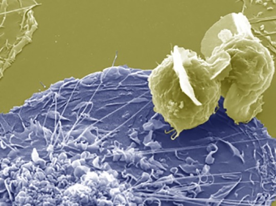
Infection by Fusion
Fusing HIV-infected CD4 T cells with macrophages – cells of the immune system that are a reservoir for HIV in tissue – infects them effectively, providing insight into the mechanisms of persistent infection
Read the published research paper here
Image from work by Rémi Mascarau and colleagues
Institut de Pharmacologie et Biologie Structurale (IPBS), Université de Toulouse, Centre National de la Recherche Scientifique, Université Toulouse III - Paul Sabatier (UPS), Toulouse, France
Image originally published with a Creative Commons Attribution 4.0 International (CC BY 4.0)
Published in Journal of Cell Biology, March 2023
You can also follow BPoD on Instagram, Twitter and Facebook
#science#biomedicine#biology#hiv#immune system#macrophages#t cells#cd4#electron microscopy#scanning electron microscopy#viral infections
7 notes
·
View notes
Text
An example of induced aerenchyma occurs in maize (corn; Zea mays) (Figure 24.19).
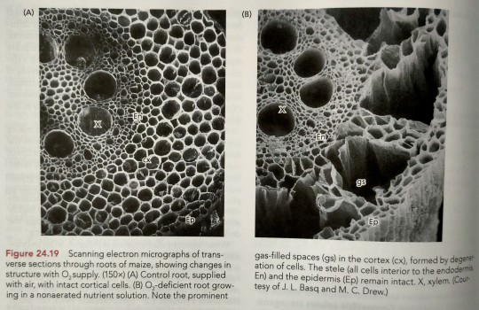
"Plant Physiology and Development" int'l 6e - Taiz, L., Zeiger, E., Møller, I.M., Murphy, A.
#book quotes#plant physiology and development#nonfiction#textbook#corn#maize#zea mays#trypophobia#scanning electron micrograph#microscopy#aerenchyma#plant cells
10 notes
·
View notes
Text
Scanning electron microscopy is awesome and I personally think the images it produces are gorgeous but objectively speaking I feel like it doesn't do any favors at all for the "scary" cultural image of insects, because I mean, here's a closeup of a carpet beetle in its true colors:

And here's an SEM image that comes up for carpet beetles on google:

And the thing about SEM images is that they aren't "photographs;" they are computer scans. They're 3-d digital models generated by scanning an object at the molecular level. Color is not preserved by this process, and if it were all the specimens would look like metal anyway (I'll explain this is in a moment), so images like this had to be colored artificially. This isn't done to recreate the true colors, but to make different body parts more visible as study material, resulting in scientific images of wacky blueberry fleas:

The subtly varying transparency levels of living tissues are completely lost as well, which is why the fine hairs of insects stand out more like cactus thorns in SEM imagery, and tardigrades look like opaque leathery things with no eyes:

...Even though a tardigrade actually has eyes, they're just under the surface of a crystal clear exoskeleton:

Another thing that probably contributes to the uncanniness of SEM images is also the fact that they can only show us embalmed corpses encased in liquid metal.

It's not possible to do this fine level of scanning "instantaneously" enough for it to work on anything that's still moving, so even when you see scanning electron images of animals in various lifelike poses, it's because they're preserved specimens that were carefully positioned, or they were live specimens basically "flash frozen" by a sudden dehydration process, mummified so fast they never knew it. Many specimens are then "sputter coated," meaning they're sprayed with a thin (like microns thin) layer of liquid gold, platinum or other fine metal in order for the electrons to perfectly bounce off of every subatomic detail and produce that perfect scan. So this is a live fruit fly:

And this is a fruit fly mummy with probably some sort of chrome plating:

5K notes
·
View notes
Text
youtube
This video shows how a Scanning Electron Microscope works? And how to use a virtual scanning electron microscope to acquire a good image. It is a great learning and teaching tool. You can access the virtual TEM at https://myscope-explore.org/virtualSE... developed by Microscopy Australia and Thermofisher Scientific.
#scanning#electronmicroscope#electronmicroscopy#electronbeam#image#images#nano#lotus#butterfly#lens#electrons#education#digitalliteracy#onlinelearning#microscope#microscopy#teaching#fiber#forensics#hair#arthropods#wool#superhydrophobic#waterrepellent#e-learning#virtuallearning#digitaltools#digitaleducation#openeducationalresources#openeducation
1 note
·
View note