#scanning electron micrograph
Explore tagged Tumblr posts
Visit Tumblr Blog
Explore Tumblr blogs with no restrictions, modern design and the best experience.
Last Seen Tumblr Blogs


Fun Fact
There are dozens of funny blogs to kill time on Tumblr.
Text
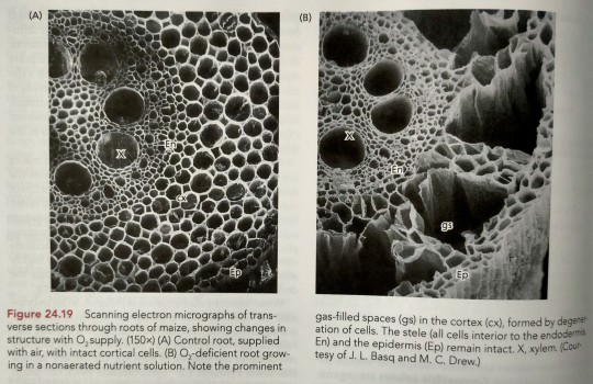
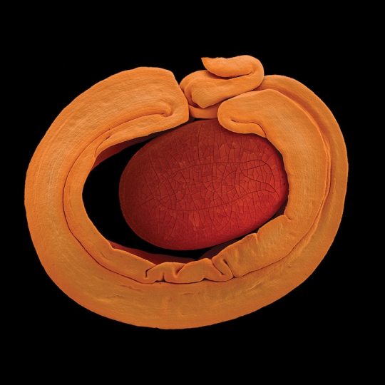
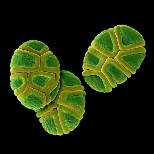
An example of induced aerenchyma occurs in maize (corn; Zea mays) (Figure 24.19).
"Plant Physiology and Development" int'l 6e - Taiz, L., Zeiger, E., Møller, I.M., Murphy, A.
#book quotes#plant physiology and development#nonfiction#textbook#corn#maize#zea mays#trypophobia#scanning electron micrograph#microscopy#aerenchyma#plant cells
10 notes
·
View notes
Text

'a scanning electron micrograph of the glochidium of an endangered pearl mussel [dwarf wedgemussel, alasmidonta heterodon] reveals "jaws" with which it will attach to its fish host' in pearls: a natural history - american museum of natural history + the field museum (2001)
68 notes
·
View notes
Text

Eduard Gübelin. Scanning electron micrographs of opals, 17150x, ca. 1975.
220 notes
·
View notes
Text
Lily Orchard recently started using the term tardigrade as an insult because she would really like to call her detractors a different word and tardigrade sort of sounds similar, I guess. This has inspired me put together a little educational aid:

And just in case you need a little more help spotting the difference, here's a higher res image:

The original tardigrade scanning electron micrograph is from Steve Gschmeissner/Science Photo Library
#lily orchard#lily orchard critical#lily posting#Lily's not cool enough to be a tardigrade#stockholm#retardigrade
36 notes
·
View notes
Text

A recent study in Nature Communications suggests that by 24 months, signs of heightened immune activity in most long-COVID patients will have returned to normal levels. Pictured above is a composite colored scanning electron micrograph of immune cells—including a single macrophage, two dendritic cells, and numerous white blood cells—involved in a cytokine storm, a life-threatening immune disorder. Cytokines are important for normal immune response, but when too many are released simultaneously it can be harmful.
COMPOSITE MICROGRAPH BY STEVE GSCHMEISSNER, SCIENCE PHOTO LIBRARY
#steve gschmeissner#photographer#national geographic#composite micrograph#science photo library#nature communications#long-covi#immune cells#cytokine storm#immune disorder#health#nature
13 notes
·
View notes
Text

Bacterial vaginosis (BV) affects nearly a third of all women in the U.S., and for over half of them, it's a recurring infection. BV has long been treated as a women's health issue—an imbalance of the vaginal microbiome. However, a groundbreaking new study suggests it's actually an STI and that treating male partners could dramatically cut recurrence rates.
Learn more about bacterial vaginosis and what experts say about new approaches to treatment: https://on.natgeo.com/43uYsFK
Pictured below, a scanning electron micrograph shows Lactobacillus bacteria, the beneficial microbes that help ward off harmful bacteria.
2 notes
·
View notes
Text

GODFLESH "Selfless" cover source
False-colour scanning electron micrograph (SEM) of human nerve cells growing on the surface of an integrated circuit (silicon chip). The picture was taken in 1985 in the course of research into biohybrid circuits, & demonstrates how much smaller organic circuits are than man-made ones. Biohybrid circuits are possible electronic devices of the future that would combine organic & inorganic components.
The neurons grow on a Motorola 16000 chip, which was in use on the first Apples (pre-Macintosh) in early 80s, and e.g. on Meade telescopes up until late 90s.
I can't credit the researcher / institute on this, but there's more photographs from the same set here — I figure the band must've been flicking through the same pics back then, and any of these could've as well ended up the cover of Selfless in an alternate timeline: https://www.sciencephoto.com/contributor/syp/
— Luke
33 notes
·
View notes
Text
Book Review: In Search of Stardust
In Search of Stardust: amazing micrometeorites and their terrestrial imposters, by Jon Larsen. I’ll give this a three out of five stars. It has information, just not at all the info I was looking for. In short: I wanted information on micrometeorites. I got, mostly, a photo gallery instead.
I mean, you would think from the title that this book would have not just info on what micrometeorites look like, but how to go about finding them, and maybe even a story or two on why one would get into such a pursuit in the first place. That’s what I was looking for, for the best reason of all. Writing material!
After all, if objects fallen from the sky are the stuff of folklore and fantasy, wouldn’t it be interesting to have a modern mage or cunning woman taking advantage of scientific knowledge and microscopes to pluck up grains of stardust for their spellwork? Wouldn’t it be cool to get in types of magic that can ordinarily only be cast under a shooting star - but a clever wizard has figured out the key factor is really micrometeorite dust in the air. And then weaponizes that knowledge to lay a smackdown no one saw coming.
All of this would be awesome. But it’s not info you’ll get in this book.
What is in this book? A bare minimum of info on how the author got his photos. (Seriously, there’s not enough to even try to replicate the procedure.) The fact that this was a several-year project with samples from all seven continents. A page on how to verify a micrometeorite. (Highly technical, and yet still too sparse in scientific detail.) A few interesting pages on how where the meteorite hits, going with or against Earth’s rotation, determines if it has a soft landing as an unmelted lump or a hard one and melts into a glassy sphere.
And lots, lots of photographs. Scanning electron micrographs to show internal structure. Color pics of all kinds of micrometeorites, from cryptocrystalline to olivine to glass. Some extraterrestrial spherules that are not, technically, micrometeorites. Nearly forty pages of anthropogenic spherules; things that look like micrometeorites but are mostly caused by human activity. Sparks from grinding wheels, welding drops, urban road dust, fireworks, and more. After that come some terrestrial lookalikes (mineral grains and fulgurites are interesting), microtektites, and some oddities that include Darwin glass, volchovites, ooids, pisoids, and Pele’s tears.
All told, if you want tiny scraps of info and a lot of visual examples, check this book out of a library. If you want real meat on things that fall from the sky... better luck elsewhere.
2 notes
·
View notes
Text

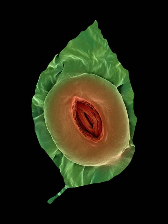
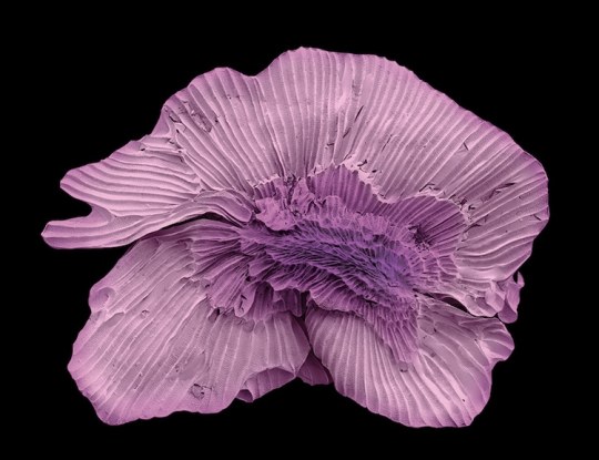
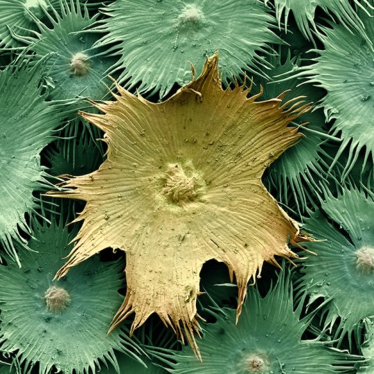
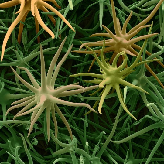
Rob Kesseler is a visual artist and Emeritus Professor of Arts, Design & Science at Central Saint Martins, London. For the past twenty years he has worked with botanical scientists and molecular biologists around the world to explore the living world at a microscopic level. Using a range of complex microscopy processes he creates multi-frame composite images of plant organs.
Using scanning electron microscopy and a mix of microscopic, scientific, digital, and manual processes, artist Rob Kesseler develops coloured micrographs of the intricate patterns within pollen and seed grains, plant cells, and leaf structures. The photographs feature specifics of cellular composition that are undetectable without magnification.
Kesseler tells that as a child, his father gifted him a microscope, marking a pivotal moment in his creative career. “What the microscope gave me was an unprecedented view of nature, a second vision,” he writes, “and awareness that there existed another world of forms, colours and patterns beyond what I could normally see.” The artist says his use of color is inspired by the time he spends researching and observing, and that just like nature, he employs it to attract attention.
36 notes
·
View notes
Text
Electron Micrographs
They're not exactly microbes, but today at my college I got to use the scanning electron microscope to look at some objects. I brought my flash drive and got the lab manager to save the images to it.
Would you guys be interested in seeing these non-microbe micrographs? I'd probably categorize them into a few separate posts and explain what's in them. It'd probably be about 4 posts.
#microbes in hats#microbe posting#interest poll#why do i have to type a title for the poll and also WHY CANT I PASTE INTO IT#rude
4 notes
·
View notes
Text

Science Photo Library
Dragonfly head. Colored scanning electron micrograph (SEM). Dragonflies belong to the scientific order Odonata, meaning toothed ones, because of their powerful serrated jaws. Dragonfly jaws can open as wide as their entire head, allowing them to eat large prey. When they hunt, the large compound eyes of a dragonfly allow it to sort its visual field, like a grid. Keeping their prey in the same section of the grid helps with accuracy when intercepting something mid-flight.
2 notes
·
View notes
Text
Experimentally, we usually observe changes in the crystal shape – a clear sign that the additive is binding to the crystal surface and changing the relative rates of growth of different crystal faces (figures 10.20 and 10.21).
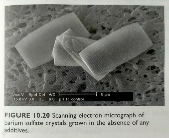

"Chemistry" 2e - Blackman, A., Bottle, S., Schmid, S., Mocerino, M., Wille, U.
#book quotes#chemistry#nonfiction#textbook#experiments#observation#crystals#microscopy#scanning electron micrograph#barium sulfate#ethylenediaminetetraphosphonate#anion
0 notes
Text
this whole matter of trying to sell things is so boring and distasteful. can you put some very bad (or very good) pattern ideas/requests in my ask to take my mind off it? please and thank you. anon is on.
I do want to start kicking these collections out the door on a two-week schedule (/shill) so the more nonsense ideas milling around of my brain the better, for once. Some off the top of my head:
Dressing Table – High Femme/Old Queen/Pickled Egg boudoir kitsch. Perfume bottles and roses in bell jars and Persian cat figurines and Art Nouveau vanity sets etc.
Viscera – Offal, just offal.
Diatoms, Animalcules, etc. These would especially be fun if I can figure out how to design round motifs (I've done one and it was just okay-looking and very hard) to mimic a microscope view.
Pollen grains – In a similar vein, my mother was a palynologist and I'd love to do a set of black-background motifs in the style of scanning electron micrographs.
Philately – I was a terrible stamp collector as a child (catastrophically disorganized from the word go), but I have a "postage stamp" border treatment I've been wanting to use.
Cryptids & Company – Mostly I just liked the title, "Putative Extinctions and 'Living Fossils'" is probably more the thing. Cryptids-as-such being culture-bound, I'd either want to limit myself to white-people cryptids (so like the loch ness monster and whom else? mothman?), which is it's own kind of weird, or solicit design/editorial assistance from someone qualified.
Turtle Island Food Science – American Thanksgiving is my problematic favourite holiday, and maybe that specific title is too dippy-white-Liberal to fly, but I do want to do a collection of Indigenous North American cultivated plants at some point.
Rune Stones – I was a teenage, twenty-something neopagan twenty years ago, it's true, but I still think this would be cute.
Things people actually buy at vendor markets – and it's just like, a knockoff Stanley mug, American junk food smuggled up from North Dakota, MLM leggings, essential oil jewellery and pickles
2 notes
·
View notes
Text
The origins of colour patterns in fossil insects revealed by maturation experiments
Published 20th September 2023
Researchers perform thermal maturation experiments on extant beetles to show how melanin-based monochromatic colour patterns are preserved in insect fossils.

Colour patterning in the elytra of Harmonia axyridis with experimental maturation

Scanning electron micrographs of untreated and matured cuticle of Harmonia axyridis

Experimentally induced change in cuticle thickness boxplots
source:
11 notes
·
View notes
Text

Lily, you're never going to make tardigrade an insult, this is never going to be a thing. People LOVE tardigrades! Just look at these little fellas go:

Credit: My Microscopic World
Besides, you saying that Twitter's full of tardigrades only makes the tardigrades sound bad.
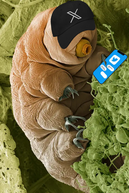
The original image from Steve Gschmeissner/Science Photo Library is a colorized scanning electron micrograph of a tardigrade; microscopic sized Twitter/X.com paraphernalia added separately.
14 notes
·
View notes
