
We highlight collections, exhibits, and events from Special Collections at the University of North Carolina at Chapel Hill's Health Sciences Library, and promote the history of the health sciences.
Don't wanna be here? Send us removal request.
Statistics
We looked inside some of the posts by unchsl and here's what we found interesting.
Average Info
Notes Per Post
262
Likes Per Post
212
Reblog Per Post
49
Reply Per Post
1
Time Between Posts
4 days
Number of Posts By Type
Photo
17
Last Seen Tumblr Blogs


Fun Fact
Tumblr was the first site to host the blog for President Barack Obama in 2011.
Photo

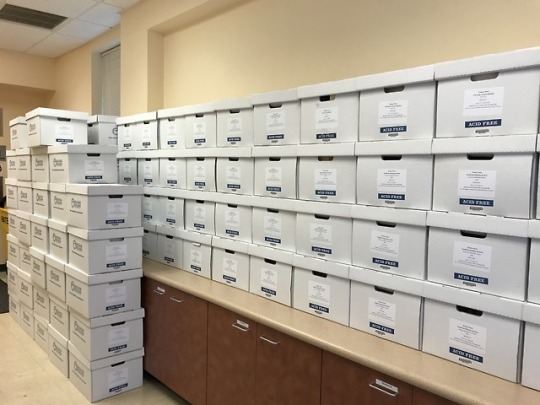


And just like that...they’re gone.
This morning, our last large shipment of boxes from the NYAM Collection of International Medical Theses left the building for the Library Service Center in Durham. Au revoir! À la prochaine!
Don’t worry, though! We will happily bring boxes back to the library for researchers.
13 notes
·
View notes
Photo
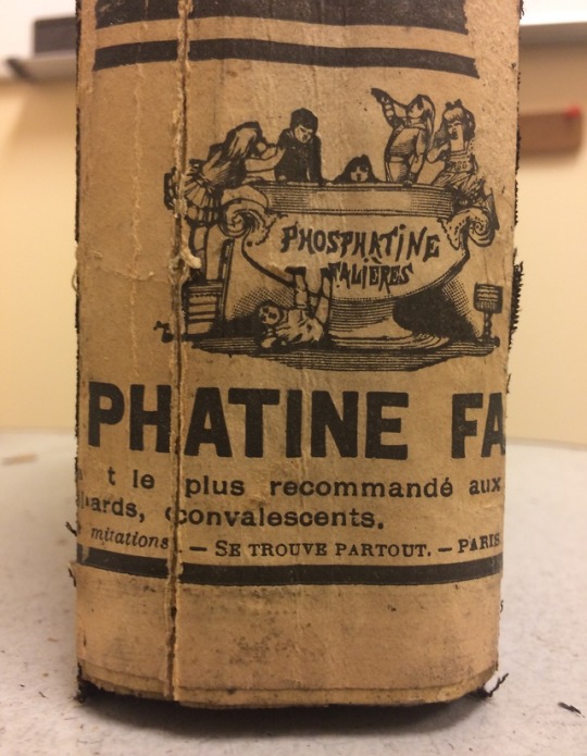
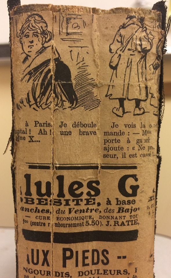
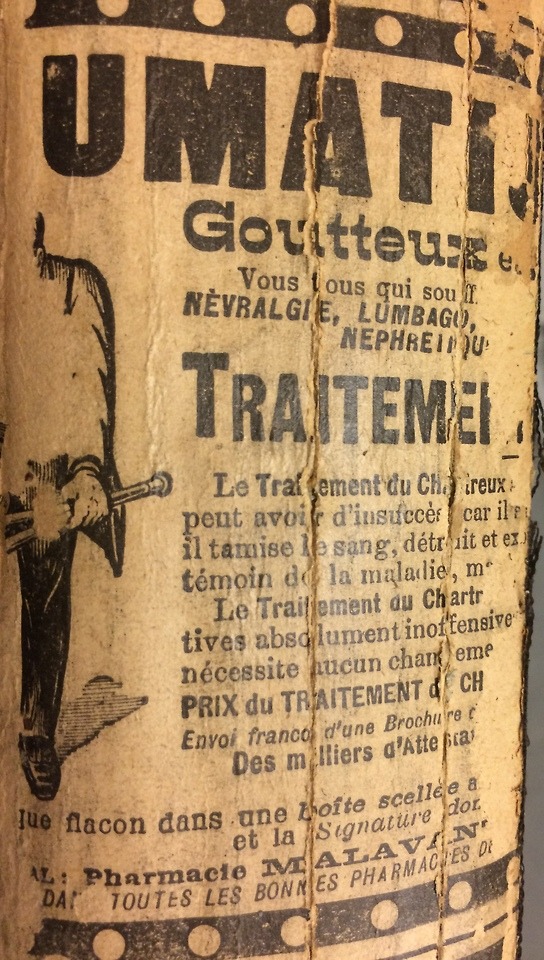
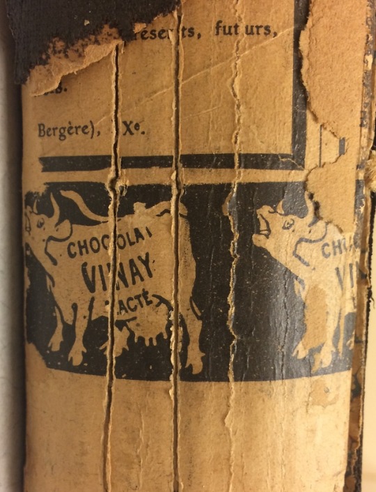
Madame X and Milk Chocolate
Most of our posts about the NYAM collection are about the contents of medical theses, but sometimes the containers are pretty neat too.
While we were processing some bound volumes dated 1889-1899, we found that their leather outer spines had become detached, revealing the backing paper. Discoveries like this are fairly common, as bookbinders in the past frequently made use of waste paper in parts of the binding that were expected to remain covered.
Here are some of the more interesting bindings we found. They are all newspaper advertisements from the first two decades of the 20th century, and likely all came from Parisian paper L’Illustration.
Respectively, they advertise (1) Phosphatine Falières, a dietary phosphate supplement for children, (2) an unknown gastrointestinal panacea sold by “Madame X,” (3) the “Carthusian Treatment” for gout, arthritis, and rheumatism, and (4) Chocolat Vinay lacté, a vintage brand of milk chocolate.
For more about dubious pharmaceutical advertising in the early 20th century (and that mysterious, omnipresent “Madame X”), see our previous post from February 18th, 2018.
#NYAMParis#advertising#clickbait#early 20th century#history of medicine#vintage advertisements#bookbinding#library science#digitization#HSL#pharmacy#chocolate
41 notes
·
View notes
Photo
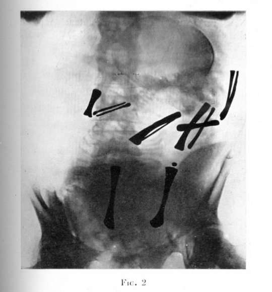
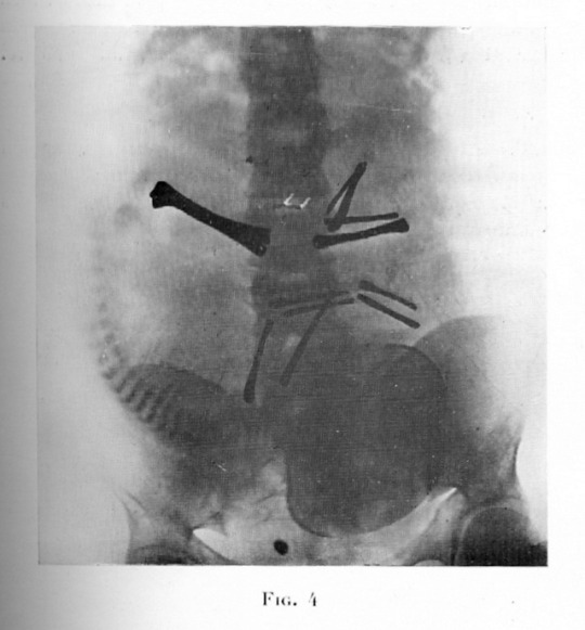
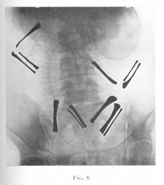
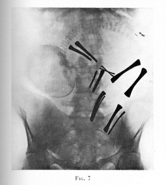
Fluoroscopic images of fetuses in utero, with position of arm and leg bones highlighted, c.1932. Fluoroscopy (continuous x-rays), while diagnostically useful, can result in lengthy exposure to radiation and may be harmful to developing fetuses; these risks were not yet fully understood in the 1930′s.
Source: Dr. Yves Torchaussé, Contribution a l’étude de la radiographie du foetus in utero (Faculté de Médecine de Paris, 1932).
64 notes
·
View notes
Photo

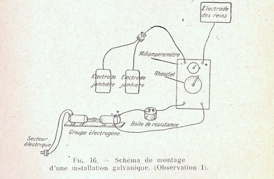

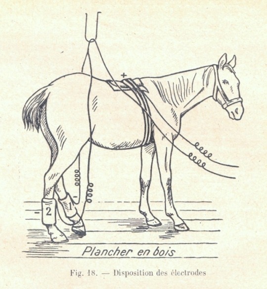
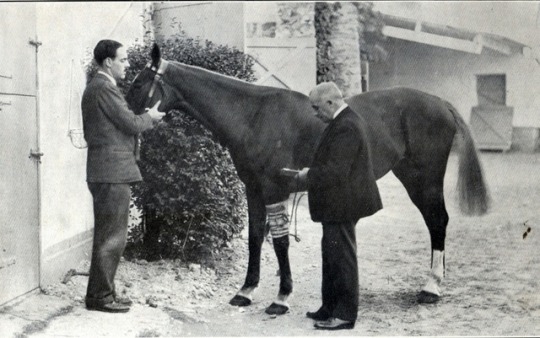
Ride the Lightening
The year: 1927.
The patient: a Bay foal, descended from “the best stock of France”
The problem: myelitis, a nervous system disorder causing the rear legs to list to one side, making the foal barely able to run
The unconventional treatment: electrotherapy
Sold by his breeder for the “ridiculous” price of 3000 francs or “about one-twentieth of his value,” the subject of this veterinary thesis from our NYAM Collection was considered a lost cause due to his rare and mysterious nervous condition---that is, until his veterinarian tried a controlled application of galvanic currents to stimulate his spinal neurons.
Now, I can find no evidence of similar treatments being tried by reputable veterinarians today, and I’m not qualified to speak to the efficacy of galvanotherapy. However, the author of the thesis gives the following account of his quadrupedal patient’s extraordinary progress two years after treatment:
“In 1929, he became the property of a trainer, who, thanks to his good care, led him to run in Saint-Cloud and Tremblay, several times in Saint-Cloud. He makes in these races a very satisfactory impression.”
The veterinarian acknowledges that myelitis in foals has been documented to heal on its own over time; however, he attributes the cure in this case to electrotherapy, and is not shy about taking the credit. He writes:
“Had this foal not become an equine experiment under my direction, he would surely have been shot on the spot.”
So early 20th-century veterinary medicine gave this racehorse a second chance and a happy ending.
Source: Jean-René Soutou, La galvanothérapie et l’ionothérapie électrique en médecine vétérinaire. École Nationale Vétérinaire d’Alfort, 1930.
#veterinary medicine#equestrian#horse#NYAMParis#galvanotherapy#electrotherapy#1920's#history of medicine#early 20th century
10 notes
·
View notes
Photo
Good Boy, Getting Better
A canine patient receives a blood transfusion in this photo from a veterinary medicine thesis in our NYAM Collection.
Source: Pierre André, De la Transfusion sanguine chez le Chien: son application thérapeutique. École Nationale Vétérinaire d’Alfort, 1933.
6 notes
·
View notes
Photo
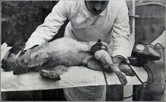
Taking a dog’s blood pressure, c.1930.
Source: Paul Julien LaFaye, La tension artérielle en clinique vétérinaire. École Nationale Vétérinaire d’Alfort, 1932.
10 notes
·
View notes
Photo
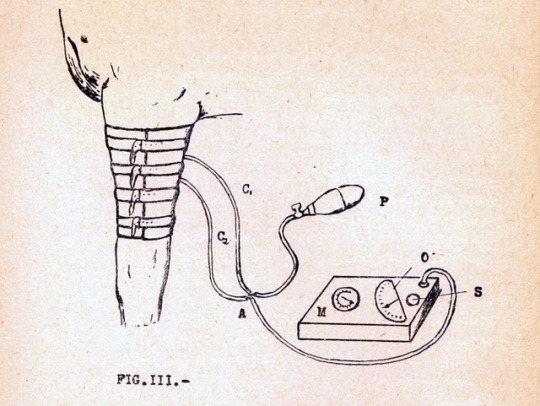
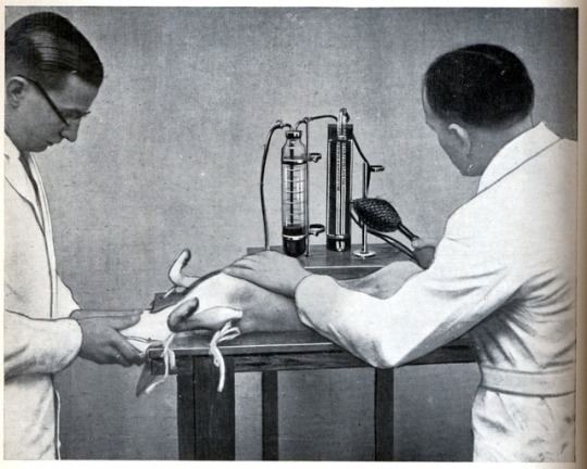
A blood pressure cuff for horses, from a veterinary thesis in our NYAM Collection.
Source: Paul Julien LaFaye, La tension artérielle en clinique vétérinaire. École Nationale Vétérinaire d’Alfort, 1932.
18 notes
·
View notes
Photo

A foal in utero, from a veterinary thesis in our NYAM Collection. This illustration shows an unusual positioning of the foal’s forelegs likely to result in dystocia, or difficult labor.
Source: Louis-Alexandre Champagne, Dystocies par déplacement ou changement de rapports de l’utérus. École Nationale Vétérinaire d’Alfort, 1925.
12 notes
·
View notes
Photo


Tri-Pawd
Diagrams from a veterinary thesis in our NYAM Collection showing the muscular anatomy of a dog’s rear and front legs.
Source: Jean-Albert Ledru, De l’Amputation des Membres dans la contiguïté chez les carnivores domestiques. École Nationale Vétérinaire d’Alfort, 1931.
9 notes
·
View notes
Photo
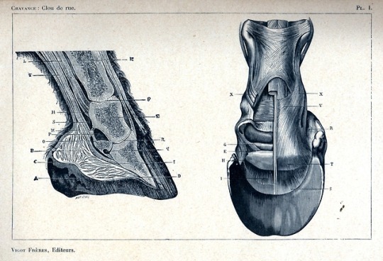
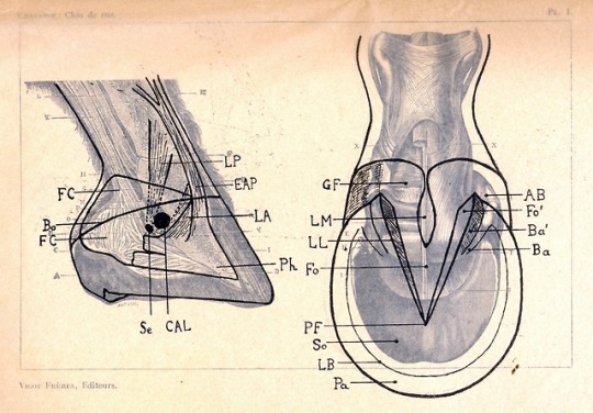
Anatomical diagrams of a horse’s hoof from a veterinary medicine thesis in the NYAM Collection.
Source: Jean-Henri Chavance, Des Traumatismes pénétrants de la région plantaire chez le cheval. École Nationale Vétérinaire D’Alfort: 1930.
12 notes
·
View notes
Photo

All of these empty shelves used to hold boxes of theses from the New York Academy of Medicine Collection of International Medical Theses waiting to be cleaned and processed. How times have changed!
9 notes
·
View notes
Photo
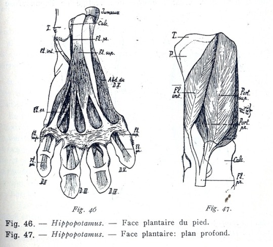
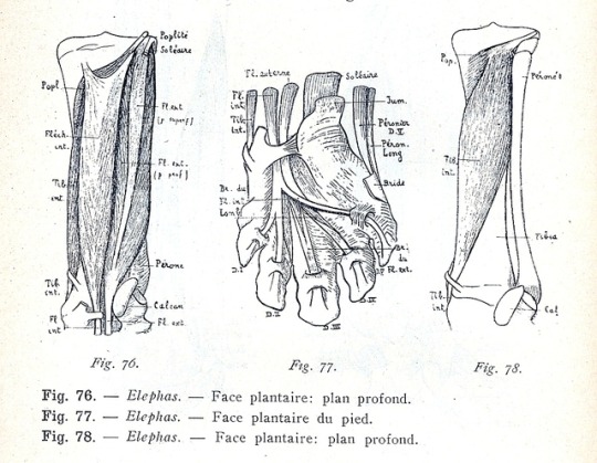
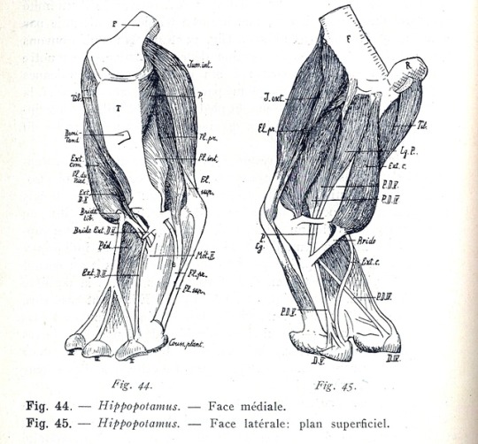
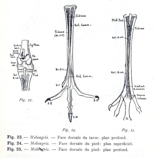
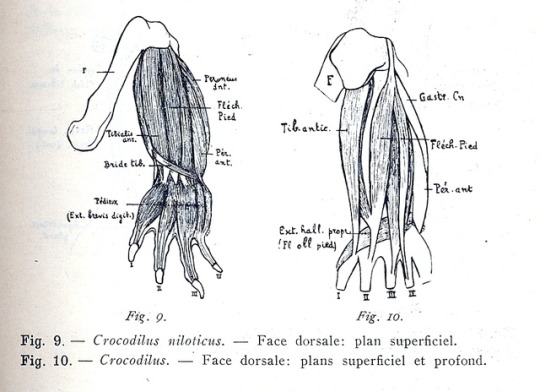
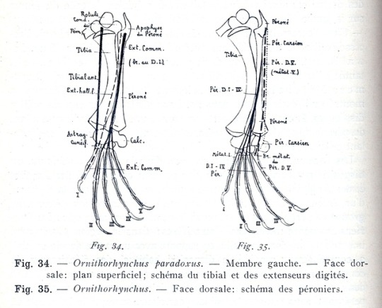
Diagrams showing the comparative musculature and bone structure of various animals, from a veterinary medicine thesis in our NYAM Collection. Shown above are the legs of a hippopotamus, an elephant, a turkey (meleagris), a crocodile, and a platypus (ornithorhynchus).
Source: Marcel-Arsène Petit, Recherches et considérations sur la myologie comparée de la région jambiére. École Nationale Vétérinaire D’Alfort: 1925.
#NYAMParis#hippopotamus#anatomy#history of medicine#platypus#animals#biology#veterinary medicine#lovely bones
9 notes
·
View notes
Photo
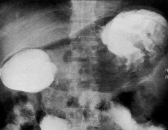
Abdominal x-ray of a patient diagnosed with Ménétrier's Disease, a condition characterized by excessive growth in the mucous membranes of the stomach. The patient ingested a hydro-soluble contrast medium, causing the overgrown gastric folds to show up as ripples inside the stomach (right).
Source: Jean-Pierre Melki, Avantages des opicifiants hydrosolubles dans l'examen radiologique du tractus oeso-gastro-intestinal. Faculté de Médecine Xavier-Bichat, Paris: 1971. Part of the New York Academy of Medicine Collection of International Medical Theses.
5 notes
·
View notes
Photo
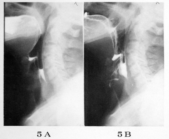
Radiograph of a patient swallowing water with a soluble radiopaque substance mixed into it, c.1971.
Source: Jean-Pierre Melki, Avantages des opicifiants hydrosolubles dans l'examen radiologique du tractus oeso-gastro-intestinal. Faculté de Médecine Xavier-Bichat, Paris: 1971.
10 notes
·
View notes
Photo

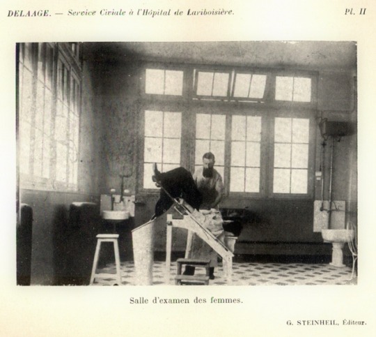
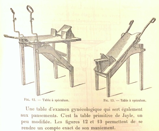
Two types of adjustable gynecological examination chair in use at l'Hôpital de Lariboisière at the turn of the nineteenth century.
Source: Léopold Pierre Delaage, Description et Foncionnement du Service Civiale a l’Hôpital de Lariboisière. Faculté de Médecine de Paris, 1901. Part of the New York Academy of Medicine Collection of International Medical Theses, 1801-1981
#nineteenth century#NYAMParis#upside-down#obstetrics and gynecology#history of medicine#19th-century medicine#badly censored#looks uncomfortable#medical examination
11 notes
·
View notes
Photo
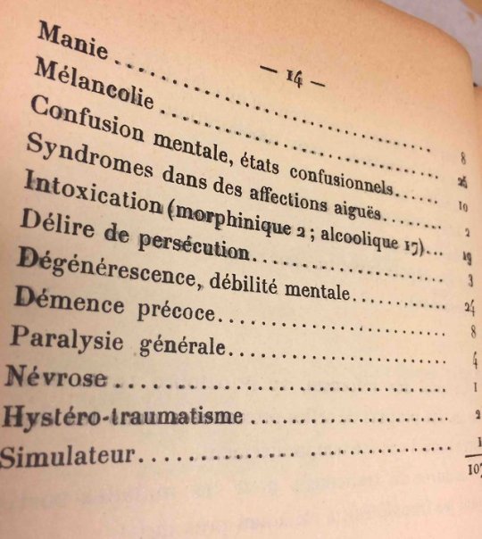
Mental Ill-Health in the Great War
***This post mentions suicide and hallucinatory episodes***
Today we’re featuring two medical theses from our NYAM Collection that contain amazingly detailed narrative accounts of diagnosing and treating mentally ill soldiers in WWI. Unsurprisingly, many of the soldiers described in these documents display symptoms the modern reader will associate with post-traumatic stress disorder (PTSD), like paranoia, hyperarousal, insomnia, nervousness, and immersive flashbacks, nightmares, and hallucinations. In the first quarter of the twentieth century, however, most medical professionals were just beginning to explore the idea that trauma could, by itself, dramatically alter the human mind.
These accounts are frequently poignant, like the story of an infantryman who was imprisoned for belligerence toward a superior officer. When interviewed by a doctor following a suicide attempt, he described experiencing a vivid hallucination of his own funeral:
…with perfect clarity, he saw his coffin, the procession, his comrades whom he recognized. No less clearly, he heard speeches and funeral marches. It is the terror of this hallucination and others he does not remember that drove him to attempt suicide. He suffered from not being able to tell reality from unreality, but he fully admits now that he has been a victim of alcohol.
After this, he was allowed to return to his unit, and to the trenches of WWI, on his own request. Substance abuse (mostly alcohol, but sometimes morphine) is a common theme in this series of case studies; I have a hard time telling whether that’s because it was truly rampant in this population or because doctors were primed to ask about alcohol use and to perceive it as a catalyst for erratic behavior.
In the case of another infantryman (a teetotaler, as the doctor is careful to mention), a specific traumatic event is cited as the origin of a psychological crisis:
On February 16, 1915, a shell exploded near him, killing three nearby men and knocking him down. He gets up, almost deaf, and is taken to Sens. Deafness becomes complete and a concussion is diagnosed. ...Towards the end of March, he enters a hospital in Orléans. He then displays fears that seem to be due to auditory hallucinations. He thinks he's being chased by someone, by a madman... [In the evening] he does not get undressed, [but] simply stretches out on his bed, and at night awakens with a start, searching the room for someone [he is convinced is] there. He is evacuated to the asylum. He is calm, reasonable, but says that he is troubled because he always believes that someone is roaming around him. One evening, an hour after bedtime, he is seized with a bout of agitation with threats of violence... He thinks himself in the trenches in the presence of the enemy; he converses with comrades, gives orders... all with eyes wide open, without taking notice of those around him...
The official diagnosis in this case was hystéro-traumatisme or ‘traumatic hysteria’, a diagnostic sub-category of hysteria proposed by Charcot in the nineteenth century.
The account ends with him being sent home on convalescent leave, making him one of a fortunate few soldiers who were actually removed from combat for serious mental distress. Soldiers like him were frequently accused of malingering or cowardice; some were sent back into battle with little or no treatment, and others were treated with ineffective and aggressive methods.
However, the medical theses in our collection also bear witness to an incipient shift in professional attitudes toward mental illness in the early twentieth century. For example, out of 107 cases of so-called ‘war psychoneurosis’, thesis author Francis Boucherot found only one to have been ‘faking it’ (simulateur). It would be decades before most doctors in France and elsewhere embraced the idea of recognizing, treating, and managing the effects of trauma instead of attributing them to deception, addiction, or hysteria.
Sources:
Francis-Marie-Auguste Boucherot, Contribution a l’Étude des Maladies mentales dans l’Armée en temps de Guerre. Faculté de Médecine de Paris, 1915. (Source of image)
Marius Albert Dumesnil, Délires de Guerre: Influence de la guerre sur les formes des psychoses chez les militaires. Faculté de Médecine de Paris, 1916. (’P.F’, ‘A.A’)
Crocq, Marc-Antoine, and Louis Crocq. “From Shell Shock and War Neurosis to Posttraumatic Stress Disorder: A History of Psychotraumatology.” Dialogues in Clinical Neuroscience 2.1 (2000): 47–55.
Bogousslavsky, Julien, and Laurent Tatu. “French Neuropsychiatry in the Great War: Between Moral Support and Electricity.” Journal of the History of the Neurosciences 22.2 (2013): 144-154.
#NYAMParis#war is hell#mental health#ptsd#WWI#early 20th century#war medicine#hallucinations#psychiatry#history of medicine#suicide#mental ill health
11 notes
·
View notes
Photo
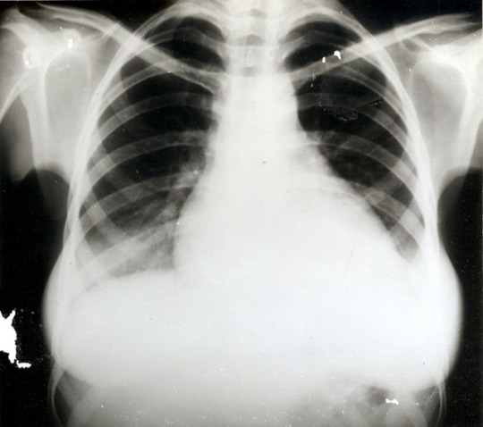
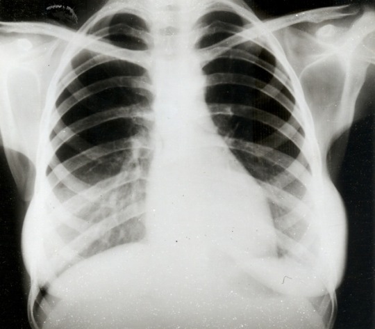
Losing Heart
Radiographs of a pregnant lupus patient’s heart before and after treatment with immunosuppressive drugs. Myocarditis visible in the first x-ray is dramatically reduced in the second, taken two months later.
Source: Marc Terdjman, Traitement immunodepresseur (cyclophosphamide) suivi d’une interruption chirurgicale de grossesse chez une femme lupique au cours d’une poussee aigue de la malade: a propos d’une observation. Faculté de Médecine de Paris, 1971. Part of the New York Academy of Medicine Collection of International Medical Theses, 1801-1981
12 notes
·
View notes