#Histopathology
Text
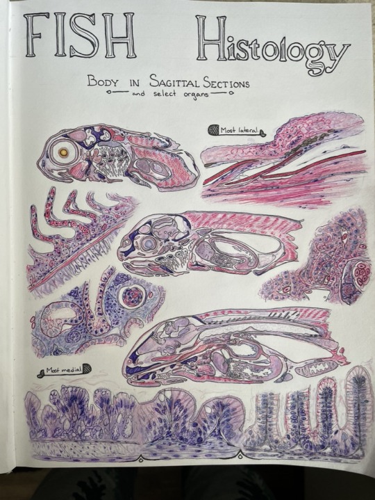


I cannot believe I didn't post this one here! Drawn last year, the biggest page of my fish anatomy and histology atlas.
Technically, transverse sections are more useful for looking at symmetry, so you can see if something is bilateral, but sagittal sections are just so cute! I love seeing a whole fish on a slide.
#fish#histology#anatomy#histopathology#histopath#fish doctor#veterinary#medicine#veterinary medicine#vet student#vetblr#art#pencil#pen#h&e staining#aquatic#port
111 notes
·
View notes
Text
Instead of flowers or trinkets my good friends give me formalin-soaked lumps and eyeballs
#I just got a box of old specimens and I am so excited to sort through them for interesting pathology#Pathology#Histopathology
30 notes
·
View notes
Text
Human herpesvirus 6
“Light micrograph of infected T-lymphocytes with typical so-called inclusion bodies (HE staining). Inclusions in HHV-6 infection (dark blue dots).” - via Wikimedia Commons (original description translated from German using Google Translate)
#human betaherpesvirus 6a#herpesvirus#hhv#hhv-6#human herpesvirus 6#histopathology#micrograph#t lymphocytes#t cells#infectious diseases#immunology#fertility#infertility#wikipedia#wikipedia pictures#nature#wikimedia commons#medicine#medical#herpesviridae#medical microbiology#virology#virus#viruses#diseases#herpesvirales#roseolovirus#betaherpesvirus#orthoherpesviridae#he staining
19 notes
·
View notes
Text

Late night study 🩷✨
#anatomy#pathology#pathologic anatomy#medicine#medicine student#draws#medical draw#histology#histopathology#cells#medical student#medicine mood#study#study time#studyblr#colors#pastel#pink#pink aesthetic#tablet#microscope#instagram#cardiology#neurology#nephrology#brain#veterinary medicine#veterinary medicine student#Bucharest#romania
28 notes
·
View notes
Text
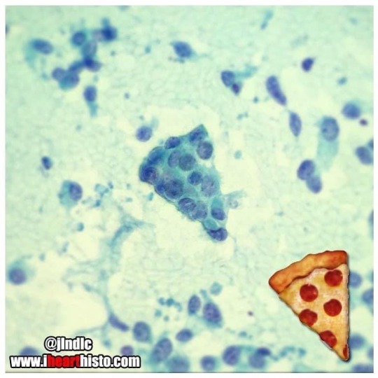
🍕Happy Pizza Day 🍕
Sometimes cells remind me of food.
These are epithelial cells from an adenocarcinoma of the lung.
i♡histo
An adenocarcinoma is a tumor that is derived from the epithelial cells of glandular tissues. The pizza was obtained from a nodule on the lung. An endoscope with an ultrasound attached to it was used to guide a needle directly into the nodule and some cells were then sucked out for observation. The technique is called endoscopic ultrasound fine needle aspiration or EUS/FNA for short. Aspirates like this allow pathologists to determine whether the cells in the nodule looked normal or cancerous so they can recommend appropriate treatment and/or surgery to remove the tumor.
#histology#science#pathology#med school#med student#ihearthisto#vet science#vet school#anatomy#premed#pizza#pizzaday#cells#dentistry#dental school#medlab#histopathology#biomedical science
57 notes
·
View notes
Text

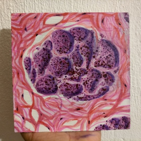

Lobular carcinoma of the breast 🧫💓
Ah yes, una pintura de una imagen histologica ajsjjs. Desde que empecé el semestre y vi por primera vez una laminilla quise pintar algo de ello y pues aproveché el carcinoma de mama (qué era la imagen histologica favorita de mi maestro favorito) para pintarsela de regalo por el fin del semestre sjsjsjs. Le gustó aunque al principio pensó que era un glomérulo, ya después dijo que sí parecía glándula jaksjaja
Él es mi tío aunque no lo sepa jajaja
4 notes
·
View notes
Text
The vet called me a little while ago with the histopathology results. IT WASN’T CANCER! I was so scared that the mass was malignant and am so grateful that it wasn’t. That’s the good news. The bad news? Still no definitive answer. The doctor is going to continue looking for answers, see if she can find any similar cases.
The report is under the cut for anyone interested in reading it.
Chloe has an appointment tomorrow morning at 11 to have her sutures removed, and her blood count, temperature, and weight rechecked.
History:
Proximal medial aspect of right hind limb. Fever of unknown origin for past year, mild stomatitis, proactively treated with azithromycin in case of Bartonella. Borderline anemic. Mass rapidly appeared over the weekend with a flattened plaque on the dorsal aspect. Purulent discharge was noted today.
-
Received: A 4.5 cm x 3.0 cm skin biopsy, with a 3.5 cm x 3.3 cm ulcerated mass.
HISTOPATH REPORT:
MICROSCOPIC FINDINGS:
Haired skin: Ulcerative dermatitis and cellulitis, necrotizing, pyogranulomatous and lymphoplasmacytic, chronic, locally extensive, marked
COMMENTS:
Histologic findings revealed severe ongoing inflammation and granulation tissue formation. No neoplastic populations are observed. Areas of inflammation extend to the deep specimen margin. The extensive degree of necrosis present is suggestive of an infarction. The underlying cause is not evident in this sample. Possible causes include a venomous bite, previous trauma, infection, or a chemical/thermal burn that caused vasculitis, leading to subsequent ischemic necrosis. Special stains are pending to further rule out fungal and acid-fast agents. Results will be forwarded in an addendum. Aerobic culture and sensitivity on fresh tissue may be indicated to further rule out an antibiotic-resistant pyoderma.
MICROSCOPIC DESCRIPTION:
Haired skin and subcutis: An extensive area of the epidermis and dermis has undergone coagulative necrosis, admixed with abundant eosinophilic necrotic coagulum, layers of poorly preserved neutrophils, edema, and streaming nuclear debris. Surrounding areas of necrosis are coalescing aggregates of numerous macrophages, lymphocytes, plasma cells, and fewer neutrophils, admixed with granulation tissue. In less affected areas, anagen hair follicles and sebaceous glands are intact.
***************** ADDENDUM COMMENTS - *********************
GMS and Fite-Faraco stains did not reveal the presence of fungal or acid-fast bacterial etiologic agents.
6 notes
·
View notes
Text
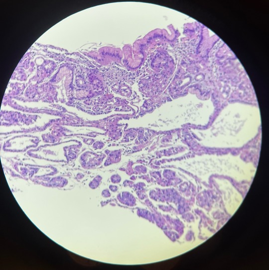
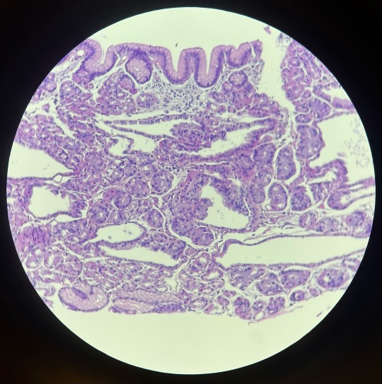
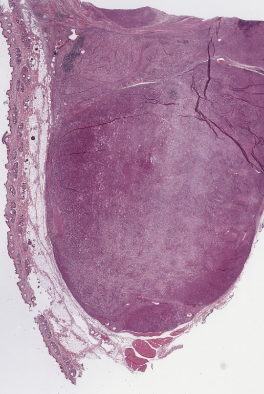
Stomach fundus with gland dilation
2 notes
·
View notes
Text
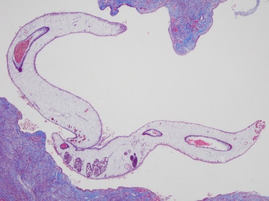
Scar Over?
The Chinese liver fluke is a flatworm found in freshwater environments. Around 15 million people in East Asia are currently infected by this parasite, often due to eating undercooked fish. Setting up camp in the bile ducts, the flatworm feeds on bile and causes liver scarring and cancer – although we don't understand exactly how. Here in cross-section we see two parasitic worms (white) surrounded by the scarred bile duct tissue (purple) of a mouse. Scientists developed genetically modified mice that capture the features of human disease when infected by the flatworm. Using these mice, they discovered that the Chinese liver fluke releases molecules that activate the immune system and cause inflammation. This rallies cells called myofibroblasts to the infection site, where they trigger scarring. This new understanding could help researchers develop more effective treatments for Chinese liver fluke infections and even ways to prevent the long-term damage they cause.
Written by Henry Stennett
Image from Key Laboratory of Zoonosis Research, Ministry of Education; College of Veterinary Medicine, Jilin University, Changchun, China
Research by Yuru Wang, Xu Zhang and Xiaocen Wang et al, Key Laboratory of Zoonosis Research
Image originally published with a Creative Commons Attribution 4.0 International (CC BY 4.0)
Published in PLOS Neglected Tropical Diseases, January 2023
You can also follow BPoD on Instagram, Twitter and Facebook
8 notes
·
View notes
Text


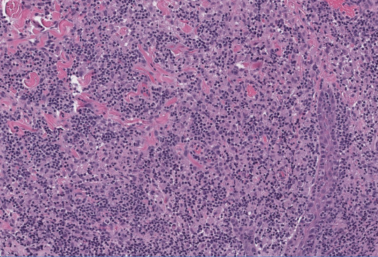
Canine Histopathology
2 notes
·
View notes
Text
Week Recap (Nov. 27 - Dec. 3, 2022)
Part 1
Beginning of Microbiology section
Tried to study for the week (Except on thursday siniksik ba naman yung lab sa finals week edi sana nagrereview kami diba)
Crammed the rest of the subjects the night before finals
Didn't finish reading and woke up with a headache during finals
Already on the acceptance stage even if the grades are not out yet
┈┈┈┈․° ☣ °․┈┈┈┈
Part 2
#1st sem#medtech#mls#medical technology#medical laboratory science#studyblr#medblr#uniblr#schoolblr#college#university#final exams#molecular biology and diagnostics#Clinical bacteriology#microbiology#hematology#clinical chemistry#histopathology#dec 2022
2 notes
·
View notes
Photo

Eloquently put by • @pathologeek 🎃 Halloween is next Monday! This will be our last post about diseases that may have inspired Halloween characters. At least for this year. So last but not least: 👰 A Monster’s Bride 👰 Today we’ll review a skin / hair condition commonly depicted on Frankenstein’s bride and also seen on Lily Munster: the streak of white hair. A medical condition that may have inspired this characteristic hair is known as Poliosis circumscripta. For starters, this nothing spooky, evil, or even different about people who have Poliosis. And you shouldn’t judge anyone with it in any way. Poliosis is defined as a localized patch of white hairs, and it is actually a clinical sign not an specific diagnosis. This is important to point out, because it can be associated with multiple diseases, from genetic syndromes to autoimmune diseases and even neoplastic entities or as a side effect to a topical or systemic drug. Above you can review some slides that show the microscopic characteristics of white versus black hair, and also some examples of skin diseases that can manifest themselves as poliosis. While we opened with a reference to the classic brides of Frankenstein’s monster, this feature is common in several beloved Halloween characters, as well as in novels such as The Mallens by Catherine Cookson, Rogue in the X-Men, and even Cruella de Vil in 101 Dalmatians. All of which would make excellent characters to dress up as this old Hallows Eve!!! #vitiligo #alopecia #halonevus #biology #poliosis #histology #pathologist #pathology #surgicalpathology #histopathology #medicine #medstudent #medicaleducation #pathologeek #pathologyresidents #medicalresident #medschool #medicalstudent #premed #pathologyassistant #histo #pathologylab #laboratory #HALLOWEEN https://www.instagram.com/p/CkOcWU5jb0zmSOcFXDO8EszjCV7gqHwZkhuJww0/?igshid=NGJjMDIxMWI=
#vitiligo#alopecia#halonevus#biology#poliosis#histology#pathologist#pathology#surgicalpathology#histopathology#medicine#medstudent#medicaleducation#pathologeek#pathologyresidents#medicalresident#medschool#medicalstudent#premed#pathologyassistant#histo#pathologylab#laboratory#halloween
2 notes
·
View notes
Text
Histology And Cytology: An Overview of Microscopic Cell And Tissue Analysis Industry
Histology refers to the study of tissues at a microscopic level. It involves the examination of cell and tissue structure and how they are organised to form different body parts. Cytology is a sub-discipline of histology that focuses specifically on the microscopic evaluation of individual cells. Together, these fields provide crucial insights into normal and abnormal cellular structures and functions.
Tools and Techniques Used in Histology Cytology
A variety of tools and techniques are used by Histology And Cytology to examine cells and tissues. Light microscopes are essential for magnifying samples up to 1000x. Chemical staining helps distinguish between different tissue components by imparting specific colors like haematoxylin and eosin (H&E). Immunohistochemistry uses labeled antibodies to detect targeted proteins in situ. Enzyme histochemistry localizes enzyme activities in tissue slices. Electron microscopy such as transmission electron microscopy (TEM) provides ultrastructural details up to 200,000x magnification. Frozen sectioning rapidly prepares fresh tissue biopsies for histological evaluation. Collection techniques like fine needle aspiration biopsy (FNAB) extract cellular samples safely and accurately.
Histological Examination of Tissues
Histologists study tissue morphology to understand cellular architecture and specialization. Epithelial tissues that line and cover organs display characteristic shapes when squamous, cuboidal or columnar. Connective tissues composed of cells and extracellular matrix vary in density from loose areolar to dense regular tissues like bone. Muscle tissues exhibit repeating striations or spindle-like bundles of myofibrils. Nervous tissues have fibrous processes called neurons and neuroglia supporting cells. Examining normal tissue organization serves as a diagnostic baseline for comparison with pathological states.
Cytological Evaluation of Bodily Fluids
Histology And Cytology involves microscopically analyzing cells collected from various body fluids. Sputum screening diagnoses respiratory diseases via expectorated mucus samples. Urine cytology checks for abnormal or cancerous cells shed in urine which may indicate bladder or kidney problems. Cerebrospinal fluid cytology evaluates cells in CSF to help diagnose inflammatory versus infectious conditions of the brain and spinal cord. Serous cavity fluids from body cavities are also examined cytologically. Perhaps the most well-known application is Pap smear testing of cervical cells to detect pre-cancerous changes or cervical cancer at early treatable stages.
Get more insights on Histology And Cytology
Alice Mutum is a seasoned senior content editor at Coherent Market Insights, leveraging extensive expertise gained from her previous role as a content writer. With seven years in content development, Alice masterfully employs SEO best practices and cutting-edge digital marketing strategies to craft high-ranking, impactful content. As an editor, she meticulously ensures flawless grammar and punctuation, precise data accuracy, and perfect alignment with audience needs in every research report. Alice's dedication to excellence and her strategic approach to content make her an invaluable asset in the world of market insights.
(LinkedIn: www.linkedin.com/in/alice-mutum-3b247b137 )

#Histology And Cytology#Histopathology#Cell Biology#Microscopy#Tissue Analysis#Cellular Morphology#Staining Techniques#Immunohistochemistry#Pathology#Diagnostic Techniques
0 notes
Text

Call for Speaker/Delegate/Poster
Attend the CME/CPD accredited 14th World Gastroenterology, IBD & Hepatology Conference from December 17-19, 2024, in Dubai, UAE & Virtual.
WhatsApp us: https://wa.me/442033222718?text=
Register here: https://gastroenterology.universeconferences.com/registration/
#Gastroenterology#IBD#PancreaticHealth#BiliaryHealth#Hepatology#GlutenFreeDiet#Histopathology#PancreaticCancer#AbdominalConcerns#GastroPractice#Pancreatitis#Digestion#PancreaticResearch
0 notes
Text


Ever heard of histopathology? Explore the intricate world of histopathology with a simple swipe!
Learn more>> https://www.sriramakrishnahospital.com/
#histopathology#sriramakrishnahospital#coimbatore#healthcare#multispecialityhospital#healthandwellness#medical#hospital#patientcare#bookyourappointment
0 notes
Text
#biopsy#pathology#breastcancer#histology#cytology#biopsia#medicine#surgery#cancer#cytopathology#pathologylab#cyto#histopathology#doctor#medicalstudent#medical#cancerdemama#breastsurgeon#mastologia#senologia#mastofamilia#medicallab#cirujanomastologo#cirugiaoncoplastica#luchacontraelcancerdemama#ecografiamamaria#mamografia#oncoplasticsurgery#paaf#ultrasonidomamario
0 notes
Last Seen Blogs

anasghareib
A.G.

automaticstudentpersonaee-blog
Untitled

mostly-funnytwittertweets
previously funnytwittertweets

kinkynavlia
Hello slave how willing do you wanna serve me how
