#pleuritical
Text
remember in "waterworks" when kim steps out of saul's office and looks to her left and fucking jesse is standing there?? idk if ill ever feel that concussive hit of dopamine again. i felt like a goddamn funko pop. rose to my feet like a streaming platform subscriber in the theaters at "avengers: endgame" and audibly cheered. guys were blowing out their pleuritic cavities in their local amc yelling because spiderman came out of a portal, meanwhile i was making up world cup chants because a man in his 40s stood in the dark with a beanie pretending to be 19. its literally never going to get better than that.
#syd squeaks#especially after she had looked to her left earlier at that alaska sign and I was like 'hey guys I will kill myself im so serious'#breaking bad#bcs
130 notes
·
View notes
Text

cantautoresimo
II Cesare raccontato nella canzone Alice di Francesco De Gregori non è altri che un diciassettenne Cesare Pavese innamorato di una cantante-ballerina alla quale chiese e ottenne un appuntamento alle sei del pomeriggio davanti all'ingresso principale del caffè dove lei stessa lavorava.
Alle sei in punto Pavese è li ma la ragazza non si fa viva. Passano le ore e Cesare è sempre li ad aspettare.
Agitato e speranzoso, deciso a non mollare.
Verso le 11 inizia a piovere molto e Pavese non ha l'ombrello, ma resta li immobile ad aspettare.
A mezzanotte però si arrende e completamente fradicio e con il cuore in mille pezzi torna a casa.
Il giorno dopo scoprirà che la ballerina era si lì alle 6 quel giorno, ma davanti all'uscita secondaria dove l'attendeva un altro spasimante più fortunato. Quella serata di pioggia costò al poeta una pleurite che lo tenne a letto per tre mesi facendogli perdere buona parte dell'anno scolastico.
Il 27 agosto di 73 anni fa moriva suicida Cesare Pavese
• «Perdono tutti e a tutti chiedo perdono. Va bene? Non fate troppi pettegolezzi»
42 notes
·
View notes
Text
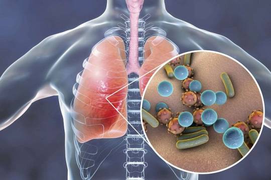
Pneumonia In Children And Adults

Introduction
Pneumonia stands as a prevalent respiratory infection, exerting a significant burden on global public health. Its impact extends beyond mere morbidity, contributing to substantial healthcare costs and socioeconomic consequences. This discussion aims to elucidate the general nature of pneumonia, encompassing its pathophysiology, clinical presentation, diagnostic modalities, treatment strategies, complications, and preventive measures. By indulging into these factors, we aim to provide a better understanding of pneumonia’s complexity and underscore the importance of timely recognition and management.
Pathophysiology

Pneumonia ensues from the infiltration of infectious agents, including bacteria, viruses, fungi, and less commonly, parasites, into the lower respiratory tract. Upon inhalation or aspiration of these pathogens, they gain access to the alveoli, where they incite an inflammatory response. This inflammatory cascade triggers the release of pro-inflammatory cytokines and chemokines, recruiting immune cells to the site of infection. Neutrophils, macrophages, and lymphocytes converge to eradicate the invading pathogens, leading to the characteristic consolidation and exudate formation within the affected lung tissue. As the infection progresses, alveolar edema, impaired gas exchange, and parenchymal damage ensue, culminating in the clinical manifestations of pneumonia.
Clinical Presentation

The clinical presentation of pneumonia encompasses a spectrum of symptoms, ranging from mild respiratory complaints to life-threatening respiratory failure. Common symptoms include cough, productive sputum production, fever, chills, pleuritic chest pain, dyspnea, tachypnea, and systemic manifestations such as malaise and fatigue. The severity of symptoms varies depending on factors such as the underlying pathogen, the extent of lung involvement, the host’s immune status, and comorbidities. In pediatric populations, pneumonia may present with nonspecific symptoms such as feeding difficulties, lethargy, and irritability, posing diagnostic challenges. Conversely, elderly individuals may exhibit atypical presentations characterized by confusion, hypothermia, and exacerbations of underlying chronic conditions.
Diagnostic Modalities

The diagnosis of pneumonia hinges on a comprehensive clinical assessment, augmented by various diagnostic modalities to confirm the presence of pulmonary infection and reveal its etiology. A thorough history and physical examination provide invaluable insights into the patient’s symptomatology, risk factors, and clinical trajectory. Symptomatic findings such as crackles, wheezes, and diminished breath sounds may aid in localizing the site of infection and assessing disease severity. Radiographic imaging, notably chest X-rays and computed tomography (CT) scans, serves as the cornerstone of pneumonia diagnosis, revealing characteristic radiographic findings such as airspace opacities, lobar consolidation, and interstitial infiltrates. Laboratory investigations, including complete blood count (CBC), C-reactive protein (CRP), and procalcitonin levels, may corroborate the clinical suspicion of pneumonia and guide therapeutic decisions. Additionally, microbiological testing of respiratory specimens through techniques such as sputum culture, blood cultures, and polymerase chain reaction (PCR) assays facilitates pathogen identification and antimicrobial susceptibility testing, thereby informing targeted therapy.
Treatment Strategies

The management of pneumonia hinges on prompt initiation of empiric antimicrobial therapy tailored to the likely causative pathogen(s) and disease severity. Antibiotics represent the mainstay of treatment for bacterial pneumonia, with the choice of agent dictated by factors such as local antimicrobial resistance patterns, patient age, comorbidities, and recent antibiotic exposure. Commonly prescribed antibiotics include beta-lactam agents (e.g., penicillins, cephalosporins), macrolides, fluoroquinolones, and combination regimens for severe or healthcare-associated infections. Conversely, viral pneumonia necessitates supportive care measures, given the limited efficacy of antiviral agents in most cases. Influenza-associated pneumonia may benefit from neuraminidase inhibitors such as oseltamivir, while respiratory syncytial virus (RSV) pneumonia may warrant ribavirin therapy in select cases. Adjunctive therapies such as oxygen supplementation, bronchodilators, and corticosteroids may mitigate respiratory distress and improve clinical outcomes, particularly in severe or hypoxemic patients. The duration of antimicrobial therapy varies depending on factors such as the causative pathogen, clinical response, radiographic resolution, and the presence of complications. Close monitoring of clinical parameters and serial imaging studies guide the decision-making process, enabling clinicians to tailor therapy to individual patient needs.
Complications

Pneumonia harbors the potential for various complications, ranging from mild to life-threatening sequelae, necessitating vigilant monitoring and timely intervention. Common complications include pleural effusion, empyema, lung abscess, respiratory failure, septic shock, and acute respiratory distress syndrome (ARDS). Pleural effusion denotes the accumulation of fluid within the pleural space, secondary to inflammation or impaired lymphatic drainage, manifesting as dyspnea, pleuritic chest pain, and dullness to percussion on physical examination. Empyema represents a purulent collection within the pleural cavity, often complicating bacterial pneumonia and necessitating drainage via thoracentesis or chest tube placement. Lung abscesses manifest as circumscribed cavities containing necrotic debris and pus within the lung parenchyma, triggered by persistent fever, productive cough, and hemoptysis. Respiratory failure ensues from impaired gas exchange and alveolar hypoventilation, caused by worsening hypoxemia, hypercapnia, and respiratory acidosis, necessitating mechanical ventilation and intensive care support. Septic shock represents a life-threatening complication of severe pneumonia, characterized by systemic inflammatory response syndrome (SIRS) and end-organ dysfunction, requiring aggressive fluid resuscitation, vasopressor therapy, and broad-spectrum antibiotics. ARDS denotes a severe form of acute lung injury, characterized by diffuse alveolar damage, refractory hypoxemia, and bilateral infiltrates on chest imaging, necessitating lung-protective ventilation and supportive care in the intensive care unit (ICU). The occurrence of complications portends a poor prognosis and underscores the need for early recognition and intervention to mitigate adverse outcomes.
Preventive Measures

Preventing pneumonia entails a broad approach encompassing vaccination, infection control measures, and health promotion strategies aimed at reducing the risk of respiratory infections and their sequelae. Vaccination stands as a cornerstone of pneumonia prevention, targeting common bacterial and viral pathogens implicated in pneumonia pathogenesis. Vaccines such as the pneumococcal conjugate vaccine (PCV13) and pneumococcal polysaccharide vaccine (PPSV23) confer protection against Streptococcus pneumoniae, the leading bacterial cause of pneumonia, particularly in high-risk populations such as young children, older adults, and immunocompromised individuals. Influenza vaccination remains paramount in mitigating influenza-associated pneumonia and reducing disease transmission, underscoring the importance of annual vaccination campaigns targeting vulnerable populations. Additionally, adherence to infection control measures, including hand hygiene, respiratory etiquette, and environmental sanitation, plays a pivotal role in reducing the spread of respiratory pathogens in healthcare settings and the community at large. Health promotion efforts aimed at smoking cessation, optimizing nutrition, and addressing underlying comorbidities such as chronic obstructive pulmonary disease (COPD), asthma, and immunodeficiency bolster immune resilience and mitigate pneumonia risk. Furthermore, early identification and management of predisposing factors such as malnutrition, homelessness, and overcrowded living conditions attenuate pneumonia susceptibility and enhance overall health outcomes.
Conclusion
In conclusion, pneumonia emerges as a formidable respiratory infection, posing significant challenges to global public health. Its diverse etiology, clinical manifestations, diagnostic modalities, treatment modalities, complications, and preventive measures underscore the nature of pneumonia management. Timely recognition and intervention are imperative in mitigating the morbidity and mortality associated with pneumonia, necessitating a collaborative approach among healthcare providers, public health authorities, and policymakers. By fostering a comprehensive understanding of pneumonia’s manifest and implementing evidence-based strategies, we can strive towards reducing its burden and improving patient outcomes. Through ongoing research, education, and advocacy efforts, we can envision a future where pneumonia-related morbidity and mortality are substantially diminished, paving the way for enhanced respiratory health and well-being worldwide.
In managing pneumonia, compassion, empathy, and a holistic approach are essential alongside clinical expertise. Striving for excellence in knowledge and practice allows us to enhance respiratory medicine and patient outcomes.
As we address pneumonia and broader cardiovascular health complexities, let’s remain committed to optimal patient care. Together, we can impact lives positively and foster a healthier future.
Medical students encounter significant academic challenges during their studies, balancing coursework, clinical rotations, research, and personal commitments. Expert Academic Assignment Help offers tailored assistance to meet their needs, providing study materials, tutoring, assignment help, and exam preparation. Beyond academics, it fosters a supportive environment for mentorship and guidance. In essence, Expert Academic Assignment Help is a valuable resource for medical students, empowering them to excel academically and develop into competent healthcare professionals. Contact us at [email protected] for professional assistance
#assignment help#medical students#healthcare#nursing school#nursing student#medicine#medical help#academic assignments#university student#medical university#university life#university#student#student life#study blog#study inspiration#studyblr community#studyblr#study motivation#medication#medical student#medical school#medicare#writing#writers on tumblr#writerscommunity#writeblr#online writing#academic writing
2 notes
·
View notes
Text

Asked my Discord group for some ponies to do random headcanons on. I'll probably do more of these. It was so much fun! Headcanons under the cut!
1.) Vapor Trail is deaf and has a cochlear implant to help her hear! She was born deaf and was always treated the same by her peers. Despite her disability, she not only managed to complete her Wonderbolt training, she was a full fledged Wonderbolt for multiple years!
2.) Coluratura is quite the foodie! Ever since she was a child, she loved the diverse flavors that food could have! When not on tour, she often finds herself at her favorite restaurant (which is how she met her partner, Saffron Masala.)
3.) Pleurite is the first Changeling to be accepted into the Wonderbolt Academy! He's well known by his kingdom to be one of the fastest Changelings around!
4.) I've decided that Twilight is able to grow a bitchin' beard >:) Most unicorns, regardless of gender, are able to grow a small, whispy beard on their chin. But Twilight, about mid-forties, is able to grow a full and complete beard. She usually keeps it shaved, but sometimes she likes to see how long she can go without shaving.
If you like what I do, please like, reblog and follow! Maybe even consider commissioning me (info in my pinned post.)
36 notes
·
View notes
Text
Another Thing my bad leg is my right so i use my cane in my left hand and thats all well and good except my pleuritic lung is Also on the left side so until it clears up i cant really use my cane without aggravating it 😟😟😟😟
3 notes
·
View notes
Text
I am concerned that the alternative brand for my infusion is not going to be as effective as what I received previously. I am starting to develop random synovitis, rashes, funky blood counts, pleuritic pains, and some of the other symptoms I get in the early stages of disease flares. I never had these symptoms on the other infusion. I was on cellcept, methotrexate, and high dose steroids before starting the infusion and my doctors stopped them all because the infusion worked so well. I really don’t want to go back to those :(
14 notes
·
View notes
Text
Myocarditis in Emergency Practice
Myocarditis, an inflammatory condition affecting the heart's myocardial tissues, is a significant cause of sudden cardiac death and dilated cardiomyopathy. With diverse etiologies ranging from viral and immune-mediated causes to toxic exposures, diagnosing and managing myocarditis can be challenging. In this blog post, we will explore the important points regarding the etiology, pathophysiology, presentation, diagnostic testing, and treatment options for myocarditis, with a focus on the perspective of emergency physicians.

Myocarditis can be caused by infectious agents (bacterial, parasitic, viral), immune-mediated conditions, and toxic exposures. Viral causes include enteroviruses, influenza, hepatitis viruses, HIV, herpes viruses, and Parvo B-19. Immune-mediated causes include systemic lupus erythematosus (SLE), scleroderma, and giant cell types. Toxic agents such as doxorubicin, antiretroviral medications, clozapine, and cocaine can also trigger myocarditis.
Myocarditis follows a three-step process. In the acute phase, infectious, autoimmune, or toxic agents directly damage cardiac myocytes. Subsequent myocyte destruction triggers immune system activation and secondary inflammation. In the later stages, the immune system mistakenly attacks the myocytes themselves, leading to progressive myocardial damage.
Myocarditis presents with a wide range of symptoms, necessitating a high index of suspicion for timely diagnosis. Symptoms may include dyspnea, palpitations, orthopnea, and chest pain. Dyspnea is the most common presenting symptom, while chest pain can vary from pleuritic to anginal. Patients may exhibit symptoms of congestive heart failure, ranging from fatigue and peripheral edema to cardiovascular collapse. Skin manifestations can be present in cases triggered by medication exposure.
Diagnostic testing for myocarditis overlaps with other cardiopulmonary evaluations. Electrocardiogram (ECG) abnormalities, such as sinus tachycardia, ST-segment elevations, T-wave inversions, AV blocks, widened QRS durations, or prolonged QT intervals, may be observed. Troponin assays may be elevated, but their absence does not rule out myocarditis. Additional blood tests, including CBC, CRP, and ESR, are often abnormal but nonspecific. Imaging studies like chest radiography and echocardiography can provide valuable information.
TThe treatment of myocarditis primarily focuses on supportive care to prevent further damage to the heart. Stabilizing the patient's ABCs (airway, breathing, circulation) is the priority. Supplemental oxygen and non-invasive positive pressure ventilation may be required for hypoxia or pulmonary edema. Heart failure therapy, including diuretics and nitroglycerin, can be administered if systemic perfusion allows. Cardiac dysrhythmias may necessitate treatment with antidysrhythmic medications. Antimicrobial therapy is required for cases associated with bacterial or parasitic infections. In severe cases, advanced interventions such as intra-aortic balloon pumps, extracorporeal membrane oxygenation (ECMO), or ventricular assist devices (VADs) may be necessary.
Myocarditis presents a complex diagnostic and management challenge for emergency physicians. The diverse etiologies, varied clinical presentations, and overlapping diagnostic tests make timely diagnosis crucial. Supportive care, stabilization, and targeted interventions are key elements of treatment. While further research is needed to refine diagnostic and therapeutic approaches, understanding the etiology, pathophysiology, presentation, and treatment options can aid emergency physicians in effectively managing myocarditis cases.
#emergency medicine#acute care#emergency#health & fitness#biology#emergency physician#foamed#myocarditis
2 notes
·
View notes
Text
Reviewing questions:
Tx for yersinia pestis (plague) = gentamicin 2 mg/kg
Yersinia pestis is a gram-negative coccobacillus that causes the plague. Early recognition and treatment of pneumonic plague is imperative as it carries a high mortality rate. Aminoglycosides are the first line agents for Y. pestis infections, with tetracyclines and fluoroquinolones being alternative agents. Antibiotic resistance does not appear to be common among isolates, but it is classically resistant to penicillins, ampicillin, and first-generation cephalosporins.
A gram stain often shows gram-negative coccobacilli. Wayson's stain (fuchsin-methylene blue and ethyl alcohol-phenol), shows bipolar staining often referred to as a "safety-pin" appearance. Its rapid disease progression, lethality, and ability to be transmitted by aerosolization have raised concerns about its potential use as a biological weapon. Such agents are deemed "Category A" agents by the Centers for Disease Control due to their high mortality and easy dissemination. Similar agents include Variola major (smallpox), Bacillus anthracis (anthrax), Clostridium botulinum toxin (botulism), Francisella tularensis (tularemia), Filoviruses (Ebola, Marburg), and Arenaviruses (Lassa, Junin, and related viruses).
There are 3 major clinical syndromes associated with Yersinia pestis: Bubonic plague - skin lesions at the site of flea bites that range from asymptomatic to necrotic lesions
Septicemic plague - a bubo might be present, associated with gastrointestinal symptoms developing into multiorgan failure.
Pneumonic plague - within hours-days from inhalation of infected droplets, patients present with sudden onset dyspnea, high fevers, pleuritic chest pain, and cough. This is rapidly fatal unless prompt antibiotics are given. Mortality rate is 100% if untreated, and 50% if treated.
Tx for anthrax = ciprofloxacin + clindamycin + anthrax immunoglobulin
Inhalational anthrax usually occurs in textile and tanning industries among workers handling contaminated animal wool, hair, and hides. Inhalational anthrax begins abruptly, usually 1 to 3 days (range, 1 to 60 days) after inhaling anthrax spores, which are 1 to 5 µm in diameter. The number of spores needed to cause inhalational anthrax varies. As evidenced by anthrax cases in the United States in 2001, fewer spores of weapon-grade anthrax may be required to cause inhalational anthrax. Patients with inhalational anthrax present initially with nonspecific symptoms, including a low-grade fever and an unproductive cough. They may report substernal discomfort early in the illness. After initial improvement, inhalational anthrax progresses rapidly, causing hemorrhagic mediastinitis and rapid clinical deterioration. The following symptoms may be present: high fever, severe shortness of breath, tachypnea, cyanosis, profuse diaphoresis, hematemesis, and chest pain, which may be severe enough to mimic acute myocardial infarction. Treatment of inhalational anthrax without meningitis includes ciprofloxacin plus clindamycin plus anthrax immunoglobulin or raxibacumab.
Bottom Line: Raxibacumab is a human monoclonal antibody intended for the prophylaxis and treatment of inhaled anthrax. A common therapeutic combination is raxibacumab plus ciprofloxacin, which are given intravenously. Antibiotics are usually taken for 60 days by people who have been exposed to anthrax, because it can take spores that long to germinate. Septicemic anthrax refers to overwhelming infection by anthrax bacilli. This form of anthrax may complicate inhalational anthrax. The anthrax bacilli multiply in the blood and proliferate to outnumber red blood cells. Another name for anthrax is black blood, which refers to the very dark color of the blood of animals or humans with overwhelming septicemic anthrax. Because humans are relatively resistant to invasion by Bacillus anthracis, most cases of septicemic anthrax occur following inhalational anthrax. The number of organisms released from the liver or spleen into the bloodstream overwhelms host defenses and produces massive amounts of lethal toxin that cause shock and death.
It is believed that amniotic fluid embolism causes a catastrophic inflammatory response to various substances within the amniotic fluid when it enters the maternal circulation. It usually occurs during labor or in the immediate postpartum period, although it can theoretically occur any time during pregnancy when there is a potential exposure (ie, trauma).
Amniotic fluid embolism can cause sudden, rapid cardiopulmonary collapse and DIC, often leading to complete cardiopulmonary arrest. The presentation is variable, depending on the amount of exposure, and can include hypotension, dyspnea, hemorrhage, altered mental status, pulmonary edema, cardiac arrest, and death.
There is no definitive diagnostic test. Rather, the diagnosis is based on clinical suspicion and exclusion of other causes. Treatment is entirely supportive and includes cardiac life support, pressors, blood products, oxygen, airway support, and cesarean section, depending on the timing and status of the fetus. The mortality is widely variable, but severe cases have high rates of mortality and long-term sequelae.
Pulmonary artery hypertension is often treated with prostaglandins, such as epoprostenol.
2 notes
·
View notes
Text
Woke up at 3AM and tried to get myself back to sleep but it just wasn’t working. Blew my nose a bunch (my anxiety is telling me I shouldn’t ever tell anyone this) and now I feel a lot better but I’m still not sleepy. What if I should go to the toilet? Maybe that’ll help? But I’m scared of bugs and I’m scared I’ll see one. I’m really comfy in bed but i also feel so energetic at the same time it’s so weird
Ive realised im using this blog as a journal and i have no regrets about that.
I have really weird left sided chest pain and I’m scared now. Pleuritic chest pain is scary. Hmm maybe I should go to a and e? Maybe it’s positional - one sec I’ll go try a bunch of positions it’s a lot better on my sides.
I feel so weird byee
0 notes
Text
Quand les blattes remplacent les fourmis dans la trophobiose : un nouveau modèle de comportement vital majeur chez des familles d'hémiptères (cicadelles, fulgores...) a été découvert grâce à une sy...
See on Scoop.it - EntomoNews
The mutualistic interspecific relationships of trophobiosis between trophobiont planthoppers (Hemiptera, Fulgoromorpha) providing food to the host called xenobiont, are reviewed.
When Cockroaches Replace Ants in Trophobiosis: A New Major Life-Trait Pattern of Hemiptera Planthoppers Behaviour Disclosed When Synthesizing Photographic Data
Diversity, 01.03.2023
Thierry Bourgoin, Ilia Gjonov, Albena Lapeva-Gjonova, Sonia Roger, Jérôme Constant, Gernot Kunz et Michael R. Wilson
[Image] Graphical Abstract
-------
NDÉ
Traduction
Les relations mutualistes interspécifiques de trophobiose entre les trophobiontes (Hemiptera, Fulgoromorpha) fournissant de la nourriture à l'hôte appelé xénobiont, sont passées en revue. Le degré de relations interspécifiques entre ces symbiotes varie d'occasionnel ou de courte durée (quelques heures à quelques jours) à des relations plus longues, avec des trophobiontes laissés libres de s'échapper (type optobiotique) par le xénobionte, ou maintenus enfermés dans des nids ou des abris de fourmis (type cryptobiotique). Sur 267 cas collectés, 126 sont de nouvelles observations illustrées. La trophobiose occasionnelle est documentée dans 13 familles de cicadelles et semble être assez générale chez Fulgoromorpha, bien qu'elle soit signalée pour la première fois chez Dictyopharidae, Eurybrachidae, et Nogodinidae.
Les xénobiontes associés aux cicadelles sont signalés chez les fourmis et d'autres hyménoptères, lépidoptères et blattodea, mais aussi chez les mollusques et même chez de petits vertébrés gekkonides. Les Tettigometridae semblent être exclusivement entretenus par les fourmis, tandis que les Fulgoridae sont beaucoup plus souvent entretenus par les blattes (40 %) que par les fourmis (27 %).
La trophobiose à long terme se produit toujours avec les fourmis, les cryptobiotiques étant signalés chez les Cixiidae, Delphacidae, Tettigometridae, Meenoplidae, Flatidae et Hypochthonellidae, tandis que les optobiotiques restent limités aux tettigometrides. De nouvelles observations étho-écologiques sur 92 espèces de tettigometrides actuellement décrites permettent de mettre l'accent sur les Tettigometridae fréquentés par les fourmis.
32 espèces différentes (35%) sont désormais connues pour être fréquentées par les fourmis. En Bulgarie, où quatorze espèces sont présentes, la trophobiose se produit avec au moins cinq espèces d'entre elles (36%). Chez les tettigometrides, la sous-socialité, la sessilité et la vie souterraine semblent être des facteurs clés permettant des relations plus complexes avec les fourmis. Cependant, la taille de la cicadelle et donc la quantité de nourriture (gouttes de miellat) est probablement aussi un facteur important. Cela pourrait expliquer de nombreuses nouvelles observations de fulgoridés de grande taille et souvent isolés avec des blattes.
Le tapotement des ailes antérieures des trophobiontes par les blattes, les papillons de nuit, ou du substrat de l'écorce par les geckos a été observé, mais les comportements de palpation antennaire par les fourmis sont les plus fréquemment observés chez les tettigometrides, bien que ce ne soit pas le cas chez les plus grandes espèces. Chez les tettigometrides, des sécrétions spécifiques des glandes tégumentaires (allomones) des pleurites de l'abdomen pourraient également jouer un rôle dans leurs associations mutualistes à long terme, voire même compléter l'action des kairomones du miellat dans la trophobiose des sauterelles en général.
Mots clés :
trophobiose ; comportement ; fourmis ; blattes ; cicadelles ; Tettigometridae ; Fulgoridae
0 notes
Text
Make the Diagnosis: A Solitary, Poorly-Defined Pulmonary Nodule in a Febrile Patient#Diagnosis #Solitary #PoorlyDefined #Pulmonary #Nodule #Febrile #Patient
Presentation
A 67-year-old man presented to the ED appearing ill with complaints of productive cough, dyspnea, pleuritic chest pain, and fever for the past 5 days. He noted that dyspnea had been present for years; he had been on low-dose rituximab as management therapy for pulmonary alveolar proteinosis for 8 years. On physical examination, the patient had a temperature of 103°F (39°C). Lab…

View On WordPress
0 notes
Text
Vertebral Osteomyelitis Masquerading as Pulmonary Embolism

Clinical entities known to mimic the presentation of pulmonary embolism (PE) include pneumonia, asthma, bronchitis, chronic obstructive airway disease (COPD) flare, congestive heart failure, acute myocardial infarction, primary pulmonary artery sarcoma, and nephrolithiasis. Only two cases of vertebral osteomyelitis mimicking PE are found in the literature. We report a rare case of lumbar vertebral osteomyelitis presenting with pleuritic chest pain and shortness of breath, and therefore causing delayed diagnosis.
Read more about articles: https://lupinepublishers.com/clinical-community-medicine/fulltext/vertebral-osteomyelitis-masquerading-as-pulmonary-embolism.ID.000123.php
Read more Lupine Publishers Google Scholar articles:
https://scholar.google.com/citations?view_op=view_citation&hl=en&user=vfbCW-wAAAAJ&cstart=20&pagesize=80&citation_for_view=vfbCW-wAAAAJ:ruyezt5ZtCIC
0 notes
Text
Pneumonia In Children And Adults

Introduction
Pneumonia stands as a prevalent respiratory infection, exerting a significant burden on global public health. Its impact extends beyond mere morbidity, contributing to substantial healthcare costs and socioeconomic consequences. This discussion aims to elucidate the general nature of pneumonia, encompassing its pathophysiology, clinical presentation, diagnostic modalities, treatment strategies, complications, and preventive measures. By indulging into these factors, we aim to provide a better understanding of pneumonia’s complexity and underscore the importance of timely recognition and management.
Pathophysiology

Pneumonia ensues from the infiltration of infectious agents, including bacteria, viruses, fungi, and less commonly, parasites, into the lower respiratory tract. Upon inhalation or aspiration of these pathogens, they gain access to the alveoli, where they incite an inflammatory response. This inflammatory cascade triggers the release of pro-inflammatory cytokines and chemokines, recruiting immune cells to the site of infection. Neutrophils, macrophages, and lymphocytes converge to eradicate the invading pathogens, leading to the characteristic consolidation and exudate formation within the affected lung tissue. As the infection progresses, alveolar edema, impaired gas exchange, and parenchymal damage ensue, culminating in the clinical manifestations of pneumonia.
Clinical Presentation

The clinical presentation of pneumonia encompasses a spectrum of symptoms, ranging from mild respiratory complaints to life-threatening respiratory failure. Common symptoms include cough, productive sputum production, fever, chills, pleuritic chest pain, dyspnea, tachypnea, and systemic manifestations such as malaise and fatigue. The severity of symptoms varies depending on factors such as the underlying pathogen, the extent of lung involvement, the host’s immune status, and comorbidities. In pediatric populations, pneumonia may present with nonspecific symptoms such as feeding difficulties, lethargy, and irritability, posing diagnostic challenges. Conversely, elderly individuals may exhibit atypical presentations characterized by confusion, hypothermia, and exacerbations of underlying chronic conditions.
Diagnostic Modalities
The diagnosis of pneumonia hinges on a comprehensive clinical assessment, augmented by various diagnostic modalities to confirm the presence of pulmonary infection and reveal its etiology. A thorough history and physical examination provide invaluable insights into the patient’s symptomatology, risk factors, and clinical trajectory. Symptomatic findings such as crackles, wheezes, and diminished breath sounds may aid in localizing the site of infection and assessing disease severity. Radiographic imaging, notably chest X-rays and computed tomography (CT) scans, serves as the cornerstone of pneumonia diagnosis, revealing characteristic radiographic findings such as airspace opacities, lobar consolidation, and interstitial infiltrates. Laboratory investigations, including complete blood count (CBC), C-reactive protein (CRP), and procalcitonin levels, may corroborate the clinical suspicion of pneumonia and guide therapeutic decisions. Additionally, microbiological testing of respiratory specimens through techniques such as sputum culture, blood cultures, and polymerase chain reaction (PCR) assays facilitates pathogen identification and antimicrobial susceptibility testing, thereby informing targeted therapy.
Treatment Strategies

The management of pneumonia hinges on prompt initiation of empiric antimicrobial therapy tailored to the likely causative pathogen(s) and disease severity. Antibiotics represent the mainstay of treatment for bacterial pneumonia, with the choice of agent dictated by factors such as local antimicrobial resistance patterns, patient age, comorbidities, and recent antibiotic exposure. Commonly prescribed antibiotics include beta-lactam agents (e.g., penicillins, cephalosporins), macrolides, fluoroquinolones, and combination regimens for severe or healthcare-associated infections. Conversely, viral pneumonia necessitates supportive care measures, given the limited efficacy of antiviral agents in most cases. Influenza-associated pneumonia may benefit from neuraminidase inhibitors such as oseltamivir, while respiratory syncytial virus (RSV) pneumonia may warrant ribavirin therapy in select cases. Adjunctive therapies such as oxygen supplementation, bronchodilators, and corticosteroids may mitigate respiratory distress and improve clinical outcomes, particularly in severe or hypoxemic patients. The duration of antimicrobial therapy varies depending on factors such as the causative pathogen, clinical response, radiographic resolution, and the presence of complications. Close monitoring of clinical parameters and serial imaging studies guide the decision-making process, enabling clinicians to tailor therapy to individual patient needs.
Complications

Pneumonia harbors the potential for various complications, ranging from mild to life-threatening sequelae, necessitating vigilant monitoring and timely intervention. Common complications include pleural effusion, empyema, lung abscess, respiratory failure, septic shock, and acute respiratory distress syndrome (ARDS). Pleural effusion denotes the accumulation of fluid within the pleural space, secondary to inflammation or impaired lymphatic drainage, manifesting as dyspnea, pleuritic chest pain, and dullness to percussion on physical examination. Empyema represents a purulent collection within the pleural cavity, often complicating bacterial pneumonia and necessitating drainage via thoracentesis or chest tube placement. Lung abscesses manifest as circumscribed cavities containing necrotic debris and pus within the lung parenchyma, triggered by persistent fever, productive cough, and hemoptysis. Respiratory failure ensues from impaired gas exchange and alveolar hypoventilation, caused by worsening hypoxemia, hypercapnia, and respiratory acidosis, necessitating mechanical ventilation and intensive care support. Septic shock represents a life-threatening complication of severe pneumonia, characterized by systemic inflammatory response syndrome (SIRS) and end-organ dysfunction, requiring aggressive fluid resuscitation, vasopressor therapy, and broad-spectrum antibiotics. ARDS denotes a severe form of acute lung injury, characterized by diffuse alveolar damage, refractory hypoxemia, and bilateral infiltrates on chest imaging, necessitating lung-protective ventilation and supportive care in the intensive care unit (ICU). The occurrence of complications portends a poor prognosis and underscores the need for early recognition and intervention to mitigate adverse outcomes.
Preventive Measures

Preventing pneumonia entails a broad approach encompassing vaccination, infection control measures, and health promotion strategies aimed at reducing the risk of respiratory infections and their sequelae. Vaccination stands as a cornerstone of pneumonia prevention, targeting common bacterial and viral pathogens implicated in pneumonia pathogenesis. Vaccines such as the pneumococcal conjugate vaccine (PCV13) and pneumococcal polysaccharide vaccine (PPSV23) confer protection against Streptococcus pneumoniae, the leading bacterial cause of pneumonia, particularly in high-risk populations such as young children, older adults, and immunocompromised individuals. Influenza vaccination remains paramount in mitigating influenza-associated pneumonia and reducing disease transmission, underscoring the importance of annual vaccination campaigns targeting vulnerable populations. Additionally, adherence to infection control measures, including hand hygiene, respiratory etiquette, and environmental sanitation, plays a pivotal role in reducing the spread of respiratory pathogens in healthcare settings and the community at large. Health promotion efforts aimed at smoking cessation, optimizing nutrition, and addressing underlying comorbidities such as chronic obstructive pulmonary disease (COPD), asthma, and immunodeficiency bolster immune resilience and mitigate pneumonia risk. Furthermore, early identification and management of predisposing factors such as malnutrition, homelessness, and overcrowded living conditions attenuate pneumonia susceptibility and enhance overall health outcomes.
Conclusion
In conclusion, pneumonia emerges as a formidable respiratory infection, posing significant challenges to global public health. Its diverse etiology, clinical manifestations, diagnostic modalities, treatment modalities, complications, and preventive measures underscore the nature of pneumonia management. Timely recognition and intervention are imperative in mitigating the morbidity and mortality associated with pneumonia, necessitating a collaborative approach among healthcare providers, public health authorities, and policymakers. By fostering a comprehensive understanding of pneumonia’s manifest and implementing evidence-based strategies, we can strive towards reducing its burden and improving patient outcomes. Through ongoing research, education, and advocacy efforts, we can envision a future where pneumonia-related morbidity and mortality are substantially diminished, paving the way for enhanced respiratory health and well-being worldwide.
In managing pneumonia, compassion, empathy, and a holistic approach are essential alongside clinical expertise. Striving for excellence in knowledge and practice allows us to enhance respiratory medicine and patient outcomes.
As we address pneumonia and broader cardiovascular health complexities, let’s remain committed to optimal patient care. Together, we can impact lives positively and foster a healthier future.
Email [email protected] to discover how we can support your academic and professional goals. Wishing you ongoing success in your medical journey.
#assignment help#medical students#healthcare#nursing student#medicine#medical help#medical school#health and wellness#online writing#nursing school#nurses#nursing#public health#heart disease#pharmacy colleges#pharmacy student#pharmacy technician#pharmacy
0 notes
Text
Dolore Toracico nel Bambino : cause, rimedi e cosa fare
Nuovo post pubblicato su https://wdonna.it/dolore-toracico-nel-bambino-cause-rimedi-e-cosa-fare/114493?utm_source=TR&utm_medium=Tumblr&utm_campaign=114493
Dolore Toracico nel Bambino : cause, rimedi e cosa fare

Il dolore toracico può essere un sintomo comune nei bambini e può essere causato da una varietà di condizioni. In alcuni casi, il dolore toracico può essere grave e richiedere una valutazione medica immediata. In altri casi, il dolore può essere meno grave e può essere trattato a casa.
Le cause comuni di dolore toracico nei bambini includono:
Infezioni respiratorie: come il raffreddore o l’influenza, che possono causare dolore o fastidio nella parte anteriore del torace a causa della tosse o della difficoltà a respirare.
Asma: una condizione respiratoria che può causare respiro affannoso e dolore al torace durante gli attacchi di asma.
Reflusso gastroesofageo: il reflusso di cibo o succhi gastrici dallo stomaco nell’esofago può causare bruciore di stomaco e dolore al torace.
Traumi: come una caduta o un colpo al torace, che può causare dolore o fastidio.
Altre condizioni mediche: come la pleurite (infiammazione della membrana che riveste i polmoni), la pericardite (infiammazione del sacco che circonda il cuore) o l’angina (dolore al petto causato da una ridotta apporto di sangue al cuore).
Se il tuo bambino ha un dolore toracico grave o improvviso, o se il dolore è accompagnato da sintomi come difficoltà a respirare, febbre alta o tosse grave, dovresti cercare assistenza medica immediata. In caso contrario, il dolore toracico può essere trattato a casa con il riposo, il bere molti liquidi e il prendere farmaci da banco per il dolore e il mal di gola, se necessario. Se il dolore persiste o peggiora, dovresti consultare il medico del tuo bambino
Infezioni respiratorie
Le infezioni respiratorie sono comuni nei bambini e possono essere causate da virus o batteri. I sintomi comuni di un’infezione respiratoria includono tosse, raffreddore, febbre, dolore al petto e difficoltà a respirare.
Le infezioni respiratorie più comuni nei bambini sono il raffreddore e l’influenza. Il raffreddore è causato da virus respiratori e di solito è lieve, con sintomi che includono naso che cola, mal di gola e starnuti. L’influenza è causata da un virus respiratorio e può essere più grave del raffreddore, con sintomi come febbre alta, mal di testa, dolori muscolari e stanchezza.
Altre infezioni respiratorie comuni nei bambini includono:
Bronchite: infiammazione dei bronchi, che sono i tubi che portano l’aria ai polmoni. La bronchite può essere causata da un’infezione virale o batterica e può causare tosse con catarro e difficoltà a respirare.
Polmonite: infiammazione dei polmoni, che può essere causata da un’infezione virale o batterica. La polmonite può causare febbre alta, tosse con catarro, dolore al petto e difficoltà a respirare.
Sinusite: infiammazione dei seni nasali, che può causare congestione nasale, mal di testa e dolore al viso.
Le infezioni respiratorie nei bambini di solito guariscono da sole, ma alcune forme di infezione possono essere gravi e richiedere un trattamento medico. Se il tuo bambino ha sintomi gravi o persistenti, o se ha difficoltà a respirare, dovresti consultare il medico del tuo bambino. Il medico può prescrivere farmaci per il trattamento dell’infezione e per alleviare i sintomi.
Reflusso Gastroesofageo
Il reflusso gastroesofageo (GERD) è una condizione in cui il contenuto dello stomaco torna indietro nell’esofago, causando bruciore di stomaco e dolore al petto. Il GERD è comune nei bambini e può essere fastidioso, ma di solito non è grave.
I sintomi del GERD nei bambini possono includere:
Bruciore di stomaco
Dolore al petto o fastidio
Tosse cronica o persistente
Rigurgito o vomito
Difficoltà a mangiare o perdita di appetito
Reflusso durante il sonno (che può causare apnea del sonno o difficoltà a respirare durante il sonno)
Le cause del GERD nei bambini possono includere:
Una dieta ricca di cibi acidi o piccanti
Obesità
Fumo passivo
Assunzione di alcuni farmaci (come gli steroidi)
Il trattamento del GERD nei bambini di solito consiste nell’evitare gli alimenti che peggiorano i sintomi, come cibi acidi o piccanti, e nell’assumere farmaci per ridurre l’acidità dello stomaco. Se i sintomi del tuo bambino sono gravi o persistenti, dovresti consultare il medico del tuo bambino per una valutazione. Il medico può prescrivere farmaci per il trattamento del GERD o suggerire altre opzioni di trattamento.
Trauma
Un trauma toracico può essere un’emergenza medica grave che può causare danni a organi vitali come il cuore, i polmoni e i vasi sanguigni. I bambini possono sperimentare un trauma toracico a causa di un incidente, come una caduta o un colpo al torace, o a causa di una lesione da taglio o da punta.
I sintomi di un trauma toracico nei bambini possono includere:
Dolore al petto o fastidio
Difficoltà a respirare o respiro affannoso
Tosse con sangue o catarro
Stanchezza o debolezza
Vertigini o svenimento
Se il tuo bambino ha subito un trauma toracico grave o sospetti che abbia subito un trauma toracico, dovresti cercare assistenza medica immediata. Il medico del tuo bambino eseguirà una valutazione completa e potrebbe ordinare radiografie, ecografie o altre procedure per determinare l’entità del danno e il trattamento appropriato.
Il trattamento del trauma toracico nei bambini dipenderà dalla gravità del danno e potrebbe includere il ricovero in ospedale, il supporto respiratorio e altre terapie. Se il tuo bambino ha subito un trauma toracico grave, il medico del tuo bambino potrebbe anche consigliare una valutazione da parte di uno specialista in trauma o di un chirurgo toracico.
0 notes
Text
Should we get our vaccines against Pneumonia?
Should we get our vaccines against Pneumonia?
Pneumonia is a parenchymal infection of the lungs that presents with acute or sub–acute onset of fever, productive cough, pleuritic chest pain, localized râles, and radiological signs of consolidation. There are three categories of this disease:
Community-acquired pneumonia (CAP)
Healthcare- associated pneumonia (HCAP)
Nosocomial pneumonia.
It is a worldwide leading cause of emergency visits,…

View On WordPress
0 notes
Last Seen Blogs

snark-and-smash
TomboyGalore

sarah-nicole
Bad Outfit, Great Lipstick

joe-shottek
Untitled

mermarve
A Suspitious Doodle

juaraslot
Untitled