#primary olfactory cortex
Explore tagged Tumblr posts
Visit Tumblr Blog
Explore Tumblr blogs with no restrictions, modern design and the best experience.
Last Seen Tumblr Blogs


Fun Fact
Forty percent of Tumblr users are between the ages of 18 to 25.
Text
Smell is the most direct of the senses. Aromas must literally enter the body before they can be consciously identified. With every inhalation, molecules travel through the thin craggy pathways that begin at the nostrils and head toward the brain. They speed past the olfactory cleft, a narrow opening toward the top of the nose. They hit the olfactory receptors, which are housed on the hairlike tips of the millions of neurons that peek through a gold-hued mucous membrane called the olfactory epithelium.
Every human has around 350 different types of these receptors, which are unique proteins on both the left and the right side of the upper nostrils. These receptors are the gateway to the complex dance of perception. They connect to the smell molecules upon arrival and then transfer signals toward the brain by chemical impulse. Every human has between six and eight million neurons in the nose to do just that. These signals are fired rapidly, by many neurons at a time, forming a pattern not unlike a line of musical notes, or the HTML coding of a webpage. When combined, the brain interprets the signals as a smell, an “odor image”.
These patterns are both complicated and minute. Scientists have found that if the chemical structure of two smells are identical except for just one carbon atom, the patterns sent in response are nonetheless distinguishably altered. Nonanoic acid, for example, is a nine-carbon chain that yields the salty smell of cheese. Decanoic acid, with only one carbon atom added to its structure, however, smells rancid, like sweat.
These patterned signals travel on pathways made by neurons, which snake from the nose through a thin sheet of bone called the cribriform plate, and are deposited in the olfactory bulb, which lies toward the bottom of the brain. The bulb takes these patterns, like reading the score of a piano concerto or lyrics to a lullaby, and sends them farther on to the olfactory cortex. The cortex, in turn, relays an interpretation to other parts of the brain like the thalamus, which deals in conscious perception, and the limbic areas, for emotional response.
— Season to Taste: How I Lost My Sense of Smell and Found My Way (Molly Birnbaum)
#book quotes#molly birnbaum#season to taste: how i lost my sense of smell and found my way#neuroscience#neurology#chemistry#biology#human biology#senses#smell#brain#neurons#olfactory receptors#olfactory epithelium#cribiform plate#olfactory bulb#primary olfactory cortex#thalamus#limbic system
12 notes
·
View notes
Text
Preserved in our archive
A research letter from 2022 highlighting the effects of even "mild" covid on the brain.
Dear Editor,
A recent study published in Nature by Douaud and colleagues1 shows that SARS-CoV-2 infection is associated with longitudinal effects, particularly on brain structures linked to the olfactory cortex, modestly accelerated reduction in global brain volume, and enhanced cognitive decline. Thus, even mild COVID-19 can be associated with long-lasting deleterious effects on brain structure and function.
Loss of smell and taste are amongst the earliest and most common effects of SARS-CoV-2 infection. In addition, headaches, memory problems, confusion, or loss of speech and motility occur in some individuals.2 While important progress has been made in understanding SARS-CoV-2-associated neurological manifestations, the underlying mechanisms are under debate and most knowledge stems from analyses of hospitalized patients with severe COVID-19.2 Most infected individuals, however, develop mild to moderate disease and recover without hospitalization. Whether or not mild COVID-19 is associated with long-term neurological manifestations and structural changes indicative of brain damage remained largely unknown.
Douaud and co-workers examined 785 participants of the UK Biobank (www.ukbiobank.ac.uk) who underwent magnetic resonance imaging (MRI) twice with an average inter-scan interval of 3.2 years, and 401 individuals testing positive for SARS-CoV-2 infection between MRI acquisitions (Fig. 1a). Strengths of the study are the large number of samples, the availability of scans obtained before and after infection, and the multi-parametric quantitative analyses of serial MRI acquisitions.1 These comprehensive and automated analyses with a non-infected control group allowed the authors to dissect consistent brain changes caused by SARS-CoV-2 infection from pre-existing conditions. Altogether, the MRI scan processing pipeline used extracted more than 2,000 features, named imaging-derived phenotypes (IDPs), from each participant��s imaging data. Initially, the authors focused on IDPs involved in the olfactory system. In agreement with the frequent impairment of smell and taste in COVID-19, they found greater atrophy and indicators of increased tissue damage in the anterior cingulate cortex, orbitofrontal cortex and insula, as well as in the ventral striatum, amygdala, hippocampus and para-hippocampal gyrus, which are connected to the primary olfactory cortex (Fig. 1b). Taking advantage of computational models allowing to differentiate changes related to SARS-CoV-2 infection from physiological age-related brain changes (e.g. decreases of brain volume with aging),3 they also explored IDPs covering the entire brain. Although most individuals experienced only mild symptoms of COVID-19, the authors detected an accelerated reduction in whole-brain volume and more pronounced cognitive declines associated with increased atrophy of a cognitive lobule of the cerebellum (crus II) in individuals with SARS-CoV-2 infection compared to the control group. These differences remained significant when 15 people who required hospitalization were excluded. Most brain changes for IDPs were moderate (average differences between the two groups of 0.2–2.0%, largest for volume of parahippocampal gyrus and entorhinal cortex) and accelerated brain volume loss was “only” observed in 56–62% of infected participants. Nonetheless, these results strongly suggest that even clinically mild COVID-19 might induce long-term structural alterations of the brain and cognitive impairment.
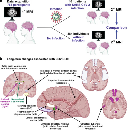
The study provides unique insights into COVID-19-associated changes in brain structure. The authors took great care in appropriately matching the case and control groups, making it unlikely that observed differences are due to confounding factors, although this possibility can never be entirely excluded. The mechanisms underlying these infection-associated changes, however, remain to be clarified. Viral neurotropism and direct infection of cells of the olfactory system, neuroinflammation and lack of sensory input have been suggested as reasons for the degenerative events in olfactory-related brain structures and neurological complications.4 These mechanisms are not mutually exclusive and may synergize in causing neurodegenerative disorders as consequence of COVID-19.
The study participants became infected between March 2020 and April 2021, before the emergence of the Omicron variant of concern (VOC) that currently dominates the COVID-19 pandemic. During that time period, the Alpha and Beta VOCs dominated in the UK and all results were obtained from individuals between 51 and 81 years of age. It will be of great interest to clarify whether Omicron, that seems to be less pathogenic than other SARS-CoV-2 variants, also causes long-term brain damage. The vaccination status of the participants was not available in the study1 and it will be important to clarify whether long-term changes in brain structure also occur in vaccinated and/or younger individuals. Other important questions are whether these structural changes are reversible or permanent and may even enhance the frequency for neurodegenerative diseases that are usually age-related, such as Alzheimer’s, Parkinson’s or Huntington’s disease. Previous findings suggest that cognitive disorders improve over time after severe COVID-19;5 yet it remains to be determined whether the described brain changes will translate into symptoms later in life such as dementia. Douaud and colleagues report that none of top 10 IDPs correlated significantly with the time interval between SARS-CoV-2 infection and the 2nd MRI acquisition, suggesting that the observed abnormalities might be very long-lasting.
Currently, many restrictions and protective measures are relaxed because Omicron is highly transmissible but usually causes mild to moderate acute disease. This raises hope that SARS-CoV-2 may evolve towards reduced pathogenicity and become similar to circulating coronaviruses causing mild respiratory infections. More work needs to be done to clarify whether the current Omicron and future variants of SARS-CoV-2 may also cause lasting brain abnormalities and whether these can be prevented by vaccination or therapy. However, the finding by Douaud and colleagues1 that SARS-CoV-2 causes structural changes in the brain that may be permanent and could relate to neurological decline is of concern and illustrates that the pathogenesis of this virus is markedly different from that of circulating human coronaviruses. Further studies, to elucidate the mechanisms underlying COVID-19-associated neurological abnormalities and how to prevent or reverse them are urgently needed.
REFERENCES (Follow link)
#public health#wear a mask#covid 19#pandemic#covid#wear a respirator#mask up#still coviding#coronavirus#sars cov 2#long covid#covid conscious#covid is airborne
27 notes
·
View notes
Text
Interesting Papers for Week 16, 2025
Cortical VIP neurons as a critical node for dopamine actions. Bae, J. W., Yi, J. H., Choe, S. Y., Li, Y., & Jung, M. W. (2025). Science Advances, 11(1).
Confidence regulates feedback processing during human probabilistic learning. Ben Yehuda, M., Murphy, R. A., Le Pelley, M. E., Navarro, D. J., & Yeung, N. (2025). Journal of Experimental Psychology: General, 154(1), 80–95.
Neural Encoding of Direction and Distance across Reference Frames in Visually Guided Reaching. Caceres, A. H., Barany, D. A., Dundon, N. M., Smith, J., & Marneweck, M. (2024). eNeuro, 11(12), ENEURO.0405-24.2024.
Bifurcation Enhances Temporal Information Encoding in the Olfactory Periphery. Choi, K., Rosenbluth, W., Graf, I. R., Kadakia, N., & Emonet, T. (2024). PRX Life, 2(4), 043011.
The origin of color categories. Garside, D. J., Chang, A. L. Y., Selwyn, H. M., & Conway, B. R. (2025). Proceedings of the National Academy of Sciences, 122(1), e2400273121.
Oppositional and competitive instigation of hippocampal synaptic plasticity by the VTA and locus coeruleus. Hagena, H., & Manahan-Vaughan, D. (2025). Proceedings of the National Academy of Sciences, 122(1), e2402356122.
Dissociable Effects of Urgency and Evidence Accumulation during Reaching Revealed by Dynamic Multisensory Integration. Hoffmann, A. H., & Crevecoeur, F. (2024). eNeuro, 11(12), ENEURO.0262-24.2024.
Allocentric and egocentric spatial representations coexist in rodent medial entorhinal cortex. Long, X., Bush, D., Deng, B., Burgess, N., & Zhang, S.-J. (2025). Nature Communications, 16, 356.
Limitation of switching sensory information flow in flexible perceptual decision making. Luo, T., Xu, M., Zheng, Z., & Okazawa, G. (2025). Nature Communications, 16, 172.
Sparse high-dimensional decomposition of non-primary auditory cortical receptive fields. Mukherjee, S., Babadi, B., & Shamma, S. (2025). PLOS Computational Biology, 21(1), e1012721.
Memory-based predictions prime perceptual judgments across head turns in immersive, real-world scenes. Mynick, A., Steel, A., Jayaraman, A., Botch, T. L., Burrows, A., & Robertson, C. E. (2025). Current Biology, 35(1), 121-130.e6.
Multisensory integration of social signals by a pathway from the basal amygdala to the auditory cortex in maternal mice. Nowlan, A. C., Choe, J., Tromblee, H., Kelahan, C., Hellevik, K., & Shea, S. D. (2025). Current Biology, 35(1), 36-49.e4.
Exploration in 4‐year‐old children is guided by learning progress and novelty. Poli, F., Meyer, M., Mars, R. B., & Hunnius, S. (2025). Child Development, 96(1), 192–202.
Aberrant auditory prediction patterns robustly characterize tinnitus. Reisinger, L., Demarchi, G., Obleser, J., Sedley, W., Partyka, M., Schubert, J., Gehmacher, Q., Roesch, S., Suess, N., Trinka, E., Schlee, W., & Weisz, N. (2024). eLife, 13, e99757.4.
Retinal ganglion cells encode the direction of motion outside their classical receptive field. Riccitelli, S., Yaakov, H., Heukamp, A. S., Ankri, L., & Rivlin-Etzion, M. (2025). Proceedings of the National Academy of Sciences, 122(1), e2415223122.
Dynamics of visual object coding within and across the hemispheres: Objects in the periphery. Robinson, A. K., Grootswagers, T., Shatek, S. M., Behrmann, M., & Carlson, T. A. (2025). Science Advances, 11(1).
Single-neuron spiking variability in hippocampus dynamically tracks sensory content during memory formation in humans. Waschke, L., Kamp, F., van den Elzen, E., Krishna, S., Lindenberger, U., Rutishauser, U., & Garrett, D. D. (2025). Nature Communications, 16, 236.
Information sharing within a social network is key to behavioral flexibility—Lessons from mice tested under seminaturalistic conditions. Winiarski, M., Madecka, A., Yadav, A., Borowska, J., Wołyniak, M. R., Jędrzejewska-Szmek, J., Kondrakiewicz, L., Mankiewicz, L., Chaturvedi, M., Wójcik, D. K., Turzyński, K., Puścian, A., & Knapska, E. (2025). Science Advances, 11(1).
A language model of problem solving in humans and macaque monkeys. Yang, Q., Zhu, Z., Si, R., Li, Y., Zhang, J., & Yang, T. (2025). Current Biology, 35(1), 11-20.e10.
Offline ensemble co-reactivation links memories across days. Zaki, Y., Pennington, Z. T., Morales-Rodriguez, D., Bacon, M. E., Ko, B., Francisco, T. R., LaBanca, A. R., Sompolpong, P., Dong, Z., Lamsifer, S., Chen, H.-T., Carrillo Segura, S., Christenson Wick, Z., Silva, A. J., Rajan, K., van der Meer, M., Fenton, A., Shuman, T., & Cai, D. J. (2025). Nature, 637(8044), 145–155.
#neuroscience#science#research#brain science#scientific publications#cognitive science#neurobiology#cognition#psychophysics#neurons#neural computation#neural networks#computational neuroscience
8 notes
·
View notes
Text
Brain pathways: an information superhighway
Understanding the neural network
The brain, our body's conductor, is a complex network of billions of interconnected neurons. These neurons communicate with each other via specialized pathways known as nerve tracts. These pathways are essential for transmitting sensory, motor and cognitive information throughout the body.

Main brain pathways
1. The pyramidal pathway
- Role: The pyramidal pathway is primarily responsible for the voluntary control of movement. It connects the primary motor cortex to the motor neurons in the spinal cord, enabling the initiation and control of precise skeletal muscle movements.
- Components: It comprises the corticospinal bundle and the corticobulbar bundle.
- How it works: Nerve signals from the motor cortex travel down this pathway to activate the muscles concerned.
2. Sensory pathways
- Role: These pathways transmit sensory information from the body to the brain.
- Types of sensitivity:
o Tactile sensitivity: Allows us to perceive touch, pressure and vibration.
o Thermal sensitivity: Allows us to perceive heat and cold.
o Deep sensitivity: Allows us to perceive the position of limbs in space (proprioception) and joint movements.
o Pain sensitivity: Allows us to perceive pain.
- Pathway: Sensory information is transmitted by peripheral nerves to the spinal cord, then back to the brain via various ascending pathways.
3. Specific sensory pathways
- Visual: transmits visual information from the retina to the occipital lobe.
- Auditory: Transmits auditory information from the inner ear to the temporal lobe.
- Olfactory pathway: transmits olfactory information from olfactory receptors to the olfactory bulb.
- Taste pathway: transmits taste information from the taste buds to the taste cortex.
4. Proprioception pathway
- Role: Proprioception is the sense that enables us to know our body's position in space.
- How it works: Proprioceptive receptors in muscles, tendons and joints constantly send information to the brain about the state of muscle contraction, joint angle and limb position.
- Importance: Proprioception is essential for movement coordination, balance and posture.
Nerve pathway disorders
Damage to or dysfunction of these pathways can lead to a variety of neurological disorders, such as :
- Hemiplegia: Paralysis of one side of the body.
- Paresthesia: Sensation of numbness or tingling.
- Ataxia: Loss of coordination of movements.
- Blindness: Loss of vision.
- Deafness: Loss of hearing.
In conclusion
Brain pathways are complex networks that ensure communication between the brain and the body. Understanding how they work is essential for grasping the mechanisms underlying many physiological and pathological processes.
Go further
#nerve pathways#brain#neurology#neuroscience#pyramidal pathway#sensitivity#proprioception#nervous system#neuroanatomy#brain health#neurological disorders#brain anatomy#neurons#synapses#cerebral cortex#spinal cord#peripheral nerves#senses#perception#movement#coordination
2 notes
·
View notes
Text
Especially because I mega appreciate you as a human being, I feel the need to share some neat & fun science facts related to pregnancy impulse cravings and how they're set up for leading to nurturing early infancy into childhood, because all of the human biology components of social bonding and early childhood cognitive development are WILDLY fascinating.
Olfactory senses are a big component of how all mammals operate, including humans. They're also a big part how mammals recognize someone familiar from someone who isn't. While mice do this as their primary form of socialization & recognition, it's less pronounced in human social interactions compared to things like sight — but it still links directly to the sensory part of your brain.
During pregnancy it's important to not only be able to recognize the new child as an "us" when core reflexes in the mother's brain has to make survival decisions based on "us vs. them" patterns. So, boosted oxytocin helps to amplify the bonding to "us" & the defensiveness against "them" to prioritize protection of the child.
When you're building a tiny new person inside of you, they're literally an extension of your body, so your body builds to learn how to treat it as the same as YOU even when it's outside. This is why newborns don't recognize themselves as different than their mothers in early infancy, and also allows their brains to develop without needing to activate their own survival stress response when they're around their parents.
This is what forms the foundation of social bonding & trust, but also what allows children to learn about dangerous things without their survival response kicking in when their guardian is close by. We get smarter by understanding things rather than reacting out of discomfort, and this is what allows for that and why human children get to take longer to develop understanding of their environment, rather than surviving on their own super rapidly.
So, when it comes to the mother recognizing her child in survival reflexes without the time for the prefrontal cortex making a calculated decision, and the child unable to activate its own fear stress responses, it's important to also have that deep sensory feedback wired up properly. So, pregnancy rewires the mother's brain to be able to do that… which will change your sensory recognition and contributes to atypical food cravings.
Combine that with all of the other hormonal changes taking place with estrogen levels, the need to supply your body with the components necessary to build a tiny human, and all of those signals being dialed up to 11 and that big fat burger craving NOW is why I think your descriptions of the hedonism are absolutely brilliant, and look forward to more.
Other details that're related: Mammals all having this cognitive development to offspring in common with one another is why we all experience some common things:
• Newborn babies smell particularly good to us, because that links to the sense that helps us bond with them. Just like how a person can smell the difference between "puppy breath" & "dog breath" which you just know if you've had dogs. Those same things are true with humans and is how pets like cats, dogs, & other mammals "understand" to be gentle and more tolerant with babies — even with their reflexive reactions.
• Since sight is much more of a factor for human social recognition, behaviour, & interaction than smell — we also have fixed action patterns to think that creatures with mammal baby proportions for their eyes, face, and body ratios look more cute and want to favourably attach to them from a social bonding drive (which is what Disney intentionally used to design animated characters to be more appealing psychologically to audiences, which directly influenced all of anime as well — so yay for fan fiction about characters being a part of this as well).
And one additional PSA-type related bit:
• As oxytocin plays such a huge part of this, it's also important to note that the "us/them" biases in the brain that this ties to can misalign. It's one of the deepest survival fixed action pattern biases in the brain, and for some mothers this instead makes it extremely difficult to connect to their child because of brain chemistry things. You're not broken or wrong if anything like that happens — which feels important to know & remember early on from a technical perspective — as that's also exactly what can make it EXTRA difficult to talk to anyone about negative feelings if they do occur, and can make that a spiral amplified by tons of other hormonal changes especially after birth. Having an educated partner will ensure that they know how to look out for those things and can make sure you're both well cared for no matter what. They're perfect for doing that because they're an external "them" that you had to learn was an "us" and that's exactly what the social bonding is all about and why it's not hard locked.
• While knowing something like this cognitively helps — that type of "knowing" is still all happening in the prefrontal cortex, not the scent sensory "knowing" where that has the strongest influence. It takes time to find the thing to recondition your sensory brain through whatever it's struggling with if that clicks a particular trigger in an unexpected way. Luckily — just like the body is doing all the cool sensory neurology stuff to influence external social behaviour — our brains as humans are designed to do use external social behaviour for that type of reprogramming from an intentional external perspective as well (why I mentioned the anime style thing and fan fiction, because we can design ways to increase our love of something in lots of ways).
Basically, humans are rad, and I really look forward to seeing all sorts of misc updates of you making pregnancy sexy & fun — 'cause if anybody on tumblr dot com could do that, it'd definitely be you.
pregnancy brain is like thinking with your dick but for hedonism
243 notes
·
View notes
Text
Artemisia p. 1 :: Nervine

[Photo cred:: Native Here Nursery:: Artemisia Douglasiana]
KEY:
Underlined green text = links
Pink = anagrams [orthographically, phonetically or syntactically]
Bold = emphasis for discussion
Italic = credit
Note: Please read the links as well
Artemisia is a rhizomatous weed belonging to the Asteraceae family (Daisy family). While there are several species of Artemisia, the one I will be highlighting is Mugwort (Artemisia Vulgaris). Most of my experience is with the wild American species Artemisia Douglasiana and Artemisia Suksdorfii. Mugwort thrives in disturbed soils, and does its deepest work in disturbed bodies. Its aggressive rhizomes can overwhelm and kill roots of surrounding plants. I describe the leaves as flame shaped, with a silver back. Its volatile oil content is why Mugwort has a lengthy list of uses throughout the world.
First, I want to give some backstory on the name of Artemisia. Artemisia stems from Greek goddess Artemis. Delve into the article Becoming Classical Artemis: A Glimpse at the Evolution of the Goddess as Traced in Ancient Arcadia as means to gain more understanding about Mugwort’s functionality both physically, and spiritually- thru the characteristics of Goddess Artemis.
Goddess Artemis’s original prehistoric form was that of an animal-shape. She “appeared as a goddess of wild beasts, especially those who live in remote places in wooded mountains,” and also went by the alias ‘Kallisto.’
Below are some notes to further illustrate Artemis’s origins
Kallisto ~ bear-nymph::
teen girls considered ~little bears::
Artemis Orthia ~ bear figurine//Orthia, meaning straight, upright, in proper order [[anagram Orthia::THOR::ROTH::AORTA]]
Artemis @ Kyrena ~ sacred law of pregnant women to offer sacrifice to the Bear before giving BEARth [[birth]]
Protectress of little girls and maidens until the age of marriage; responsible for safe delivery of children
Now, I’m taking you deeper into the bold words from above. To start, it seems in the article that the primary animal goddess Artemis took form in was a bear. This is important because I learned from an #Oracle that the midbrain, our forward most portion of the brain stem, likens itself into the shape of a bear...funny how that works. I consider this key to grasp how and *where* Mugwort behaves when administered to a person. Succinctly, the midbrain has many functions, and is a continuation of the olfactory cortex responsible for memory [[dream]] processing and smell. The midbrain is considered “archipallium” in origin- which is to say phylogenetically the eldest region of the brain’s cerebral cortex. In the same way, Mugwort is called “the oldest of plants” in Charm of the Nine Worts. Mugwort’s classification as a nervine is due to easing anxiety, nervousness, and primal fears held in the midbrain. The source of this stress may exist due to trauma, karma, *generational or otherwise.
To take things further into the nervine properties of Mugwort, lets step into its signature silver-flame leaves. The Force of the metal Silver is alkhemically attributed to the Moon; on the periodic table Silver is #47. Native silver is mostly found in earth’s crust, and alloyed::allied with gold, argentite, and chlorargyrite. Silver is rarely found standalone- it is usually coupled with metals such as Copper (Venus), Gold (Sun), Lead (Saturn), and Zinc. Therefore, I surmise that Mugwort pairs well with other plants that are ruled by Venus (Copper), Sun (Gold), and Saturn (Lead). A Mugwort person may also need to increase the Zinc in their diets in tandem with the herb. I recommend oysters and fish for ZInc, but I will discuss that in another article.
In alchemy, refined Oil of Silver “cleanses the receptor sites deep within the limbic system, lowers accumulated stress hormones long enough for the brain to generate new neural pathways that strengthen positive thinking” when ingested. Kymia Arts.
Mugwort’s “flame” shaped leaves makes known the ‘temperature’ and tastes of Mugwort. Warming, pungent, and bitter. This warmth, according to Sajah Popham, is a rare quality in a nervine plant, as most of them are cooling. In this way, Mugwort is not only associated with the Moon, but also fiery sister Venus. With the heat of Venus, it is no coincidence that Mugwort responds well when burned and smoked. It has been recorded several times as a smokeable herb, and even a Kanabyss replacement. In Traditional Chinese Medicine, practitioners use Mugwort in Moxibustion. The special preparation of a flammable Moxa releases fragrant, medicinal oils. According to TCM, this heat warms up various channels in the body and dispels cold, promotes blood circulation, strenghtens Qi, and more. I also say experiment with taking dried or fresh Mugwort in steam rooms and sauna.
Below is the mental/psychological picture of one who benefits from Mugwort.
Excerpt from Sajah Popham’s Mugwort Materia Medica:
- Dyslexic, difficulty recognizing words but has complex thoughts - Disorders of sleep and imagination - Body seems to be asleep, but mind is awake - When they sleep deep they have vivid dreams
.... - Lies awake in bed for long periods of time, thinking, imagining - Epileptic seizures, sensitivity to light, sleep disorders - Irritation of the nervous system
…
“… Dorothy Hall profiles the Mugwort person as being “highly intelligent with complex thoughts that are difficult to describe, speech disorders and dyslexia, highly elevated senses, sensitivity to light and sound, with great difficulty getting deep sleep. It is suited to people in whom the intuitive, psychic, psychological, creative, and artistic side of the mind is highly developed, but who have trouble with expression, or with the world around them.” These are all qualities that we would associate with an excess of vata, or wind/ tension, to connect this description to our energetic qualities above.
In addition to the higher mental qualities of Mugwort, I consider Her to be a Dream Modulator. She has the ability to lessen or increase frequency of dreams. As a dreamer myself, I noticed deeper sleep and reduced dreaming while taking mugwort. However, the dreams I *did* have- the messages provided me with clarity. In an individual who dreams infrequently, or desires to dream more, Mugwort opens them up to the dream world, and increases their dream intensity.
Dream Modulation Notes:
- Soothes the hyperactive & nightmarish dreamer trapped in the astral; allows them to get real rest
- Increases dream activation in those disconnected from the dream realm - Opens one to wisdom when dreaming - Allows for messages to really get the thru the mesencephalon (aka Midbrain) and heal
#Artemisia#Materia Medica#Ma'ateria Meda#Mugwort#PCOS#Nervine#Alkhemy#SIlver#Bear#Venus#Moon#my words#herbalism
15 notes
·
View notes
Text
Drugs & Behavior Week 1
Intro
drugs: chemical compounds administered to produce a desired change in the body.
pharmacology:
(a) study of drug action on the body AKA pharmacodynamics
(b) study of the fate of drugs in the body AKA pharmacokinetics
psychoactive drugs: affect behavior by altering the functions of the nervous system.
psychopharmacology: study of how drugs affect the nervous system and behavior.
What You Will Need to Know for the Exam
Difference between the Central Nervous System (CNS) and Peripheral Nervous System (PNS), components of each system
Difference between somatic and autonomic nervous systems
Difference between sensory and motor function, definitions of each
Definition of plasticity
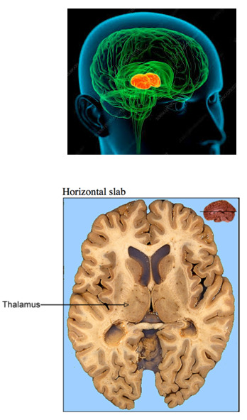
Explain the three different cuts to view brain structures: horizontal, sagittal, coronal
Three brain divisions based on surface features: cerebrum, cerebellum, brain stem
Three brain divisions based on brain development: forebrain, midbrain, hindbrain
Components of forebrain:
A. End brain
1. Cortex
2. Corpus callosum
3. Limbic system
4. Basal ganglia
5. Olfactory bulb
B. Between brain
1. Thalamus
2. Hypothalamus
Components of midbrain:
A. Tectum
B. Tegmentum
Components of hindbrain:
A. Pons
B. Cerebellum
C. Medulla
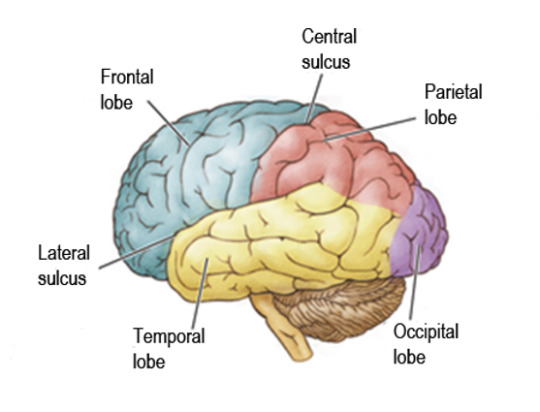
Divisions of the cortex:
1. Frontal lobe
2. Temporal lobe
3. Parietal lobe
4. Occipital lobe
Difference between sulci and gyri (i.e. which is a bump and which is a groove?)
Three areas of cortex:
1. primary motor cortex
2. primary sensory cortex
3. association cortex
Which brain structure can be severed as a last-resort treatment for severe epilepsy?
Structures of the limbic system:
1. Amygdala
2. Hippocampus
3. Cingulate cortex
Structures of basal ganglia:
1. Striatum
2. Globus pallidus
3. Nucleus accumbens
Difference between thalamus and hypothalamus, including locations and their major functions
What is the function of the pineal gland?
Structures of midbrain:
1. Substantia nigra
2. Ventral tegmental area (VTA)
3. Periaqueductal gray (PAG)
What happens when there is cerebellar dysfunction? Is there complete paralysis?
What is the medulla’s role in respiration?
What drugs suppress respiratory control?
How does the medulla act as a vomiting center?
Definition of a ventricle; know the four ventricle locations
Why is CSF so important?
What causes hydrocephalus?
Difference between cranial and spinal nerves
How many segments does the spinal cord consist of?
Major functions of the spinal cord
How is either the gray or white matter organized in the spinal cord?
What are the functions of the Autonomic Nervous System (ANS)?
Difference between the Sympathetic and Parasympathetic divisions of ANS:
(A) Which is rest & digest vs. fight or flight?
(B) Know general body controls with each division (i.e. heart rate, digestion, etc.)
What do neurotransmitters are released by the ANS?
10 notes
·
View notes
Link
2 notes
·
View notes
Text
The Visual System Explained

What is the Visual System?
The visual system is the part of our sensory system that is responsible for our sense of sight. This complex system starts a journey with light waves that enter your eyes, then a chemical signal is sent to the primary visual cortex-this is located in the occipital lobe of the brain. Once there, the signal is then processed and converted into images we see and perceive.
The issues with the visual sensory system referred to below have nothing to do with sight damage or other ophthalmic disorders. If you are concerned your child has visual processing issues, the first step is to get their eyesight evaluated to rule out the need for glasses or other interventions.
The Visual Sensory System is responsible for:
1.) visual processing and visual perception,
2.) eye movement control
3.) color, shape, light brightness and contrast, and movement.
The visual sensory system is extremely important for information gathering and learning. Visual sensory disorders can be the culprit behind struggles in school, speech and language difficulties and learning disabilities.
The visual and vestibular systems play an important role when working with and teaching children. Remember, you can always request your school district give your child an occupational therapy evaluation. Working with a qualified occupational therapist in the school setting can help the child improve their visual processing skills and learning abilities in school activities.
A Healthy Visual System
Vision works closely with all of the other senses in our sensory system. As children, when we see a dinner our mom made, we realize we are hungry because the sight of the food made us more aware of the smell of the food (olfactory system) and our mouths may have began to water imagining the taste (gustatory system)! Or if you are walking to the car alone late at night and you see a shadow of someone walking up behind you, you become aware of your surroundings and may develop goosebumps that developed from our tactile system.
Problems With Visual Processing
Even a unproblematic visual system works in a very intricate and complicated matter. There 8 subtypes of visual processing disorder and they can appear in any combination or independently.
Subtypes of Visual Processing
1.) Visual Discrimination
2.) Visual Memory, long and short term
3.) Visual Motor Processing
4.) Visual Sequential Memory
5.) Visual Spatial Processing
6.) Visual Figure Ground
7.) Visual Form Constancy
8.) Visual Closure
Listed below are signs that your student may be having issues with their visual processing skills. I’ve specifically chose to add things you’d observe in a school setting. However, this is no where near an exhaustive list of symptoms since everyone has a unique set of circumstances and may present differently from one person to the next.

Characteristics of Malfunctioning Visual Processing In School Environments:
* Difficulty with sports and physical education
* Falling asleep in class or struggling to sit still in class
* Squints or develops headaches from reading or copying from the board
* Nausea when trying to read or do homework in the car
* Poor math skills
* May complain of pain in eyes
* Difficulty memorizing spelling words,
* May struggle to retain or recall what they have read or watched
* Work performance is disorganized
* Difficulty with writing eye-hand coordination skills like letter spacing, letter reversal or out of order, or copying, sloppy writing for their current grade level when compared to peers.
* May show difficulty completing puzzles
* Unable to judge distances accurately, this can be displayed as bumping into things easily or trouble pouring liquids
* Difficulty with fine motor skills such as cutting, art work, tracing, etc.

Children showing signs that they are having difficulty processing visual information can exhibit a hypersensitivity or hyposensitivity. Hypersensitivity means the sensory system is over reacting to visual stimuli it’s taking in. Hypo-sensitive is when the sensory system is under reacting to he visual stimuli it’s taking in. They may present with some of the following characteristics.
Hypersensitive
* Aversion to bright lights or colors
* Avoids eye contact
* Reoccurring nausea and headaches after reading or computer activities
* Frequently covers eyes
* Bumps into things
* Highly overwhelmed,easily anxious or other emotional behaviors
* Avoids group play activities
* Avoids crowds
Hyposensitive
* Spaces out
* Stares at objects or light
* Looks unusually close at objects, or out of the side of their eyes
* Enjoys bright colors
* Engages in excessive movement when reading or doing schoolwork, self soothing by increasing their vestibular stimulation

Home Based Activities To Help With Visual System Differences:
* If struggling with headaches or eye pain, limit reading and time spent watching TV at night
* Keep areas clear of clutter
* Choose either calming or stimulating colors, based on desired response from child
* Choose natural light when possible and avoid fluorescent lighting
* Encourage your child to stimulate their vestibular system by engaging in activities such as rocking, swinging, hanging upside down, etc.
* Encourage your child’s proper posture during all activities
* Help ensure your child is understanding, comprehending and retaining the learning materials
* Use large lettering
* Play fun physical games with your child like homemade obstacle courses, scavenger hunts, hide and seek, etc
* Other fun activities for you to do with your child include building a model using Lego’s, cooking with instructions, tracing, origami, etc
Advice To Caregivers:
As mentioned above, ruling out vision loss or damage is the first step before addressing any visual processing related to the sensory system. The next logical step is an occupational therapist to help you come up with case specific activities you can do with your child to best support them. When doing the activities with your child, keep it light and fun. If it loses its fun factor the child won’t want to participate and even if they still do, progress will likely stop.
#autism parenting#autism education#sensory system#visual processing disorder#occupational therapy#autism mom#vestibular#school#iep#student accommodation#eye sight#sensory processing disorder#sensoryplay#speech#language development#learning difficulties
9 notes
·
View notes
Photo

Immense pride, tinged with sadness.
For those who would like to read the full list:
1908 MECHNIKOV, ELIE
FOR THEIR WORK ON IMMUNITY
1908 EHRLICH, PAUL
FOR THEIR WORK ON IMMUNITY
1914 BARANY, ROBERT
FOR HIS WORK ON THE PHYSIOLOGY AND PATHOLOGY OF THE VESTIBULAR APPARATUS
1922 MEYERHOF, OTTO FRITZ
FOR HIS DISCOVERY OF THE FIXED RELATIONSHIP BETWEEN THE CONSUMPTION OF
OXYGEN AND THE METABOLISM OF LACTIC ACID IN THE MUSCLE
1930 LANDSTEINER, KARL
FOR HIS DISCOVERY OF HUMAN BLOOD GROUPS
1936 LOEWI, OTTO
FOR THEIR DISCOVERIES RELATING TO CHEMICAL TRANSMISSION OF NERVE IMPULSES
1944 ERLANGER, JOSEPH
FOR THEIR DISCOVERIES RELATING TO THE HIGHLY DIFFERENTIATED FUNCTIONS OF SINGLE NERVE FIBRES
1945 CHAIN, ERNST BORIS
FOR THE DISCOVERY OF PENICILLIN AND ITS CURATIVE EFFECT IN VARIOUS INFECTIOUS DISEASES
1946 MULLER, HERMANN J.
FOR THE DISCOVERY OF THE PRODUCTION OF MUTATIONS BY MEANS OF X-RAY IRRADIATION
1947 CORI, GERTY THERESA, RADNITZ
FOR THEIR DISCOVERY OF THE COURSE OF THE CATALYTIC CONVERSION OF GLYCOGEN
1950 REICHSTEIN, TADEUS
FOR THEIR DISCOVERIES RELATING TO THE HORMONES OF THE ADRENAL CORTEX, THEIR STRUCTURE AND BIOLOGICAL EFFECTS
1952 WAKSMAN, SELMAN A.
FOR HIS DISCOVERY OF STREPTOMYCIN, THE FIRST ANTIBIOTIC EFFECTIVE AGAINST TUBERCULOSIS
1953 LIPMANN, FRITZ ALBERT
FOR HIS DISCOVERY OF CO-ENZYME A AND ITS IMPORTANCE FOR INTERMEDIARY METABOLISM
1953 KREBS, HANS ADOLF
FOR HIS DISCOVERY OF THE CITRIC ACID CYCLE
1958 LEDERBERG, JOSHUA
FOR HIS DISCOVERIES CONCERNING GENETIC RECOMBINATION AND THE ORGANISATION OF THE GENETIC MATERIAL OF BACTERIA
1959 KORNBERG, ARTHUR
FOR THEIR DISCOVERY OF THE MECHANISMS IN THE BIOLOGICAL SYNTHESIS OF RIBONUCLEIC ACID AND DEOXYRIBONUCLEIC ACID
1964 BLOCH, KONRAD
FOR THEIR DISCOVERIES CONCERNING THE MECHANISM AND REGULATION OF THE CHOLESTEROL AND FATTY ACID METABOLISM
1965 JACOB, FRANCOIS
FOR THEIR DISCOVERIES CONCERNING GENETIC CONTROL OF ENZYME AND VIRUS SYNTHESIS
1965 LWOFF, ANDRE
FOR THEIR DISCOVERIES CONCERNING GENETIC CONTROL OF ENZYME AND VIRUS SYNTHESIS
1967 WALD, GEORGE
FOR THEIR DISCOVERIES CONCERNING THE PRIMARY PHYSIOLOGICAL AND CHEMICAL VISUAL PROCESSES IN THE EYE
1968 NIRENBERG, MARSHALL W.
FOR THEIR INTERPRETATION OF THE GENETIC CODE AND ITS FUNCTION IN PROTEIN SYNTHESIS
1969 LURIA, SALVADOR E.
FOR THEIR DISCOVERIES CONCERNING THE REPLICATION MECHANISM AND THE GENETIC STRUCTURE OF VIRUSES
1970 KATZ, BERNARD
FOR THEIR DISCOVERIES CONCERNING THE HUMORAL TRANSMITTERS IN THE NERVE TERMINALS AND THE MECHANISM
FOR THEIR STORAGE, RELEASE AND INACTIVATION
1970 AXELROD, JULIUS
FOR THEIR DISCOVERIES CONCERNING THE HUMORAL TRANSMITTERS IN THE NERVE TERMINALS AND THE MECHANISM
FOR THEIR STORAGE, RELEASE AND INACTIVATION
1972 EDELMAN, GERALD M.
FOR THEIR DISCOVERIES CONCERNING THE CHEMICAL STRUCTURE OF ANTIBODIES
1975 TEMIN, HOWARD M.
FOR THEIR DISCOVERIES CONCERNING THE INTERACTION BETWEEN TUMOR VIRUSES AND THE GENETIC MATERIAL OF THE CELL
1975 BALTIMORE, DAVID
FOR THEIR DISCOVERIES CONCERNING THE INTERACTION BETWEEN TUMOR VIRUSES AND THE GENETIC MATERIAL OF THE CELL
1976 BLUMBERG, BARUCH S.
FOR THEIR DISCOVERIES CONCERNING NEW MECHANISMS FOR THE ORIGIN AND DISSEMINATION OF INFECTIOUS DISEASES
1977 YALOW, ROSALYN
FOR THE DEVELOPMENT OF RADIOIMMUNOASSAYS OF PEPTIDE HORMONES
1977 SCHALLY, ANDREW V.
FOR THEIR DISCOVERIES CONCERNING THE PEPTIDE HORMONE PRODUCTION OF THE BRAIN
1978 NATHANS, DANIEL
FOR THE DISCOVERY OF RESTRICTION ENZYMES AND THEIR APPLICATION TO PROBLEMS OF MOLECULAR GENETICS
1980 BENACERRAF, BARUJ
FOR THEIR DISCOVERIES CONCERNING GENETICALLY DETERMINED STRUCTURES ON THE CELL SURFACE THAT
REGULATE IMMUNOLOGICAL REACTIONS
1984 MILSTEIN, CESAR
FOR THEORIES CONCERNING THE SPECIFICITY IN DEVELOPMENT AND CONTROL OF THE IMMUNE SYSTEM AND THE DISCOVERY OF THE
PRINCIPLE FOR PRODUCTION OF MONOCLONAL ANTIBODIES
1985 BROWN, MICHAEL S.
FOR THEIR DISCOVERIES CONCERNING THE REGULATION OF CHOLESTEROL METABOLISM
1985 GOLDSTEIN, JOSEPH L.
FOR THEIR DISCOVERIES CONCERNING THE REGULATION OF CHOLESTEROL METABOLISM
1986 COHEN, STANLEY
FOR THEIR DISCOVERIES OF GROWTH FACTORS
1986 LEVI-MONTALCINI, RITA
FOR THEIR DISCOVERIES OF GROWTH FACTORS
1988 ELION, GERTRUDE B.
FOR THEIR DISCOVERIES OF IMPORTANT PRINCIPLES FOR DRUG TREATMENT
1989 VARMUS, HAROLD E.
FOR THEIR DISCOVERY OF THE CELLULAR ORIGIN OF RETROVIRAL ONCOGENES
1994 RODBELL, MARTIN
FOR THEIR DISCOVERY OF G-PROTEINS AND THE ROLE OF THESE PROTEINS IN SIGNAL TRANSDUCTION IN CELLS
1994 GILMAN, ALFRED G.
FOR THEIR DISCOVERY OF G-PROTEINS AND THE ROLE OF THESE PROTEINS IN SIGNAL TRANSDUCTION IN CELLS
1997 PRUSINER, STANLEY B.
FOR HIS DISCOVERY OF PRIONS - A NEW BIOLOGICAL PRINCIPLE OF INFECTION
1998 FURCHGOTT, ROBERT F.
FOR THEIR DISCOVERIES CONCERNING NITRIC OXIDE AS A SIGNALING MOLECULE IN THE CARDIOVASCULAR SYSTEM
2000 GREENGARD, PAUL
FOR THEIR DISCOVERIES CONCERNING SIGNAL TRANSDUCTION IN THE NERVOUS SYSTEM
2000 KANDEL, ERIC R.
FOR THEIR DISCOVERIES CONCERNING SIGNAL TRANSDUCTION IN THE NERVOUS SYSTEM
2002 BRENNER, SYDNEY
FOR THEIR DISCOVERIES CONCERNING GENETIC REGULATION OF ORGAN DEVELOPMENT AND PROGRAMMED CELL DEATH
2002 HORVITZ, H. ROBERT
FOR THEIR DISCOVERIES CONCERNING GENETIC REGULATION OF ORGAN DEVELOPMENT AND PROGRAMMED CELL DEATH
2004 AXEL, RICHARD
FOR THEIR DISCOVERIES OF ODORANT RECEPTORS AND THE ORGANIZATION OF THE OLFACTORY SYSTEM
2006 FIRE, ANDREW Z.
FOR THEIR DISCOVERY OF RNA INTERFERENCE - GENE SILENCING BY DOUBLE-STRANDED RNA
2011 STEINMAN, RALPH M.
FOR THEIR DISCOVERIES CONCERNING THE ACTIVATION OF INNATE IMMUNITY
2011 BEUTLER, BRUCE A.
FOR THEIR DISCOVERIES CONCERNING THE ACTIVATION OF INNATE IMMUNITY
2013 SCHEKMAN, RANDY W.
FOR THEIR DISCOVERIES OF MACHINERY REGULATING VESICLE TRAFFIC, A MAJOR TRANSPORT SYSTEM IN OUR CELLS
2013 ROTHMAN, JAMES E.
FOR THEIR DISCOVERIES OF MACHINERY REGULATING VESICLE TRAFFIC, A MAJOR TRANSPORT SYSTEM IN OUR CELLS
2017 ROSBASH, MICHAEL
FOR THEIR DISCOVERIES OF MOLECULAR MECHANISMS CONTROLLING THE CIRCADIAN RHYTHM
Likud UK
75 notes
·
View notes
Text
There is strong evidence of brain-related abnormalities in COVID-191,2,3,4,5,6,7,8,9,10,11,12,13. However, it remains unknown whether the impact of SARS-CoV-2 infection can be detected in milder cases, and whether this can reveal possible mechanisms contributing to brain pathology. Here we investigated brain changes in 785 participants of UK Biobank (aged 51–81 years) who were imaged twice using magnetic resonance imaging, including 401 cases who tested positive for infection with SARS-CoV-2 between their two scans—with 141 days on average separating their diagnosis and the second scan—as well as 384 controls. The availability of pre-infection imaging data reduces the likelihood of pre-existing risk factors being misinterpreted as disease effects. We identified significant longitudinal effects when comparing the two groups, including (1) a greater reduction in grey matter thickness and tissue contrast in the orbitofrontal cortex and parahippocampal gyrus; (2) greater changes in markers of tissue damage in regions that are functionally connected to the primary olfactory cortex; and (3) a greater reduction in global brain size in the SARS-CoV-2 cases. The participants who were infected with SARS-CoV-2 also showed on average a greater cognitive decline between the two time points. Importantly, these imaging and cognitive longitudinal effects were still observed after excluding the 15 patients who had been hospitalised. These mainly limbic brain imaging results may be the in vivo hallmarks of a degenerative spread of the disease through olfactory pathways, of neuroinflammatory events, or of the loss of sensory input due to anosmia. Whether this deleterious effect can be partially reversed, or whether these effects will persist in the long term, remains to be investigated with additional follow-up.
2 notes
·
View notes
Text
Interesting Papers for Week 18, 2024
Neural circuit mechanisms for transforming learned olfactory valences into wind-oriented movement. Aso, Y., Yamada, D., Bushey, D., Hibbard, K. L., Sammons, M., Otsuna, H., … Hige, T. (2023). eLife, 12, e85756.
Stimulus-Specific Prediction Error Neurons in Mouse Auditory Cortex. Audette, N. J., & Schneider, D. M. (2023). Journal of Neuroscience, 43(43), 7119–7129.
Guinea baboons are strategic cooperators. Formaux, A., Sperber, D., Fagot, J., & Claidière, N. (2023). Science Advances, 9(43).
Perceptual learning across saccades: Feature but not location specific. Grzeczkowski, L., Shi, Z., Rolfs, M., & Deubel, H. (2023). Proceedings of the National Academy of Sciences, 120(43), e2303763120.
Continuous multiplexed population representations of task context in the mouse primary visual cortex. Hajnal, M. A., Tran, D., Einstein, M., Martelo, M. V., Safaryan, K., Polack, P.-O., … Orbán, G. (2023). Nature Communications, 14, 6687.
Mood fluctuations shift cost–benefit tradeoffs in economic decisions. Heerema, R., Carrillo, P., Daunizeau, J., Vinckier, F., & Pessiglione, M. (2023). Scientific Reports, 13, 18173.
Reliable retrieval is intrinsically rewarding: Recency, item difficulty, study session memory, and subjective confidence predict satisfaction in word-pair recall. Holm, L., & Wells, M. (2023). PLOS ONE, 18(10), e0292866.
Curiosity evolves as information unfolds. Hsiung, A., Poh, J.-H., Huettel, S. A., & Adcock, R. A. (2023). Proceedings of the National Academy of Sciences, 120(43), e2301974120.
Human perception of spatial frequency varies with stimulus orientation and location in the visual field. Kirsch, W., & Kunde, W. (2023). Scientific Reports, 13, 17656.
Dynamic neural representations of memory and space during human ambulatory navigation. Maoz, S. L. L., Stangl, M., Topalovic, U., Batista, D., Hiller, S., Aghajan, Z. M., … Suthana, N. (2023). Nature Communications, 14, 6643.
Visual event boundaries restrict anchoring effects in decision-making. Ongchoco, J. D. K., Walter-Terrill, R., & Scholl, B. J. (2023). Proceedings of the National Academy of Sciences, 120(44), e2303883120.
A thalamic-hippocampal CA1 signal for contextual fear memory suppression, extinction, and discrimination. Ratigan, H. C., Krishnan, S., Smith, S., & Sheffield, M. E. J. (2023). Nature Communications, 14, 6758.
A quantitative model of ensemble perception as summed activation in feature space. Robinson, M. M., & Brady, T. F. (2023). Nature Human Behaviour, 7(10), 1638–1651.
Quantifying decision-making in dynamic, continuously evolving environments. Ruesseler, M., Weber, L. A., Marshall, T. R., O’Reilly, J., & Hunt, L. T. (2023). eLife, 12, e82823.
Predictions and rewards affect decision-making but not subjective experience. Sánchez-Fuenzalida, N., van Gaal, S., Fleming, S. M., Haaf, J. M., & Fahrenfort, J. J. (2023). Proceedings of the National Academy of Sciences, 120(44), e2220749120.
Lateral orbitofrontal cortex integrates predictive information across multiple cues to guide behavior. Tegelbeckers, J., Porter, D. B., Voss, J. L., Schoenbaum, G., & Kahnt, T. (2023). Current Biology, 33(20), 4496-4504.e5.
Cross-modal representation of identity in the primate hippocampus. Tyree, T. J., Metke, M., & Miller, C. T. (2023). Science, 382(6669), 417–423.
Optogenetic activation of visual thalamus generates artificial visual percepts. Wang, J., Azimi, H., Zhao, Y., Kaeser, M., Vaca Sánchez, P., Vazquez-Guardado, A., … Rainer, G. (2023). eLife, 12, e90431.
Parietal-driven visual working memory representation in occipito-temporal cortex. Xu, Y. (2023). Current Biology, 33(20), 4516-4523.e5.
Neuronal Population Activity in Macaque Visual Cortices Dynamically Changes through Repeated Fixations in Active Free Viewing. Yamane, Y., Ito, J., Joana, C., Fujita, I., Tamura, H., Maldonado, P. E., … Grün, S. (2023). ENeuro, 10(10).
#neuroscience#science#research#brain science#scientific publications#cognitive science#neurobiology#cognition#psychophysics#neurons#neural computation#neural networks#computational neuroscience
12 notes
·
View notes
Text

In 1598, following in the tradition of previous Popes, Pope Clement VIII ruled that no Christian could be treated by a Jewish Doctor, thus barring Christians from seeking treatment from any Jewish Physician. Bear in mind that virtually every Pope in history had had a personal Physician who was Jewish. In May 2020, the so-called 'Palestinian Leadership' ruled that no 'Palestinian' could be treated for COVID-19 using equipment sent by the UAE that had landed on Israeli soil. Bear in mind that virtually every 'Palestinian Leader' (or a member of their family) had at some point received potentially life-saving treatment in an Israeli Hospital. This is the vile hypocrisy of Antisemitism. COVID-19, like Ebola, Dengue, Smallpox and Sars before it, will eventually fade into the background, with a potential vaccine at hand to combat it should it reoccur. Sadly, there is no known cure (or vaccine) for Antisemitism. 80 years ago, it went unchecked, and killed over 6 million men, women and children. NEVER AGAIN * For those who are finding it difficult to read the (very long) list of Jewish Nobel Prize Winners in the Medical Field on our meme, here it is. A few people have asked why Jonas Salk, who discovered the Polio vaccine is not on the list. Sadly (and totally unjustly), Salk was never awarded a Nobel Prize :
1908 MECHNIKOV, ELIE FOR THEIR WORK ON IMMUNITY 1908 EHRLICH, PAUL FOR THEIR WORK ON IMMUNITY 1914 BARANY, ROBERT FOR HIS WORK ON THE PHYSIOLOGY AND PATHOLOGY OF THE VESTIBULAR APPARATUS 1922 MEYERHOF, OTTO FRITZ FOR HIS DISCOVERY OF THE FIXED RELATIONSHIP BETWEEN THE CONSUMPTION OF OXYGEN AND THE METABOLISM OF LACTIC ACID IN THE MUSCLE 1930 LANDSTEINER, KARL FOR HIS DISCOVERY OF HUMAN BLOOD GROUPS 1936 LOEWI, OTTO FOR THEIR DISCOVERIES RELATING TO CHEMICAL TRANSMISSION OF NERVE IMPULSES 1944 ERLANGER, JOSEPH FOR THEIR DISCOVERIES RELATING TO THE HIGHLY DIFFERENTIATED FUNCTIONS OF SINGLE NERVE FIBRES 1945 CHAIN, ERNST BORIS FOR THE DISCOVERY OF PENICILLIN AND ITS CURATIVE EFFECT IN VARIOUS INFECTIOUS DISEASES 1946 MULLER, HERMANN J. FOR THE DISCOVERY OF THE PRODUCTION OF MUTATIONS BY MEANS OF X-RAY IRRADIATION 1947 CORI, GERTY THERESA, RADNITZ FOR THEIR DISCOVERY OF THE COURSE OF THE CATALYTIC CONVERSION OF GLYCOGEN 1950 REICHSTEIN, TADEUS FOR THEIR DISCOVERIES RELATING TO THE HORMONES OF THE ADRENAL CORTEX, THEIR STRUCTURE AND BIOLOGICAL EFFECTS 1952 WAKSMAN, SELMAN A. FOR HIS DISCOVERY OF STREPTOMYCIN, THE FIRST ANTIBIOTIC EFFECTIVE AGAINST TUBERCULOSIS 1953 LIPMANN, FRITZ ALBERT FOR HIS DISCOVERY OF CO-ENZYME A AND ITS IMPORTANCE FOR INTERMEDIARY METABOLISM 1953 KREBS, HANS ADOLF FOR HIS DISCOVERY OF THE CITRIC ACID CYCLE 1958 LEDERBERG, JOSHUA FOR HIS DISCOVERIES CONCERNING GENETIC RECOMBINATION AND THE ORGANISATION OF THE GENETIC MATERIAL OF BACTERIA 1959 KORNBERG, ARTHUR FOR THEIR DISCOVERY OF THE MECHANISMS IN THE BIOLOGICAL SYNTHESIS OF RIBONUCLEIC ACID AND DEOXYRIBONUCLEIC ACID 1964 BLOCH, KONRAD FOR THEIR DISCOVERIES CONCERNING THE MECHANISM AND REGULATION OF THE CHOLESTEROL AND FATTY ACID METABOLISM 1965 JACOB, FRANCOIS FOR THEIR DISCOVERIES CONCERNING GENETIC CONTROL OF ENZYME AND VIRUS SYNTHESIS 1965 LWOFF, ANDRE FOR THEIR DISCOVERIES CONCERNING GENETIC CONTROL OF ENZYME AND VIRUS SYNTHESIS 1967 WALD, GEORGE FOR THEIR DISCOVERIES CONCERNING THE PRIMARY PHYSIOLOGICAL AND CHEMICAL VISUAL PROCESSES IN THE EYE 1968 NIRENBERG, MARSHALL W. FOR THEIR INTERPRETATION OF THE GENETIC CODE AND ITS FUNCTION IN PROTEIN SYNTHESIS 1969 LURIA, SALVADOR E. FOR THEIR DISCOVERIES CONCERNING THE REPLICATION MECHANISM AND THE GENETIC STRUCTURE OF VIRUSES 1970 KATZ, BERNARD FOR THEIR DISCOVERIES CONCERNING THE HUMORAL TRANSMITTERS IN THE NERVE TERMINALS AND THE MECHANISM FOR THEIR STORAGE, RELEASE AND INACTIVATION 1970 AXELROD, JULIUS FOR THEIR DISCOVERIES CONCERNING THE HUMORAL TRANSMITTERS IN THE NERVE TERMINALS AND THE MECHANISM FOR THEIR STORAGE, RELEASE AND INACTIVATION 1972 EDELMAN, GERALD M. FOR THEIR DISCOVERIES CONCERNING THE CHEMICAL STRUCTURE OF ANTIBODIES 1975 TEMIN, HOWARD M. FOR THEIR DISCOVERIES CONCERNING THE INTERACTION BETWEEN TUMOR VIRUSES AND THE GENETIC MATERIAL OF THE CELL 1975 BALTIMORE, DAVID FOR THEIR DISCOVERIES CONCERNING THE INTERACTION BETWEEN TUMOR VIRUSES AND THE GENETIC MATERIAL OF THE CELL 1976 BLUMBERG, BARUCH S. FOR THEIR DISCOVERIES CONCERNING NEW MECHANISMS FOR THE ORIGIN AND DISSEMINATION OF INFECTIOUS DISEASES 1977 YALOW, ROSALYN FOR THE DEVELOPMENT OF RADIOIMMUNOASSAYS OF PEPTIDE HORMONES 1977 SCHALLY, ANDREW V. FOR THEIR DISCOVERIES CONCERNING THE PEPTIDE HORMONE PRODUCTION OF THE BRAIN 1978 NATHANS, DANIEL FOR THE DISCOVERY OF RESTRICTION ENZYMES AND THEIR APPLICATION TO PROBLEMS OF MOLECULAR GENETICS 1980 BENACERRAF, BARUJ FOR THEIR DISCOVERIES CONCERNING GENETICALLY DETERMINED STRUCTURES ON THE CELL SURFACE THAT REGULATE IMMUNOLOGICAL REACTIONS 1984 MILSTEIN, CESAR FOR THEORIES CONCERNING THE SPECIFICITY IN DEVELOPMENT AND CONTROL OF THE IMMUNE SYSTEM AND THE DISCOVERY OF THE PRINCIPLE FOR PRODUCTION OF MONOCLONAL ANTIBODIES 1985 BROWN, MICHAEL S. FOR THEIR DISCOVERIES CONCERNING THE REGULATION OF CHOLESTEROL METABOLISM 1985 GOLDSTEIN, JOSEPH L. FOR THEIR DISCOVERIES CONCERNING THE REGULATION OF CHOLESTEROL METABOLISM 1986 COHEN, STANLEY FOR THEIR DISCOVERIES OF GROWTH FACTORS 1986 LEVI-MONTALCINI, RITA FOR THEIR DISCOVERIES OF GROWTH FACTORS 1988 ELION, GERTRUDE B. FOR THEIR DISCOVERIES OF IMPORTANT PRINCIPLES FOR DRUG TREATMENT 1989 VARMUS, HAROLD E. FOR THEIR DISCOVERY OF THE CELLULAR ORIGIN OF RETROVIRAL ONCOGENES 1994 RODBELL, MARTIN FOR THEIR DISCOVERY OF G-PROTEINS AND THE ROLE OF THESE PROTEINS IN SIGNAL TRANSDUCTION IN CELLS 1994 GILMAN, ALFRED G. FOR THEIR DISCOVERY OF G-PROTEINS AND THE ROLE OF THESE PROTEINS IN SIGNAL TRANSDUCTION IN CELLS 1997 PRUSINER, STANLEY B. FOR HIS DISCOVERY OF PRIONS - A NEW BIOLOGICAL PRINCIPLE OF INFECTION 1998 FURCHGOTT, ROBERT F. FOR THEIR DISCOVERIES CONCERNING NITRIC OXIDE AS A SIGNALING MOLECULE IN THE CARDIOVASCULAR SYSTEM 2000 GREENGARD, PAUL FOR THEIR DISCOVERIES CONCERNING SIGNAL TRANSDUCTION IN THE NERVOUS SYSTEM 2000 KANDEL, ERIC R. FOR THEIR DISCOVERIES CONCERNING SIGNAL TRANSDUCTION IN THE NERVOUS SYSTEM 2002 BRENNER, SYDNEY FOR THEIR DISCOVERIES CONCERNING GENETIC REGULATION OF ORGAN DEVELOPMENT AND PROGRAMMED CELL DEATH 2002 HORVITZ, H. ROBERT FOR THEIR DISCOVERIES CONCERNING GENETIC REGULATION OF ORGAN DEVELOPMENT AND PROGRAMMED CELL DEATH 2004 AXEL, RICHARD FOR THEIR DISCOVERIES OF ODORANT RECEPTORS AND THE ORGANIZATION OF THE OLFACTORY SYSTEM 2006 FIRE, ANDREW Z. FOR THEIR DISCOVERY OF RNA INTERFERENCE - GENE SILENCING BY DOUBLE-STRANDED RNA 2011 STEINMAN, RALPH M. FOR THEIR DISCOVERIES CONCERNING THE ACTIVATION OF INNATE IMMUNITY 2011 BEUTLER, BRUCE A. FOR THEIR DISCOVERIES CONCERNING THE ACTIVATION OF INNATE IMMUNITY 2013 SCHEKMAN, RANDY W. FOR THEIR DISCOVERIES OF MACHINERY REGULATING VESICLE TRAFFIC, A MAJOR TRANSPORT SYSTEM IN OUR CELLS 2013 ROTHMAN, JAMES E. FOR THEIR DISCOVERIES OF MACHINERY REGULATING VESICLE TRAFFIC, A MAJOR TRANSPORT SYSTEM IN OUR CELLS 2017 ROSBASH, MICHAEL FOR THEIR DISCOVERIES OF MOLECULAR MECHANISMS CONTROLLING THE CIRCADIAN RHYTHM
Source: Likud-Herut UK
36 notes
·
View notes
Text
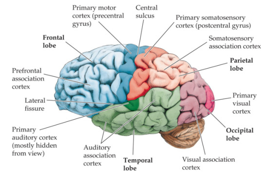
Areas of the brain and their functions (A2 level)
Key word - contralateral: the areas in the right lobe of the brain deals with the left side of the body and vice versa.
Frontal lobe: (top left of the diagram) is the part of the brain responsible for emotion expression, language, memory etc.
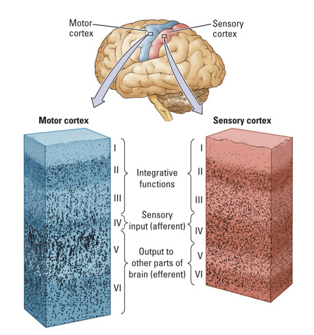
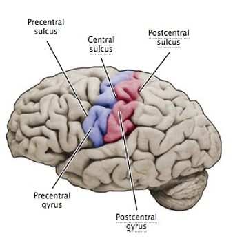
Motor cortex: Located in the rear of the frontal lobe (just before the central sulcus which separates the frontal and parietal lobe). This area is responsible for complex movements like kicking (so not things like coughing or crying). It is contralateral.
Brocas area: Located in the frontal lobe. This is responsible for speech production. Those with brocas aphasia understand speech, but cannot speak fluently.
Olfactory bulb (not necessary) Located in the frontal lobe. Respnsible for the sense of smell.
Temporal lobe: responsible for primary auditory perception (hearing).
Auditory cortex: Located in the temporal lobe. It processes auditory information. It is contralateral
Wernickes area: Located in the temporal lobe. It is responsible for understanding speech. Those with wernickes aphasia will be able to speak fluently, but their sentences will not make sense.
Spinal cord: Along with the brain it makes up the CNS, it connects the peripheral nervous system to the brain.
Cerebellum: Coordinates voluntary movements such as balance and coordination.
Occipital lobe: The visual processing centre of the brain
Parietal lobe: Main area for sense of touch
Sensory cortex (somatosensory cortex) Responsible for processing sensory information such as pressure and heat. It is contralateral.

#psychology#biopsychology#psychology a level#a level psychology#a level#a levels#study#study notes#studyblr#education#brain#brain areas#biology#information
31 notes
·
View notes
Text

Olfactory nerve is a sensory nerve (one of the shortest in our body) responsible for the sense of smell. It originates in the cerebrum itself, unlike many others. It travels from the upper part of the nasal cavity through the cribriform plate (spongy bone separating your nasal area from your skull) to the olfactory bulb (base of the frontal lobe). Then, the olfactory tract connects the olfactory bulb to the cortex. Fun fact - if there is a strong emotional aspect to your olfaction, the information first goes through the limbic system (responsible for emotional processing).
What does it do?
Olfactory nerve allows us to smell odours and detect scents. The odour itself consists of small molecules that are inhaled and recognised by olfactory receptors. Those receptors have axons (long parts of the cell) that form bundles - the fila - that go through the cribriform plate and come together to form the olfactory nerve.
What if it isn’t working properly?
Some of the signs that your olfaction is impaired might be complete or partial loss of smell, distorted sense of smell or smelling unpleasant odours without them actually being present. It has also been linked to distorted flavour perception.
What conditions might result in an impaired olfactory nerve?
Loads, actually. From simple ones like sinus infection and nasal polyps to head and neck cancer, brain tumour, diabetes, epilepsy, Parkinson’s or Alzheimer’s disease. It can also be a result of tobacco use, use of certain antibiotics, inhaling environmental toxins, experiencing a concussion or, as we all have learned, COVID. If you suspect that there is something wrong with your sense of smell, contact your primary physician for a check up!
WARNING: somewhat graphic picture below

#neuroscience#science#christmas#12 days of christmas#brain#cranial nerves#cranial nerve I#olfaction#olfactory#olfactory nerve#neurology#biology
0 notes
Text
All About the Forebrain
End Brain
Cortex
Corpus callosum
Limbic system
Basal ganglia
Olfactory bulb
Cortex
derived from Latin: “bark”
thin (3-4 mm) surface layer of the cerebral hemispheres
appears gray because of high concentration of cell bodies
cells are arranged in six layers
Other structures of the cortex:
Sulcus (plural sulci): a groove in the brain matter.
Central sulcus separates frontal lobe from parietal lobe.
Lateral sulcus separates temporal lobe from frontal and parietal lobe.
Gyrus (plural gyri): a small protrusion or bump formed by the folding of the cerebral cortex.
Physiological Divisions of the Cortex
Three “types”:
A. Primary Motor Cortex: precentral gyrus in frontal lobe
B. Primary Sensory Cortex:
somatosensory (touch, balance, joint position, pain): postcentral gyrus in parietal lobe
auditory (hearing): in temporal lobe
visual: in occipital lobe
C. Association cortex (in all lobes): accounts for about 90% of total cortex
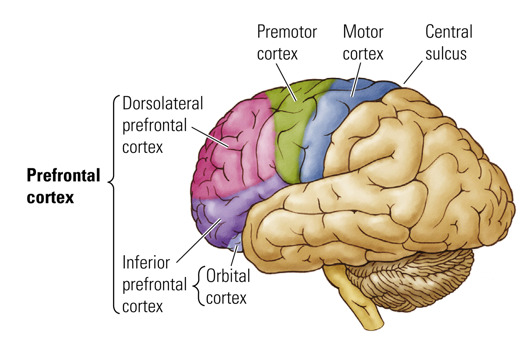
Prefrontal Cortex
- Association cortex
- Part of frontal lobe
- Functions:
planning actions
decision making
emotional control
- Impaired in many psychiatric disorders
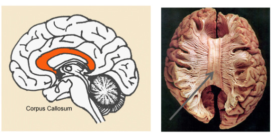
Corpus Callosum
- Latin for “tough body”
- Connects the two cerebal hemispheres
- Consists of white matter (neuronal fibers)
- Can be cut to treat severe epilepsy
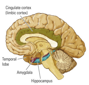
Limbic System
- from the Latin limbus: “border, edge”
- Group of structures between the cortex and brain stem
- Principal structures:
amygdala
hippocampus
cingulate cortex
- Functions:
emotion
memory (hippocampus)
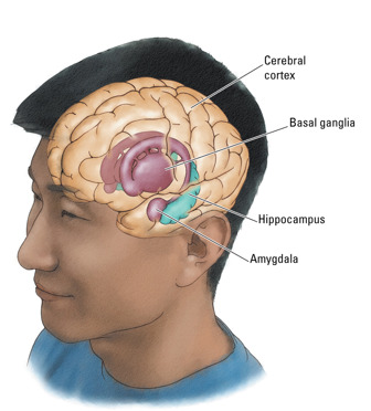
Basal Ganglia
- Latin: “large balls at the bottom”
- accumulations of the gray matter inside the hemispheres below the cortex
- Striatum (Latin: “striped mass”), globus pallidus (Latin: “pale ball”)
movement control (automatic movements, adjustment of movements)
Parkinson’s disease, Tourette’s syndrome
Nucleus accumbens (”adjacent” nucleus)
Reward, pleasure
Addiction
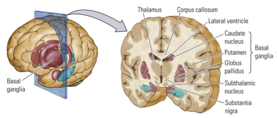
Between Brain AKA Diencephalon
Thalamus
Hypothalamus
Pineal gland
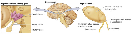
Thalamus
- Greek: “chamber”
- Gateway for channeling sensory information to the cortex
- Allows cortex to focus on selective sensory signals and diminishes significance of others
- Processing of visual, auditory, somatosensory, and gustatory information
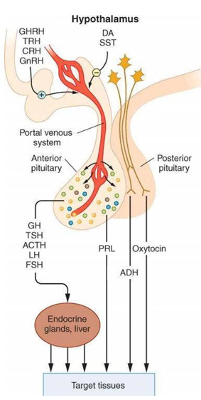
Hypothalamus
- Greek: “under” thalamus
- Regulates body temperature, salt/water balance, hunger, thirst, energy metabolism, reproductive behaviors, and emotional responses
- Controls both the autonomic nervous system & the endocrine system.
- Releases hormones through connections with pituitary gland = hypophysis
drugs = exogenous chemicals
endogenous chemicals =
hormones: carried by blood, control body organs, work from a distance
neurotransmitters: work in the synaptic cleft, help neuronal cells communicate
enzymes: change hormones & neurotransmitters
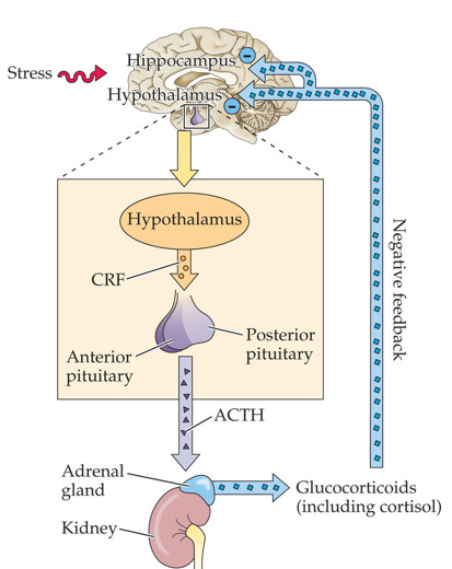
More About the Hypothalamus
- The paraventricular nucleus in the hypothalamus regulates hormonal response to stress.
- HPA axis: stress response depends on the interaction of the hypothalamus, pituitary gland, and adrenal gland.
- Excessive stress response (cortisol) leads to damaging changes in brain & other body systems.
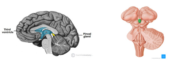
Pineal gland
- sleep control
- releases melatonin
5 notes
·
View notes