#transmission electron microscopy
Text
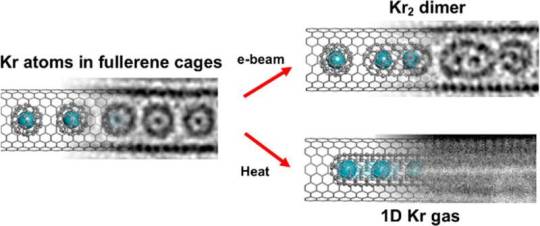
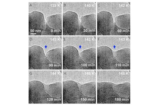
Scientists trap krypton atoms to form one-dimensional gas
For the first time, scientists have successfully trapped atoms of krypton (Kr), a noble gas, inside a carbon nanotube to form a one-dimensional gas.
Scientists from the University of Nottingham's School of Chemistry used advanced transmission electron microscopy (TEM) methods to capture the moment when Kr atoms joined together, one by one, inside a "nano test tube" container with diameter half a million times smaller than the width of a human hair. The research has been published in ACS Nano.
The behavior of atoms has been studied by scientists ever since it was hypothesized that they are the basic units of the universe. The movement of atoms has significant impact on fundamental phenomena such as temperature, pressure, fluid flow and chemical reactions.
Traditional spectroscopy methods can analyze the movement of large groups of atoms and then use averaged data to explain phenomena at the atomic scale. However, these methods don't show what individual atoms are doing at a specific point in time.
Read more.
#Materials Science#Science#Krypton#Gases#1D materials#Carbon nanotubes#Transmission electron microscopy#Carbon#Nanotubes#Nanotechnology#Atoms#University of Nottingham
18 notes
·
View notes
Text


#electron microscopy#transmission electron microscopy#nanoparticles#rare earth hydroxides#zinc tungstate#silver on zinc oxide#zinc titanate#original content
6 notes
·
View notes
Photo

Diverse Defenders
A burning question in COVID-19 research is, why do some people become severely ill while others have only mild symptoms or none at all? While studying the lungs of people as they battle SARS-CoV-2 infection might provide answers, it’s practically and ethically impossible. Researchers have therefore turned to mice, transplanting human lung tissue and immune cells into the animals before infecting them with the virus and tracking the disease. Such humanised mice have enabled researchers to conclude that the severity of infection is, at least in part, down to the variety of macrophages entering the lungs. The image shows a human macrophage containing and surrounded by SARS-CoV-2 particles (green) in one of the humanised mouse lungs. Accumulation of both pro-inflammatory and non-inflammatory macrophages in the lungs was key to ensuring a swift response to the virus while preventing the sort of out-of-control inflammation that’s associated with severe COVID-19 in humans.
Written by Ruth Williams
Picture courtesy of F Douam and D J Kenney, taken in collaboration with the Harvard Medical School Electron Microscopy Facility
Department of Microbiology, Boston University School of Medicine, Boston, MA, USA
Image originally published with a Creative Commons Attribution 4.0 International (CC BY 4.0)
Research published in Cell Reports, April 2022
You can also follow BPoD on Instagram, Twitter and Facebook
#science#biomedicine#covid_19#covid-19#sars-cov-2#macrophages#immune response#inflammation#transmission electron microscopy
15 notes
·
View notes
Text
electronmicroscopy 🧠🔬🧬
#saját#studyblr#lizystudying#myown#lizysinpain#studying#study#study vibes#electronmicroscopy#microscope#microscopy#analysis#electron#transmission#lizysbionics#lizysvibe#engineeer#engineering#engineering student#student#student vibe#university
81 notes
·
View notes
Text
A Journey into the World of Microscopy: From Humble Beginnings to High-Tech Magnification
The science of looking into the hidden invisible Microscopy has transformed our understanding of the world around us. It can explore the universe beyond the reach of our naked eyes, with complex cellular structures, red blood cells, viruses and other viruses and microorganisms taking on amazing perspectives
The history of the microscope is a fascinating story of human curiosity, scientific genius, and relentless exploration. From the humble beginnings of simple magnifying glasses to the sophistication of modern electronic microscopes, the invention of microscopes has shaped our understanding of the microscopic world
In the 1600s, Dutch opticians such as Hans and Zachary Janssen are credited with inventing the first microscope. Known for this hybrid microscope, many lenses were used to magnify objects up to 30 times.At the end of the 17th century, Antony van Leeuwenhoek, Dutch draper some changed our perception of thumbnails. Armed with a well-made single-lens microscope, and explored the hidden reaches of nature. In 1674, Leeuwenhoek discovered microorganisms in lake water, which he aptly named “animalcules”. His discovery laid the foundations of biology and inspired generations of scientists. This incredible feat allowed him to uncover a hidden universe – the first sightings of bacteria, red blood cells, and other microorganisms.
Formation of the scientific environment (17th-19th centuries): Leeuwenhoek’s discoveries boosted scientific research. Robert Hooke, an English scientist, established these developments. In 1665, his book "Micrographia" recorded his observations with a compound microscope. Notably, the term "cell" was coined by Hooke when he examined cork tissue, laying the foundation for cell biology.Microscope systems flourished throughout the 18th and 19th centuries Joseph Lister and other scientists addressed the limitations of the early lenses, introducing improvements that reduced image distortion.
Beyond the Limits of Light: The Beginning of the New Age (19th-20th century): As the 19th century progressed, the limitations of optical microscopy became apparent and scientists yearned for a tool which can go deeper into cells. This research culminated in the development of the electron microscope in the 1930s. The 20th century was revolutionary with the invention of the electron microscope. Unlike light microscopes, which use visible light, electron microscopes use electron beams to achieve much higher magnification.Formation of the scientific environment (17th-19th centuries): Leeuwenhoek’s discoveries boosted scientific research. Robert Hooke, an English scientist, established these developments. In 1665, his book "Micrographia" recorded his observations with a compound microscope. Notably, the term "cell" was coined by Hooke when he examined cork tissue, laying the foundation for cell biology.Microscope systems flourished throughout the 18th and 19th centuries Joseph Lister and other scientists addressed the limitations of the early lenses, introducing improvements that reduced image distortion.
Beyond the Limits of Light: The Beginning of the New Age (19th-20th century): As the 19th century progressed, the limitations of optical microscopy became apparent and scientists yearned for a tool which can go deeper into cells. This research culminated in the development of the electron microscope in the 1930s. The 20th century was revolutionary with the invention of the electron microscope. Unlike light microscopes, which use visible light, electron microscopes use electron beams to achieve much higher magnification.
In the 1930s, German experts Max Knoll and Ernst Ruska made the first electron microscope. This tool let us see tiny things like cells and even atoms by using electron beams, not light, getting images many times bigger. This cool invention showed us the tiny parts inside cells, viruses, and stuff too small to see before. The 1900s brought even more cool microscopes. New kinds like phase-contrast and confocal microscopy let scientists look at live cells without using stuff that could hurt them. Now, the world of looking at tiny things is getting even better. Today, we have high-tech microscopes that use computers and lasers. These let us see and even change tiny things in ways we never could before.
Modern Microscopy's Diverse Arsenal - Today, the field of microscopy boasts a diverse range of specialized instruments, each tailored to address specific scientific needs. Here's a glimpse into some remarkable examples:
Scanning Electron Microscope (SEM): Imagine a high-tech camera that captures images using a beam of electrons instead of light. That's the essence of a SEM. By scanning the surface of a sample with a focused electron beam, SEMs generate detailed information about its topography and composition. This makes them ideal for studying the intricate structures of materials like insect wings, microchips, and even pollen grains.
Transmission Electron Microscope (TEM): While SEMs provide exceptional surface detail, TEMs take us a step further. They function by transmitting a beam of electrons through a very thin sample, allowing us to observe its internal structure. TEMs are the go-to instruments for visualizing the intricate world of viruses, organelles within cells, and macromolecules like proteins.
Confocal Microscopy: Ever wished to focus on a specific layer within a thick biological sample and blur out the rest? Confocal microscopy makes this possible. It utilizes a laser beam to precisely illuminate a chosen plane within the sample, effectively eliminating information from out-of-focus regions. This allows researchers to create sharp, three-dimensional images of cells, tissues, and even small organisms.
Atomic Force Microscopy (AFM): This technique takes a completely different approach, venturing into the realm of physical interaction. AFM employs a tiny cantilever, akin to a microscopic feeler, to physically scan the surface of a sample. By measuring the minute forces between the cantilever and the sample's surface, AFM can map its topography at an atomic level. This provides invaluable insights into the properties of materials at an unimaginable scale, making it crucial for research in fields like nanotechnology and surface science.
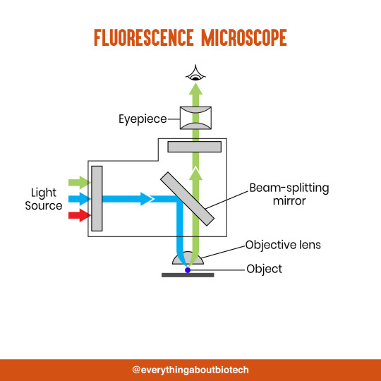
Fluorescence Microscopy: Imagine illuminating a sample with specific wavelengths of light and observing it glowing in response. That's the essence of fluorescence microscopy. This technique utilizes fluorescent molecules or tags that bind to specific structures within a cell or tissue. When excited by light, these tags emit their own light, highlighting the target structures with remarkable clarity. This allows researchers to visualize specific proteins, DNA, or even pathogens within biological samples.
Super-resolution Microscopy (SRM): Overcoming the limitations imposed by the wavelength of light, SRM techniques like STED (Stimulated Emission Depletion) and PALM (Photoactivated Localization Microscopy) achieve resolutions surpassing the diffraction limit. This allows researchers to visualize structures as small as 20 nanometers, enabling the observation of intricate cellular machinery and the dynamics of individual molecules within living cells.
Cryo-Electron Microscopy (Cryo-EM): This powerful technique takes a snapshot of biological samples in their near-life state. Samples are rapidly frozen at ultra-low temperatures, preserving their native structure and minimizing damage caused by traditional fixation methods. Cryo-EM has been instrumental in determining the three-dimensional structures of complex molecules like proteins and viruses, providing crucial insights into their function and potential drug targets.
Correlative Microscopy: Combining the strengths of multiple microscopy techniques, correlative microscopy offers a comprehensive view of biological samples. For instance, researchers can utilize fluorescence microscopy to identify specific structures within a cell and then switch to electron microscopy to examine those structures in high detail. This integrated approach provides a deeper understanding of cellular processes and their underlying mechanisms.
Light Sheet Microscopy (LSM): Imagine illuminating a thin slice of a sample within a living organism. LSM achieves this feat by focusing a laser beam into a thin sheet of light, minimizing photobleaching and phototoxicity – damaging effects caused by prolonged exposure to light. This allows researchers to observe dynamic processes within living organisms over extended periods, providing valuable insights into cellular behavior and development.
Expansion Microscopy (ExM): This innovative technique physically expands biological samples by several folds while preserving their structural integrity. This expansion allows for better resolution and visualization of intricate cellular structures that would otherwise be difficult to distinguish using traditional microscopy methods. ExM holds immense potential for studying the organization and function of organelles within cells.
Scanning Near-Field Optical Microscopy (SNOM): This innovative technique pushes the boundaries of resolution by utilizing a tiny probe that interacts with the sample at an extremely close range. SNOM can not only image the surface features of a sample with exceptional detail but also probe its optical properties at the nanoscale. This opens doors for research in areas like material science and photonics, allowing scientists to study the behavior of light at the interface between materials.
X-ray Microscopy: Stepping outside the realm of light and electrons, X-ray microscopy offers unique capabilities. By utilizing high-energy X-rays, this technique can penetrate deep into samples, making it ideal for studying the internal structure of dense materials like bones and minerals. Additionally, it allows for the visualization of elements within a sample, providing valuable information about their distribution and composition.
From revealing the building blocks of life to aiding in the development of new medicines, the microscope has played an undeniable role in shaping our scientific understanding. As technology continues to evolve, one can only imagine the future breakthroughs this remarkable invention holds in unveiling the secrets of our universe, both seen and unseen. These advancements hold the potential to revolutionize our understanding of biological processes, develop new materials with extraordinary properties, and ultimately pave the way for breakthroughs in medicine, nanotechnology, and countless other fields. As we continue to refine and develop novel microscopy techniques and the future holds immense promise for further groundbreaking discoveries that will undoubtedly revolutionize our perception of the world around us.
#science sculpt#life science#science#molecular biology#biology#biotechnology#artists on tumblr#microscopy#microscope#Scanning Electron Microscope#Transmission Electron Microscope#Confocal Microscopy#Atomic Force Microscopy#Fluorescence Microscopy#Expansion Microscopy#X-ray Microscopy#Super-resolution Microscopy#Light Sheet Microscopy#illustration#illustrator#illustrative art#education#educate yourself#techniques in biotechnology#scientific research#the glass scientists#scientific illustration#scientific advancements
6 notes
·
View notes
Text
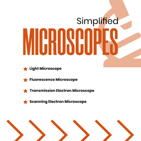


Microscopes Simplified
Light Microscope
Fluorescence Microscope
Transmission Electron Microscope
Scanning Electron Microscope
#microscope#light microscope#Fluorescence Microscope#Transmission Electron Microscope#Scanning Electron Microscope#microscopic#science#microscopy#microscopic life#biotechnology#biotech#biology#biochemistry#studyblr#molecularbiology#notes#class notes#students#science illustration#illustration#illustragram#scientific illustration#graphic design#graphicwork#study notes#study#study motivation#study tips#studygram
82 notes
·
View notes
Text
Uses of Different Types of Microscopes in Forensics
Uses of Different Types of Microscopes in Forensics
The forensic and microbiological labs include a variety of microscopes that may be used. The value of microscopes is increased by how widely they may be used and modified.
#microscope #forensicscience
(more…)

View On WordPress
#comparison microscope#comparison microscope in Forensic Science#dark-field microscope in forensic science#Fluorescent microscope#Forensic science#forensic science notes#microscopy notes#polarising microscope#Scanning Electron Microscope#stereoscopic microscope#stereoscopic microscope uses in forensic science#Transmission Electron Microscope#use of compound microscope in forensic science#use of digital microscope in forensic science#use of fluorescent microscope in forensic science#use of inverted microscope in forensic science#use of phase contrast microscopy in forensic science#use of polarising microscope in forensic science#use of scanning electron microscope#use of scanning electron microscope in forensic science#Uses of Different types of Microscope In Forensics
7 notes
·
View notes
Text
I love how these kids are multilingual, they'll have a full conversation in French and just drop "TEM, motherfucker!!" in the middle of it
0 notes
Text
Figure 7.14 shows a transmission electron micrograph of a thin section of a pea chloroplast.
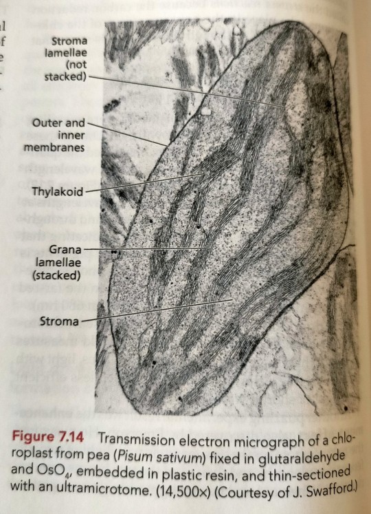
"Plant Physiology and Development" int'l 6e - Taiz, L., Zeiger, E., Møller, I.M., Murphy, A.
#book quotes#plant physiology and development#nonfiction#textbook#transmission electron micrograph#microscopy#chloroplast#stroma lamellae#membrane#thylakoid#grana lamellae#stroma#pea#pisum sativum
0 notes
Photo

Nanoscale materials represent a class of substances that approach the size of individual atoms, about 0.1 to 10 nanometers, in at least one dimension. These materials exhibit properties that are unique from larger scale substances, and they are essential to advancing a wide array of technologies, from magnetic data-storage systems to superconducting quantum computers to biomaterials and biomedical applications.
To study and use these materials, tools are required to examine them at the atomic level while minimizing any disturbance to their structure. Electron microscopy, which uses electron beams to “see” atoms, is one of the fundamental means by which the structures of nanoscale materials can be studied.
Current designs of high-resolution transmission electron microscopes require a lens to focus an electron beam on the specimen surface, said University of Illinois Chicago physicist Robert Klie.
“A side effect of this design is that the sample material is inadvertently exposed to a high magnetic field, so turning the lens off to eliminate the magnetic field when studying magnetic or superconducting materials makes atomic-resolution analysis impossible,” he said.
UIC researchers will soon have a new tool built to overcome the side effect.
Through an $4 million National Science Foundation grant, UIC will be home to the world’s first analytical, aberration-corrected and monochromated transmission electron microscope with a magnetic field-free objective lens. UIC, through the Office of the Vice Chancellor for Research, will contribute the required cost-share of 30%, or $1.7 million, toward the total instrument cost. OVCR will also contribute nearly $4M to renovate space in the Center for Structural Biology not only to house this microscope, but to refresh some of their other shared research facilities.
“The instrument acquired through this grant has a new lens design, providing a magnetic-field-free sample region and, when combined with a nearly mono-energetic electron source, allows for atomic-resolution imaging as well as chemical analysis of these critical materials,” said Klie, UIC professor of physics and the project’s principal investigator.
The instrument will also be equipped with several sample or specimen stages that allow researchers to heat, cool or expose materials to liquids or a controlled magnetic field. The monochromated electron source will also allow for new spectroscopy measurements of materials that undergo changes in structure or properties, including magnetic ordering or superconducting transition, at atomic resolution.
Students and scientists at UIC, and across the Midwest and U.S., will also benefit from the microscope acquisition, according to the researchers. The only other microscope similar to this in the world is currently at the University of Tokyo. Read more.
0 notes
Text
In an effort to understand how and why 2D interfaces take on the structures they do, researchers at the University of Illinois Urbana-Champaign have developed a method to visualize the thermally-induced rearrangement of 2D materials, atom-by-atom, from twisted to aligned structures using transmission electron microscopy (TEM). They observed a new and unexpected mechanism for this process where a new grain was seeded within one monolayer, whose structure was templated by the adjacent layer. Being able to control the macroscopic twist between layers allows for more control over the properties of the entire system.
17 notes
·
View notes
Text
Even far below freezing, ice's surface begins melting as temperatures rise
Physics is filled with mysteries. To find a few worth exploring, look no further than an ice cube. At room temperature, of course, the cube will melt before your eyes. But even far below freezing, ice can shift in barely perceptible ways that scientists are still trying to understand. Using imaging tools at the U.S. Department of Energy's (DOE) Argonne National Laboratory, researchers have detected a phenomenon known as premelting at temperatures far lower than those previously observed.
Their findings are published in the journal Proceedings of the National Academy of Sciences.
Premelting is the reason that a patch of ice can be slippery even on a frigid, clear day. Though the spot is frozen, some part at the surface is wet, an idea first posited by Michael Faraday in the mid-1800s. The idea of a premelted, liquid-like layer on ice opens up other longstanding questions about how water transforms from liquid to solid to vapor—and how, under certain conditions, it can be all three at once.
Read more.
#Materials Science#Science#Ice#Melting#Temperature#Materials characterization#Transmission electron microscopy
16 notes
·
View notes
Photo
#transmission electron microscopy#bright field#dark field#HRTEM#Fourier transform#nickel alloy#scanning transmission electron microscopy#original content
5 notes
·
View notes
Text
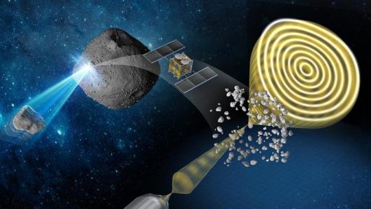
The effects of interplanetary space on asteroid Ryugu
Samples reveal evidence of changes experienced by the surface of asteroid Ryugu, some probably due to micrometeoroid bombardment.
Analyzing samples retrieved from the asteroid Ryugu by the Japanese Space Agency’s Hayabusa2 spacecraft has revealed new insights into the magnetic and physical bombardment environment of interplanetary space. The results of the study, carried out by Professor Yuki Kimura at Hokkaido University and co-workers at 13 other institutions in Japan, are published in the journal Nature Communications.
The investigations used electron waves penetrating the samples to reveal details of their structure and magnetic and electric properties, a technique called electron holography.
Hayabusa2 reached asteroid Ryugu on 27 June 2018, collected samples during two delicate touchdowns, and then returned the jettisoned samples to Earth in December 2020. The spacecraft is now continuing its journey through space, with plans for it to observe two other asteroids in 2029 and 2031.
One advantage of collecting samples directly from an asteroid is that it allows researchers to examine long-term effects of its exposure to the environment of space. The ‘solar wind’ of high energy particles from the sun and bombardment by micrometeoroids cause changes known as space-weathering. It is impossible to study these changes precisely using most of the meteorite samples that land naturally on Earth, partly due to their origin from the internal parts of an asteroid, and also due to the effects of their fiery descent through the atmosphere.
“The signatures of space weathering we have detected directly will give us a better understanding of some of the phenomena occurring in the Solar System,” says Kimura. He explains that the strength of the magnetic field in the early solar system decreased as planets formed, and measuring the remnant magnetization on asteroids can reveal information about the magnetic field in the very early stages of the solar system.
Kimura adds, “In future work, our results could also help to reveal the relative ages of surfaces on airless bodies and assist in the accurate interpretation of remote sensing data obtained from these bodies.”
One particularly interesting finding was that small mineral grains called framboids, composed of magnetite, a form of iron oxide, had completely lost their normal magnetic properties. The researchers suggest this was due to collision with high velocity micrometeoroids between 2 and 20 micrometers in diameter. The framboids were surrounded by thousands of metallic iron nanoparticles. Future studies of these nanoparticles will hopefully reveal insights into the magnetic field that the asteroid has experienced over long periods of time.
“Although our study is primarily for fundamental scientific interest and understanding, it could also help estimate the degree of degradation likely to be caused by space dust impacting robotic or manned spacecraft at high velocity,” Kimura concludes.
TOP IMAGE....Conceptual illustration of the study Credit Yuki Kimura
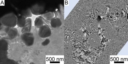
CENTRE IMAGE....Magnetite (round particles) particles cut from a Ryugu sample. (A) Bright field transmission electron microscopy image. (B) Magnetic flux distribution image obtained by electron holography. The concentric circular stripes seen inside the particles correspond to magnetic lines of force. They are called vortex magnetic domain structures and are more stable than ordinary hard disks, which can record magnetic fields for more than 4.6 billion years. (Yuki Kimura, et al. Nature Communications. April 29, 2024) Credit Yuki Kimura, et al. Nature Communications. April 29, 2024
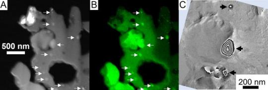
LOWER IMAGE....Iron nanoparticles distributed around pseudo-magnetite. (A) Dark-field image taken with a scanning transmission electron microscope. (B) Corresponding iron distribution image. White arrows indicate iron nanoparticles. (C) Magnetic flux distribution image of the central region of A and B. No magnetic field lines can be seen in the pseudo-magnetite, whereas concentric vortex-like magnetic domain structures can be seen inside the iron particles as shown by black arrows. (Yuki Kimura, et al. Nature Communications. April 29, 2024) Credit Yuki Kimura, et al. Nature Communications. April 29, 2024
5 notes
·
View notes
Text
MAGNUS ARCHIVES BIOLOGY ACADEMIA
requested by @adozenforks :)
- just from reading something’s scientific name, you can picture it perfectly in your mind. this is odd, as you’ve never taken any Latin before
- your DNA model twists and turns on for eternity, an endless helix of nucleotides that you can’t look away from
- you’ve computed the Punnett squares. you Knew Graham’s parents, knew their blue eyes, and it just doesn’t Make Sense that Graham’s eyes are a deep, dark brown
- you cut into the oleander leaf hoping to examine the cellular structure with transmission electron microscopy. as soon as the scalpel gets past the skin, red beads of a viscous fluid drops from the plant. if you didn’t know any better, you’d say it looked like blood
- they brought in fetal pigs to dissect during class. opening your pig, the organs are smeared with … clown makeup?
- you copy the diagrams directly from your textbook, though you think it might be an outdated copy, or maybe a misprint with incorrect proportions. you don’t remember the tibia being that Long, when you look down to check your leg it seems to match the diagram exactly. by the end of the semester, you are a perfect, textbook specimen. your Professor asks you to stay after class for some, ah, extra lab experience
#tma dark academia#tma#the magnus archives#dark academia#biology#biology academia#gothic academia#jonathan sims#martin blackwood
22 notes
·
View notes
Last Seen Blogs

paworn
Paworn

aryxa
Aryxa

aristocrat-67
Untitled

sid-cam
solo vive en mi su recuerdo.

pooltableservice
Pool Table Movers New York