
Last Seen Blogs

reachmylevel-blog1
dedicated

i-want-a-vagina-2
I Want A Vagina

ameliaegg
sweet dreams

fedoraspooky
Meanwhile, elsewhere...

imhuter2
제목 없음
Video
Learn about bacteriophage cycle. . . . Follow --------> www.thescientificreporters.com . Follow --------> www.thescientificreporters.com . Follow --------> www.thescientificreporters.com . . . #biotech #biotechnology #science #biology #research #dna #microbiology #health #biotechnologist #healthcare #medicine #pharma #lab #scientist #biochemistry #biotechusa #chemistry #molecularbiology #genetics #laboratory #cellbiology #biotechnologystudent #medical #microbiologist #innovation #lifescience #biodesign #technology #bio #bhfyp https://www.instagram.com/p/CCtaF1ujUY4/?igshid=12h7kxxx5ske9
#biotech#biotechnology#science#biology#research#dna#microbiology#health#biotechnologist#healthcare#medicine#pharma#lab#scientist#biochemistry#biotechusa#chemistry#molecularbiology#genetics#laboratory#cellbiology#biotechnologystudent#medical#microbiologist#innovation#lifescience#biodesign#technology#bio#bhfyp
0 notes
Video
#Repost Credit @nanobioart --- An #outbreak of #coronavirus infections in the #Wuhan province of #China and December 2019 is threatening to emerge as the next #pandemic. At current count, over 4556 cases and 106 deaths reported in China and approximately 100 cases in 15 other countries. According to Lancaster University researchers this will grow to 250,000 cases and the next 12 days. The alarming speed of transmission is in part because the #virus spreads through the air and by touch. Using the power of 3d #medical animation, this video covers what is a pandemic, how this coronavirus is spreading, how coronavirus attacks the body, and what are recommended precautions to take for protecting against epidemics. Mechanism of disease of coronavirus The 2019 novel coronavirus is a newly discovered strain from the coronavirus family that causes respiratory disease. Humans have long been infected by coronavirus as it is one of those responsible for the common cold. It is a contagious #viralinfection spread through inhalation or ingestion of viral droplets. As a result, coughing and sneezing and touching infected surfaces are primary sources of infection. The structure of coronavirus has multiple parts. Inside the virus lies the #genetic encoding that allows the virus to hijack human cells and turn them into virus factories. A protein encapsulates the genetic material known as the viral envelope. On the surface of the viral are S and HE proteins. The structure of 2019 novel coronavirus is a #mutation. The large S glycoproteins are used by the virus to gain entry to human cells. They attach to receptors on the cell membrane. This binding convinces the cell that the viren is not a threat allowing the virus entry. The exact mechanism for this is not known. Possibly the virus binds with the human cells’ membrane releasing its contents into the cytoplasm. Alternatively, as shown here the human cell ingests the virus in a process known as endocytosis. Meanwhile, the stress of viral production on the endoplasmic reticulum eventually leads to apoptosis or cell death. © Scientific Animations™ #nCoV2019 #sars #SARSCoV #virology #microbiology #epidemic #globalhealth #healthcar https://www.instagram.com/p/B77vgA6JAU5/?igshid=x97b4daog2qv
#repost#outbreak#coronavirus#wuhan#china#pandemic#virus#medical#viralinfection#genetic#mutation#ncov2019#sars#sarscov#virology#microbiology#epidemic#globalhealth#healthcar
0 notes
Video
#petct A procedure in which a small amount of radioactive glucose (sugar) is injected into a vein, and a scanner is used to make detailed, computerized pictures of areas inside the body where the glucose is taken up. Because cancer cells often take up more glucose than normal cells, the pictures can be used to find cancer cells in the body. Also called positron emission tomography scan. ☢️ #science #nuclearmedicine #medicine #diagnostic Credit @road_to_science_phd #biology #biochemistry #physics #mathematics #maths #chemistryteachers #water #melon #watermelon #biochemistry #biochem #bioquimica #medicalscience #biomedicalscience #cellbiology #cellularbiology #space #scicomm #scicommunity #sciencecommunication #scicommindia #scicomms #phd #phdlife #phdstudents #thescientificreporters https://www.instagram.com/p/B7vICaiJ1Hi/?igshid=s1nw378gyurf
#petct#science#nuclearmedicine#medicine#diagnostic#biology#biochemistry#physics#mathematics#maths#chemistryteachers#water#melon#watermelon#biochem#bioquimica#medicalscience#biomedicalscience#cellbiology#cellularbiology#space#scicomm#scicommunity#sciencecommunication#scicommindia#scicomms#phd#phdlife#phdstudents#thescientificreporters
0 notes
Video
Zoom out of thylakoid -> chloroplast -> plant cell . . . . #plantcell #we_love_biology_ig #biology #science #photosynthesis #green #plantart #nature #sciart #biologylife #biologynotes #zoomout #studygram #biologyislife #notes #biologyart #chloroplast #thylakoid #cinema4d #c4d #cinema4dart #c4dart #maxon a #3danimation #octane #aftereffects #animation #cinema4deveryday Credit @nanoclustering https://www.instagram.com/p/B7tOOmIpLu7/?igshid=ugeqsllyhkqo
#plantcell#we_love_biology_ig#biology#science#photosynthesis#green#plantart#nature#sciart#biologylife#biologynotes#zoomout#studygram#biologyislife#notes#biologyart#chloroplast#thylakoid#cinema4d#c4d#cinema4dart#c4dart#maxon#3danimation#octane#aftereffects#animation#cinema4deveryday
1 note
·
View note
Video
*Substrate Coenzyme Enzyme* by medical animation studio @medical_animation_anatomicg #biology #biochemistry #physics #mathematics #maths #chemistryteachers #water #melon #watermelon #biochemistry #biochem #bioquimica #medicalscience #biomedicalscience #cellbiology #cellularbiology #space #scicomm #scicommunity #sciencecommunication #scicommindia #scicomms #phd #phdlife #phdstudents #thescientificreporters https://www.instagram.com/p/B7q_M5eJ_Id/?igshid=16rybvuac8768
#biology#biochemistry#physics#mathematics#maths#chemistryteachers#water#melon#watermelon#biochem#bioquimica#medicalscience#biomedicalscience#cellbiology#cellularbiology#space#scicomm#scicommunity#sciencecommunication#scicommindia#scicomms#phd#phdlife#phdstudents#thescientificreporters
0 notes
Video
This video shows the actin protein filaments (purple) and microtubules (gold) that make up the cytoskeleton of a human epithelial breast cancer cell, taken using spinning disk confocal microscopy magnified at 60x. Since the cytoskeleton is important to how the cell moves and divides, close studies of the cytoskeleton such as this can help us understand these processes. Plus, it looks utterly baller. This video was recently a runner up in the Green Fluorescent Protein Image and Video Contest run by the The American Society for Cell Biology. 📹: Jeffrey van Haren/UCSF #biology #biochemistry #physics #mathematics #maths #chemistryteachers #biochemistry #biochem #bioquimica #medicalscience #biomedicalscience #cellbiology #cellularbiology #molecularbiology #glycolysis #scicommunity #scicomm #metabolicpathways #glycolysis #mcat #mcatprep #phdstudentlife #chemistrynotes #chemistry https://www.instagram.com/p/B7plsjtJeyB/?igshid=1wo0z3m2vt6fv
#biology#biochemistry#physics#mathematics#maths#chemistryteachers#biochem#bioquimica#medicalscience#biomedicalscience#cellbiology#cellularbiology#molecularbiology#glycolysis#scicommunity#scicomm#metabolicpathways#mcat#mcatprep#phdstudentlife#chemistrynotes#chemistry
0 notes
Video
Branching of airways and blood vessels in a mouse lung. Airways can be differentiated from the blood vessels by their mostly uniform wall thickness and more regular circular shape #fluorescence #microscopy #science #scienceiscool #imaging #lung #lungs #biology #artofmicroscopy #biotech #biology #biochemistry #physics #mathematics #maths #chemistryteachers #water #melon #watermelon #biochemistry #biochem #bioquimica #medicalscience #biomedicalscience #cellbiology #cellularbiology #space #scicomm #scicommunity #sciencecommunication #scicommindia #scicomms #phd #phdlife #phdstudents #thescientificreporters Credit @artofmicroscopy (follow them for more) https://www.instagram.com/p/B7isVaVJVSW/?igshid=11w4yiepwmc06
#fluorescence#microscopy#science#scienceiscool#imaging#lung#lungs#biology#artofmicroscopy#biotech#biochemistry#physics#mathematics#maths#chemistryteachers#water#melon#watermelon#biochem#bioquimica#medicalscience#biomedicalscience#cellbiology#cellularbiology#space#scicomm#scicommunity#sciencecommunication#scicommindia#scicomms
0 notes
Photo
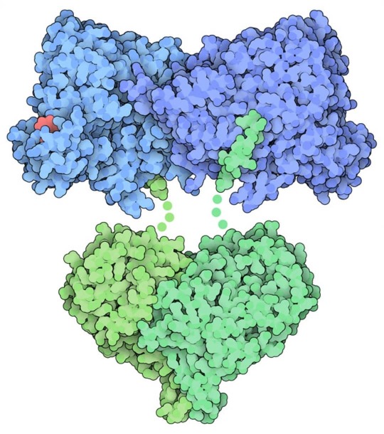
Ribonucleotide Reductase Escherichia coli ribonucleotide reductase forms a tetramer with two alpha subunits (blue) and two beta subunits (green). A nucleotide (red) is bound in a regulatory site in this structure. Ribonucleotide reductase creates the building blocks of DNA DNA and RNA are almost identical in structure, but one small difference has big consequences. A single oxygen atom, which is missing in each DNA nucleotide, distinguishes DNA from RNA. While small, this missing oxygen makes DNA more stable, making it a good molecule for long-term storage of information. On the other hand, RNA is less stable. The hydroxyl group formed by the extra RNA oxygen makes it more susceptible to hydrolysis, so RNA often acts as a biological “flash drive,” storing temporary biological data that is discarded when no longer needed. The only way of creating deoxyribonucleotides in our bodies is from ribonucleotides using the enzyme ribonucleotide reductase, which is essential for DNA synthesis and repair. #biology #biochemistry #physics #mathematics #maths #chemistryteachers #water #melon #watermelon #biochemistry #biochem #bioquimica #medicalscience #biomedicalscience #cellbiology #cellularbiology #space #scicomm #scicommunity #sciencecommunication #scicommindia #scicomms #phd #phdlife #phdstudents #thescientificreporters Ref-PDB https://www.instagram.com/p/B7fbKN_Jjm4/?igshid=hsbuirk2p51s
#biology#biochemistry#physics#mathematics#maths#chemistryteachers#water#melon#watermelon#biochem#bioquimica#medicalscience#biomedicalscience#cellbiology#cellularbiology#space#scicomm#scicommunity#sciencecommunication#scicommindia#scicomms#phd#phdlife#phdstudents#thescientificreporters
0 notes
Video
*Immunotherapy in Cancer Treatment* @aixsponza #thescientificreporters #science #scicomm #scicommgroup #stem #scg #womeninstem #chemistry - #regrann #biology #physics #scientist #science #womeninscience #womendoingscience #whatascientistlookslike #scientistswhoselfie #stemeducation #stemambassadors #womentransformingscience #techniciansofinstagram #primaryscience #primaryeducation #scientistsofinstagram #scientistselfie https://www.instagram.com/p/B7dZUAnpXLM/?igshid=2aq9e6aepo1u
#thescientificreporters#science#scicomm#scicommgroup#stem#scg#womeninstem#chemistry#regrann#biology#physics#scientist#womeninscience#womendoingscience#whatascientistlookslike#scientistswhoselfie#stemeducation#stemambassadors#womentransformingscience#techniciansofinstagram#primaryscience#primaryeducation#scientistsofinstagram#scientistselfie
0 notes
Photo

Lecturer in Metabolomics -Phd position Royal Holloway – University of London –Department of Biological Science Location: Egham Salary: 43,660 to 51,687 per annum – including London Allowance Closing Date: Monday 10 February 2020 Interview Date: Wednesday 26 February 2020 Read more about the position on thescientificreporters.com (link in bio) Follow us for job updates. DM for positions you are looking for. We will help share job openings for same. #biology #physics #scientist #science #womeninscience #womendoingscience #whatascientistlookslike #scientistswhoselfie #stemeducation #stemambassadors #womentransformingscience #techniciansofinstagram #primaryscience #primaryeducation #scientistsofinstagram #scientistselfie #biochemistry https://www.instagram.com/p/B7dZCFLJtfx/?igshid=fa5xhd2imzgp
#biology#physics#scientist#science#womeninscience#womendoingscience#whatascientistlookslike#scientistswhoselfie#stemeducation#stemambassadors#womentransformingscience#techniciansofinstagram#primaryscience#primaryeducation#scientistsofinstagram#scientistselfie#biochemistry
0 notes
Photo

Scientific Coordinator and Project Manager To know more about the job opportunities visit thescientificreporters.com (link in bio) #biology #physics #scientist #science #womeninscience #womendoingscience #whatascientistlookslike #scientistswhoselfie #stemeducation #stemambassadors #womentransformingscience #techniciansofinstagram #primaryscience #primaryeducation #scientistsofinstagram #scientistselfie #biochemistry (at Switzerland) https://www.instagram.com/p/B7dYp-pp04e/?igshid=1utkyp81nqnpg
#biology#physics#scientist#science#womeninscience#womendoingscience#whatascientistlookslike#scientistswhoselfie#stemeducation#stemambassadors#womentransformingscience#techniciansofinstagram#primaryscience#primaryeducation#scientistsofinstagram#scientistselfie#biochemistry
0 notes
Photo
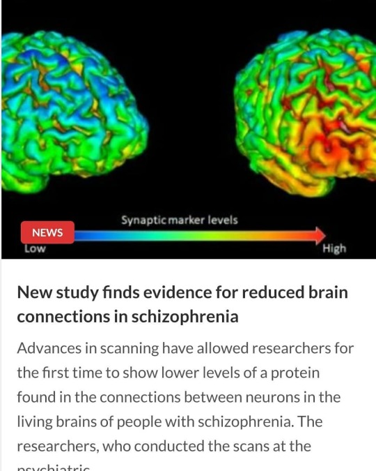
Advances in scanning have allowed researchers for the first time to show lower levels of a protein found in the connections between neurons in the living brains of people with schizophrenia Read more on thescientificreporters.com #biology #physics #scientist #science #womeninscience #womendoingscience #whatascientistlookslike #scientistswhoselfie #stemeducation #stemambassadors #womentransformingscience #techniciansofinstagram #primaryscience #primaryeducation #scientistsofinstagram #scientistselfie #hazardgirls #togetherforscience #scicomm https://www.instagram.com/p/B7aQk92psW7/?igshid=hnjiko16ghjn
#biology#physics#scientist#science#womeninscience#womendoingscience#whatascientistlookslike#scientistswhoselfie#stemeducation#stemambassadors#womentransformingscience#techniciansofinstagram#primaryscience#primaryeducation#scientistsofinstagram#scientistselfie#hazardgirls#togetherforscience#scicomm
0 notes
Video
This animation explains why we always see one side of the Moon. This phenomenon is called "Tidal Lock".⠀ Credits: @sciensationist #bacterialcolonies #study #science #biotechlife #labbiology #bacteria #thebiotechguy #stemcells #scientology #medicalbiotech #genetics #microbiolove #biochemistry #labmemes #laboratory #lablogger #biomemes https://www.instagram.com/p/B7aIUwFpZro/?igshid=1suzca0s41eqf
#bacterialcolonies#study#science#biotechlife#labbiology#bacteria#thebiotechguy#stemcells#scientology#medicalbiotech#genetics#microbiolove#biochemistry#labmemes#laboratory#lablogger#biomemes
0 notes
Photo

Important zones and nerve in your face. Doctor's & Future Doctors Keep Learning Keeping Sharing Keep supporting #doctor #medical #medico #medicine #mbbs #study #studymotivation #neetpg #fmge #usmle #medicos #biology #scientists #scicomm #neet #mci #exams #doctors #doctorslife #aiims #doc #dr #medicalschool #medschool #med #medlife #anatomy #maxillary #ophthalmic #zone #nerve #trigeminalnerve https://www.instagram.com/p/B7YTSRJJw-Q/?igshid=1x3mz5iu5iy3w
#doctor#medical#medico#medicine#mbbs#study#studymotivation#neetpg#fmge#usmle#medicos#biology#scientists#scicomm#neet#mci#exams#doctors#doctorslife#aiims#doc#dr#medicalschool#medschool#med#medlife#anatomy#maxillary#ophthalmic#zone
0 notes
Video
Gel electrophoresis is a technique used to separate DNA fragments (or other macromolecules, such as RNA and proteins) based on their size and charge. Electrophoresis involves running a current through a gel containing the molecules of interest. Based on their size and charge, the molecules will travel through the gel in different directions or at different speeds, allowing them to be separated from one another. All DNA molecules have the same amount of charge per mass. Because of this, gel electrophoresis of DNA fragments separates them based on size only. Using electrophoresis, we can see how many different DNA fragments are present in a sample and how large they are relative to one another. We can also determine the absolute size of a piece of DNA by examining it next to a standard "yardstick" made up of DNA fragments of known sizes. Separation of molecules in electrophoresis is based on charge (the thing that gets pulled) and the effective cross-section of the molecule in whatever state it finds itself – folded, unfolded, bound to other molecules, etc. A collection of DNA fragments separate by length because they are all the same type of molecule. In general, the only meaningful difference between the various fragments should be their length. However, there are some exceptions to this rule. For instance, some DNA molecules are circular (like bacterial plasmids), while others are linear. Circular DNA molecules may run differently than linear ones through a gel. Plasmids, for example, can exist in a form called "supercoiled," in which they actually move faster through a gel. - -credit @b.m.d.e, follow for interesting videos. #dynamic_science #thescientificreporters #scicomm #biology #biochemistry #physics #mathematics #maths #chemistryteachers #water #melon #watermelon #biochemistry#sciencecommunication #scicommindia #scicomms #phd #phdlife #phdstudents #biochem #bioquimica #medicalscience #biomedicalscience #cellbiology #cellularbiology https://www.instagram.com/p/B7QFtD-p_Iv/?igshid=1eqmlstz6rlbp
#dynamic_science#thescientificreporters#scicomm#biology#biochemistry#physics#mathematics#maths#chemistryteachers#water#melon#watermelon#sciencecommunication#scicommindia#scicomms#phd#phdlife#phdstudents#biochem#bioquimica#medicalscience#biomedicalscience#cellbiology#cellularbiology
13 notes
·
View notes
Photo

A poem written to highlight the blunder we humans are doing that led to Australia Bush Fire. #labmonk #crispr.daily #sciencefun #bio_edu #marinescience #microbiologist #laminar #bacterialcolonies #study #science #biotechlife #labbiology #bacteria #thebiotechguy #stemcells #scientology #medicalbiotech #genetics #microbiolove #biochemistry #labmemes #laboratory #lablogger #biomemes # scicomm https://www.instagram.com/p/B7DbltFAkAF/?igshid=1sb46muweuqfp
#labmonk#crispr#sciencefun#bio_edu#marinescience#microbiologist#laminar#bacterialcolonies#study#science#biotechlife#labbiology#bacteria#thebiotechguy#stemcells#scientology#medicalbiotech#genetics#microbiolove#biochemistry#labmemes#laboratory#lablogger#biomemes
0 notes
Video
--- Beautiful video displaying actin fibers in a live human mesenchymal stem cell.⠀ ⠀ 💭❔ Which other cell organelles are you able to recognize? Leave us your comments below.⠀ Credits: @nanolive_sa https://www.instagram.com/p/B68nrD4pFRh/?igshid=jy9c3psxjqw
0 notes