#Machine learning in metastasis
Explore tagged Tumblr posts
Visit Tumblr Blog
Explore Tumblr blogs with no restrictions, modern design and the best experience.
Last Seen Tumblr Blogs


Fun Fact
Tumblr has a 66 index score for customer satisfaction in the US.
Text
Understanding the Uncoupling of Proliferation and Metastasis in Cancer

Uncoupling of proliferation and metastasis is a critical concept in cancer research, where tumor growth and metastatic spread are regulated independently. This phenomenon challenges traditional treatment strategies that primarily target tumor size, often neglecting metastatic potential.We provides cutting-edge solutions to study and manipulate this uncoupling of proliferation and metastasis, aiming to develop therapies that specifically inhibit metastasis without affecting normal cell proliferation. By identifying unique molecular pathways driving metastasis separately from tumor growth, researchers can create targeted interventions to improve patient survival rates. With advancements in cancer biology, understanding and controlling this process could revolutionize therapeutic approaches, leading to better clinical outcomes. Mestastop continues to pioneer research in this field, offering innovative tools and expertise to combat metastatic disease effectively.
#Prevention of distant metastasis#Uncoupling of proliferation and metastasis#Primary patient metastasis prediction#Metastasis probability companion diagnostics#Machine learning in metastasis#Bedside to bench to bedside approach
0 notes
Text

Predicting Spread
Using image-based machine learning to analyse mouth cancer tumours has identified single cells bearing a combination of molecular markers disseminating from the main tumour – their presence is predictive of cancer spread (metastasis)
Read the published research paper here
Image from work by Gehad Youssef and colleagues
Blizard Institute, Queen Mary University of London, London, UK
Image originally published with a Creative Commons Attribution 4.0 International (CC BY 4.0)
Published in eLife, November 2023
You can also follow BPoD on Instagram, Twitter and Facebook
#science#biomedicine#immunofluorescence#biology#mouth cancer#cancer#oral cancer#metastasis#oncology#machine learning#prognostic indicator#sci art
4 notes
·
View notes
Text
"Listen now: we have nothing but time, so I shall not rush. The first violence I commit to you is to tell you my story. You will know before you die: My mother died when she bore me, hooked to tubes and deadlife machines. Your men had her bear me with the assistance of an exo: a machine massaged her heart, currents forced her muscles to work, a black box strapped to her chest forced breath into her lungs. All of this because you knew. You knew at that point the only protest we had was to die and deny you new bodies. “So you took me from her and I never knew her again and in the care of your stone-matrons I learned how to tear worlds to pieces. I learned the lash, and the pick, and how to read the earth you had me kill. “When I was ten, I learned that I must take these pills to survive. That I was born riddled with cancers that you would never take from me – too expensive. The recovery process too long. And your quarterly profits could never slip, as it would mean ruin for your name. “Look at me. You are why we are hideous. Your propaganda paints us as beasts – but it is your hand that shapes our flesh. It is your word that scars our skin and bids cancer grow thick in our bodies. In the names of manna and your house, you ruin millions. “Millions. There are millions of us and countless more. This is your unbecoming. Your death, below your feet. Every inch of every palace and ship and grand city you build, you build on our backs, with our labor, at the cost of our lives. It began in the mines, but it will not end there: your Baronies are as riddled with us as my body is with metastasis. “Yes, weep now. For your perfect face. Your perfect worlds. Your perfect dominion. “Do you know that I did not know what the sky was until the Ungratefuls found me? Liberation means this: not simply seeing the sky, but knowing that there is such a thing as land without stone above you. And then knowing that there are those who put you there. I always thought – we were always taught this – that we were damned and penitent. That we had transgressed against Lordgod’s perfect kingdom and must mine in penance. “How wrong I was! It was not a divine prescript that doomed us to labor, but you and others like you. How surmountable the problem became then. “This is the Ungratefuls’ gift to me. This is their gift to the rest of us – the waste, the offal of your courts and palaces. This is why even your machines fight alongside us. Because you never taught us there could be a thing like the sky. Because you thought that we would never learn. “Goodbye, grand baron. Your house burns, your coffers have been emptied, your monuments have been torn down. Your line – your sons and daughters - hang from the balconies of your own palace. “We learned, grand baron. We learned how to hope, and who to hate.” - Tyrannocleave The Ungrateful
From Lancer by Miguel Lopez and Tom Parkinson Morgan.
#Lancer RPG#karrakin trade baronies#The Ungratefuls#Tom Bloom#miguel lopez#this quote goes so insanely hard
12 notes
·
View notes
Text
Radiotherapy: A Vital Cancer Treatment Option
Radiotherapy has revolutionized cancer care, offering precision and effectiveness in treating various malignancies. As Dr. Edward Yang Tuck Loong from Parkway Cancer Centre aptly describes, radiotherapy is a cornerstone in modern oncology, playing curative, adjuvant, and palliative roles to improve patient outcomes.
What Is Radiotherapy?
Radiotherapy, also known as radiation therapy, uses high-energy X-rays to target and destroy cancer cells by damaging their DNA. This prevents tumor growth and reduces the risk of metastasis. Unlike chemotherapy, which affects the entire body, radiotherapy is localized, minimizing harm to surrounding healthy tissues.
How Radiotherapy Works
Radiation beams are meticulously shaped to focus on the tumor while sparing adjacent organs. Treatments are typically delivered over several weeks, with sessions lasting only minutes each day. For palliative care, shorter courses—sometimes a single session—are used to alleviate symptoms like pain and obstruction.
Radiotherapy in Singapore
Singapore boasts world-class oncology services with cutting-edge radiotherapy technologies such as Intensity Modulated Radiotherapy (IMRT) and stereotactic radiosurgery. The country’s radiotherapy centres are equipped with advanced linear accelerators and tomotherapy machines across public hospitals like the National Cancer Centre Singapore (NCCS) and private institutions such as Gleneagles Hospital.
Why Choose Radiotherapy in Singapore?
Singapore’s healthcare infrastructure ensures access to state-of-the-art treatments for both local and regional patients. With five dedicated radiotherapy centres offering comprehensive care, patients benefit from expert teams of radiotherapists and oncologists who specialize in various cancer types.
Types of Radiotherapy Treatments
Brain Radiotherapy
Brain radiotherapy is often used for treating brain tumors or metastases. It provides symptom relief for conditions like seizures or neurological impairments while aiming to shrink tumors effectively.
Breast Radiotherapy
Breast radiotherapy is a common treatment for early-stage breast cancer or post-surgery cases to prevent recurrence. It preserves cosmetic outcomes while delivering targeted therapy to affected areas.
Understanding Costs: Radiotherapy Cost in Singapore
The cost of radiotherapy treatment in Singapore varies depending on the type of cancer, treatment modality, and whether patients opt for public or private care. Public hospitals generally offer subsidized rates, while private centers provide tailored services at higher costs.
For example:
Public hospitals like NCCS offer affordable options for government-subsidized patients.
Private institutions such as Mount Elizabeth Hospital may have higher fees but provide premium services with shorter waiting times.
To learn more about radiotherapy cost Singapore, visit Specialist Sphere.
Choosing the Right Radiotherapy Centre
Selecting the right radiotherapy centre is crucial for optimal care. Factors to consider include:
Availability of advanced technologies like IMRT or stereotactic radiosurgery.
Expertise of the radiotherapy doctor specializing in your cancer type.
Accessibility and affordability tailored to your needs.
Singapore’s facilities are equipped with world-class technologies and multidisciplinary teams that ensure personalized care for every patient.
Final Thoughts: Why Radiotherapy Matters
Radiotherapy is a powerful tool in combating cancer, offering hope to millions worldwide. Whether you’re considering radiotherapy Singapore or exploring options for specific treatments like brain radiotherapy or breast radiotherapy, Singapore’s healthcare system ensures you receive top-tier care tailored to your needs.
If you’re seeking more information about treatment options or costs, explore Specialist Sphere for detailed insights into this life-saving therapy!
0 notes
Text
Integrated machine learning algorithms reveal a bone metastasis-related signature of circulating tumor cells in prostate cancer
http://dlvr.it/T8svCY
0 notes
Text
RNA Signaling: Illuminating Molecular Paths
Introduction to RNA Biomarkers
Ribonucleic acid RNA markers refer to specific RNA molecules found in blood, other body fluids, or tissues that are indicative of a normal or abnormal process, or of a condition or disease. These RNA molecules can act as signatures for a wide range of conditions from cancer to genetic disorders. Because RNA is more abundant and stable than proteins in body fluids, RNA biomarkers offer potential advantages over existing protein biomarkers. Additionally, advances in sequencing technologies now allow researchers to analyze RNA at an unprecedented scale and depth. This has accelerated discovery of novel RNA biomarkers and their clinical applications. Types of Ribonucleic Acid (RNA) Markers
There are several types of RNA molecules that can serve as potential biomarkers: messenger RNA (mRNA) - mRNA reflects the genes actively expressed in a cell or tissue. Disease-associated changes in gene expression can be detected through changes in mRNA levels. Analysis of mRNA expression patterns offers insights into disease mechanisms and pathways. microRNAs (miRNAs) - miRNAs are short, non-coding RNA molecules that regulate gene expression. Aberrant miRNA expression has been associated with many diseases. Circulating miRNAs show promise as minimally invasive biomarkers. long non-coding RNAs (lncRNAs) - lncRNAs are RNA molecules longer than 200 nucleotides that do not code for proteins but can regulate genes. Disease-associated changes in specific lncRNAs may serve as biomarkers. circular RNAs (circRNAs) - circRNAs are covalently closed RNA circles that are highly stable. Emerging evidence suggests circRNAs may have biomarker potential. snRNAs, snoRNAs, tRNAs - Small nuclear RNAs (snRNAs), small nucleolar RNAs (snoRNAs) and transfer RNAs (tRNAs) play important roles in gene expression and protein synthesis. Changes in levels of these non-coding RNA fractions also carry biomarker potential. Applications of RNA Biomarkers in Cancer Detection and Management Cancer biomarkers based on RNA analysis hold enormous promise, especially due to the cell-type and stage specificity of RNA expression patterns. A few examples illustrate its clinical usefulness: - Breast cancer molecular subtyping - RNA profiling helps distinguish between luminal A, luminal B, HER2-enriched and basal-like subtypes, aiding treatment selection. The 70-gene MammaPrint signature analyzes breast cancer recurrence risk. - Lung cancer diagnosis and staging - RNA signatures have been developed to distinguish benign from malignant pulmonary nodules and to detect residual disease. miRNA signatures improve staging of early-stage tumors. - Colorectal cancer screening - RNA biomarkers show potential as non-invasive alternatives to colonoscopy for population screening and detection of residual/recurrent disease after surgery. - Prostate cancer management - PCA3 and TMPRSS2-ERG gene fusions are FDA-approved RNA biomarkers to aid diagnosis and guide repeat biopsy decisions. miRNA panels may predict disease progression. - Melanoma prognosis - A proprietary RNA expression signature (DecisionDx-Melanoma) stratifies patients' risk of metastasis, informing treatment choices. - Blood-based pan-cancer screening - Highly multiplexed RNA panels analyzed in blood can detect various cancer types at early, historically difficult-to-detect stages through a non-invasive liquid biopsy. Challenges and Future Prospects
While RNA biomarkers hold great promise, key challenges remain around analytical reproducibility, standardization of detection methods, understanding functional relevance, and clinical validation before widespread clinical application. Large, well-designed prospective trials are required to establish clinical utility. Integration of multiple data types including RNA, DNA and protein promises to significantly improve disease classification and management. Machine learning approaches will enable extraction of more information from high-dimensional omics datasets. Overall, RNA-based approaches appear poised to transform disease diagnosis, prognosis prediction and therapeutic monitoring in the coming years.
In Summary, ribonucleic acid (RNA) markers represent a frontier of innovation in healthcare, offering transformative insights into disease biology and treatment response. By profiling RNA expression patterns in biological samples, researchers can uncover hidden disease signatures, identify biomarkers for early detection and prognosis, and guide treatment selection for optimal patient outcomes. Furthermore, RNA markers hold promise for monitoring disease progression, predicting therapeutic responses, and detecting treatment resistance, paving the way for more effective and personalized approaches to healthcare. As technologies for RNA marker analysis continue to advance, so too will their potential to revolutionize diagnostics, therapeutics, and patient care across a wide range of medical specialties.
0 notes
Link
0 notes
Text
Early Detection and Diagnosis: Key to Winning the Battle Against Skin Cancer
In the fight against skin cancer, early detection and accurate diagnosis stand as the most powerful weapons. With skin cancer rates on the rise globally, understanding the importance of early detection and the processes involved in diagnosing skin cancer is crucial for everyone. This comprehensive guide delves into the critical steps and advancements in the detection and diagnosis of skin cancer, empowering individuals to take charge of their skin health.
The Critical Role of Early Detection
Early detection of skin cancer significantly increases the chances of successful treatment and survival. Skin cancers detected at an early stage are often less aggressive, smaller, and more contained, making them easier to treat with less invasive methods. This early intervention can prevent the cancer from spreading to other parts of the body, a process known as metastasis, which complicates treatment and reduces survival rates.

Awareness and regular skin examinations play a pivotal role in early detection. Individuals are encouraged to perform monthly self-examinations to familiarize themselves with their skin's baseline and to detect any new or changing lesions that might be indicative of skin cancer diagnosis. Dermatologists recommend an annual skin check-up, especially for individuals with a higher risk of skin cancer, such as those with fair skin, a history of sunburns, excessive UV exposure, or a family history of skin cancer.
Advances in Skin Cancer Detection
The field of dermatology has seen significant advancements in technologies and methodologies for skin cancer detection. Dermoscopy, a non-invasive imaging technique, allows dermatologists to observe the microstructures of the skin in vivo, providing a clearer view of pigmented skin lesions that are not visible to the naked eye. This tool has become a staple in dermatological practice for the early detection of melanoma and other skin cancers.
Another groundbreaking technology is digital mole mapping, which involves taking detailed photographs of a patient's entire skin surface. These images serve as a baseline for future comparisons, enabling the early detection of new moles or changes to existing ones, crucial indicators of potential skin cancer.
Artificial intelligence (AI) and machine learning are also making strides in skin cancer detection. AI algorithms trained with thousands of images of skin lesions can assist dermatologists in diagnosing skin cancer by highlighting areas of concern and reducing human error.
The Diagnosis Process: From Observation to Confirmation
The diagnosis of skin cancer begins with a thorough skin examination. If a suspicious lesion is identified, the next step is usually a biopsy, where a small sample of the lesion is removed and sent to a laboratory for microscopic examination. The biopsy not only confirms whether the lesion is cancerous but also determines the type of skin cancer, its depth, and aggressiveness, which are crucial factors in deciding the most effective treatment plan.
The Path Forward: Awareness, Prevention, and Early Action
Prevention and early detection of skin cancer are intertwined. While it's essential to adopt preventive measures such as wearing sunscreen, seeking shade, and wearing protective clothing, these efforts must be complemented by vigilance and early action. Understanding the warning signs of skin cancer and undergoing regular skin examinations can make a profound difference in outcomes.
In conclusion, the keys to combating skin cancer lie in early detection and an accurate diagnosis. With ongoing advancements in detection technologies and a heightened awareness of the importance of regular skin checks, we can all take proactive steps to protect our skin health. Remember, the earlier skin cancer is detected, the higher the chance for a successful treatment, underscoring the critical nature of awareness and timely action in the battle against skin cancer.
Source
0 notes
Text
Advancing Cancer Diagnosis: Integrating Immunohistochemical Staining with Digital Pathology
In the realm of cancer diagnosis and research, the integration of immunohistochemical (IHC) staining with digital pathology represents a significant leap forward. This powerful combination offers a multitude of benefits that enhance accuracy, efficiency, and collaboration in cancer pathology.
Immunohistochemical staining allows for the visualization of specific proteins within tissue samples, providing valuable insights into cancer biology. By targeting proteins associated with tumor markers, proliferation, and metastasis, IHC staining aids pathologists in characterizing tumors and guiding treatment decisions.
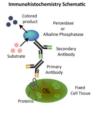
Digital pathology further elevates the capabilities of traditional microscopy by digitizing tissue slides and enabling remote access and analysis. With digital slides, pathologists can view high-resolution images on computer screens, facilitating collaboration among multidisciplinary teams and eliminating the need for physical slide transportation.

The integration of IHC staining with digital pathology offers several advantages:
Enhanced Visualization: Digital slides provide superior image quality and enable pathologists to zoom in and navigate through tissue sections with precision, enhancing visualization of staining patterns and cellular morphology.
Time Efficiency: Digital pathology streamlines workflow processes, reducing the time required for slide preparation, staining, and analysis. This efficiency translates to faster turnaround times for cancer diagnosis and treatment planning.
Data Accessibility: Digital slides can be easily archived and accessed for future reference, facilitating retrospective studies, quality assurance initiatives, and educational purposes.
Telepathology: Remote access to digital slides enables pathologists to consult with colleagues in real-time, regardless of geographical location. This capability enhances collaboration and facilitates expert consultations, particularly in underserved areas.
Machine Learning Integration: Digital pathology platforms can leverage machine learning algorithms to assist pathologists in analyzing large datasets and identifying subtle histological features, further enhancing diagnostic accuracy and efficiency.
The integration of immunohistochemical staining with digital pathology represents a transformative approach to cancer diagnosis and research. By harnessing the combined power of these technologies, healthcare providers can deliver more accurate and timely diagnoses, ultimately improving patient outcomes in the fight against cancer.
0 notes
Text
The Use Of CT Scans As A Diagnostic Tool In Ovarian Cancer Screening
Ovarian cancer is a significant health concern affecting women worldwide. Early detection plays a pivotal role in enhancing treatment outcomes and increasing survival rates.
Medical imaging techniques, such as CT scans, have been instrumental in the diagnosis and staging of various cancers.
This article explores the utility of CT scans as a diagnostic tool in ovarian cancer screening, highlighting their benefits, limitations, and potential impact on patient care. Read on.
I. Understanding Ovarian Cancer Screening:
Screening aims to identify cancer in individuals who do not exhibit any symptoms. Ovarian cancer screening is particularly challenging due to its nonspecific symptoms and the lack of reliable screening tests.
Current screening methods include transvaginal ultrasound (TVUS), CA-125 blood test, and pelvic examinations. However, these methods exhibit limitations in their accuracy and reliability.
II. The Role of CT Scans in Ovarian Cancer Diagnosis:
CT scans, or computed tomography scans, utilize X-ray technology to produce detailed cross-sectional images of the body.
In ovarian cancer diagnosis, CT scans can provide valuable information about the size, location, and characteristics of tumors.
They are especially useful in staging ovarian cancer, determining the extent of disease spread, and assessing the involvement of nearby structures.
III. Benefits of CT Scans in Ovarian Cancer Screening:
Improved Visualization: CT scans offer high-resolution images, allowing physicians to detect small lesions and abnormalities in the ovaries or surrounding tissues. This enhances the chances of early cancer detection and subsequent treatment.
Comprehensive Evaluation: CT scans provide a comprehensive evaluation of the pelvic and abdominal regions, enabling physicians to identify potential metastasis or involvement of nearby lymph nodes.
Precise Staging: Accurate staging is crucial for determining the most appropriate treatment approach. CT scans aid in determining the stage of ovarian cancer, which helps guide treatment decisions.
Treatment Planning: CT scans assist in surgical planning by providing detailed anatomical information. Surgeons can use these images to determine the optimal approach and extent of surgery.
IV. Limitations and Considerations:
Radiation Exposure: CT scans involve exposure to ionizing radiation, which can be a concern, especially if multiple scans are required. Efforts are made to minimize radiation doses while still obtaining diagnostically useful images.
False Positives and Negatives: Like any diagnostic tool, CT scans can produce false-positive or false-negative results. Additional tests or clinical assessments may be necessary to confirm or rule out suspected findings.
Cost and Accessibility: CT scans can be costly, and their availability may vary depending on geographic location and healthcare resources. Access to CT scanning facilities may be a limiting factor in some regions.
V. The Future of CT Scans in Ovarian Cancer Screening:
Researchers continue to explore advancements in CT imaging techniques, aiming to improve accuracy and reduce radiation exposure.
Emerging technologies, such as dual-energy CT and contrast-enhanced CT, show promise in enhancing the sensitivity and specificity of ovarian cancer detection.
Furthermore, artificial intelligence and machine learning algorithms are being developed to aid radiologists in interpreting CT images and detecting subtle abnormalities.
CT scans have proven to be a valuable diagnostic tool in ovarian cancer screening, offering improved visualization, comprehensive evaluation, and precise staging.
Despite limitations and considerations, CT scans provide critical information for treatment planning and guiding surgical interventions.
Continued research and advancements in imaging technology hold the potential to further enhance the role of CT scans in early ovarian cancer detection, ultimately leading to improved patient outcomes.
Discover a premier imaging provider in Australia, with conveniently located centers in Altona, Hoppers Crossing, Ocean Grove, Tarneit, and more. With a commitment to excellence, they offer unparalleled CT scan experiences. Don't delay; prioritize early cancer detection and undergo screening without hesitation.
1 note
·
View note
Text
Machine Learning in Metastasis: Transforming Cancer Research
Machine learning in metastasis is revolutionizing how we understand and tackle cancer spread. By leveraging advanced algorithms, researchers can analyze complex biological data to predict metastatic patterns and improve patient outcomes. Machine learning in metastasis provides precise insights, enabling early detection and personalized treatment plans. Mestastop specializes in innovative solutions in this domain, offering cutting-edge tools to accelerate research and enhance medical strategies. Their expertise bridges the gap between technology and healthcare, ensuring actionable results that can save lives. With the growing role of artificial intelligence in oncology, Mestastop continues to lead advancements in machine learning in metastasis, setting new standards for medical innovation.
#sample patient profile case study#small molecule drugs#treatment for distant metastasis#Machine Learning in Metastasis
0 notes
Text
TYRANNOCLEAVE THE UNGRATEFUL
“Listen now: we have nothing but time, so I shall not rush. The first violence I commit to you is to tell you my story. You will know before you die: My mother died when she bore me, hooked to tubes and deadlife machines. Your men had her bear me with the assistance of an exo: a machine massaged her heart, currents forced her muscles to work, a black box strapped to her chest forced breath into her lungs. All of this because you knew. You knew at that point the only protest we had was to die and deny you new bodies.
So you took me from her and I never knew her again and in the care of your stone-matrons I learned how to tear worlds to pieces. I learned the lash, and the pick, and how to read the earth you had me kill.
“When I was ten, I learned that I must take these pills to survive. That I was born riddled with cancers that you would never take from me – too expensive. The recovery process too long. And your quarterly profits could never slip, as it would mean ruin for your name.
“Look at me. You are why we are hideous. Your propaganda paints us as beasts – but it is your hand that shapes our flesh. It is your word that scars our skin and bids cancer grow thick in our bodies. In the names of manna and your house, you ruin millions.
Millions. There are millions of us and countless more. This is your unbecoming. Your death, below your feet. Every inch of every palace and ship and grand city you build, you build on our backs, with our labor, at the cost of our lives. It began in the mines, but it will not end there: your Baronies are as riddled with us as my body is with metastasis.
Yes, weep now. For your perfect face. Your perfect worlds. Your perfect dominion.
Do you know that I did not know what the sky was until the Ungratefuls found me? Liberation means this: not simply seeing the sky, but knowing that there is such a thing as land without stone above you. And then knowing that there are those who put you there. I always thought – we were always taught this – that we were damned and penitent. That we had transgressed against Lordgod’s perfect kingdom and must mine in penance.
How wrong I was! It was not a divine prescript that doomed us to labor, but you and others like you. How surmountable the problem became then.
“This is the Ungratefuls’ gift to me. This is their gift to the rest of us – the waste, the offal of your courts and palaces. This is why even your machines fight alongside us. Because you never taught us there could be a thing like the sky. Because you thought that we would never learn.
Goodbye, grand baron. Your house burns, your coffers have been emptied, your monuments have been torn down. Your line – your sons and daughters – hang from the balconies of your own palace.
We learned, grand baron. We learned how to hope, and who to hate.
Lancer (Core Rulebook), p. 421-422
15 notes
·
View notes
Quote
The Earth is being hammered by this incessant war – suffering from accelerated entropy – from the activities of humans strip-mining for fool’s gold on a mega-scale. And the primary agent of this sickness is raw unencumbered economics. As cliché as it’s become to use “disease” metaphors or analogies when discussing these things, I still feel that Cancer in metastasis is the best way to adequately describe what all industrial economic systems are doing to life on Earth. Both cancer and economic systems grow primarily for the sake of growing, like self-replicating expansionist programs, while they busily tear asunder, toxify, and finally destroy their host body. But aside from my wrath over all the non-human life-forms being obliterated by this “emptiness that devours”, what I really want to look t here are the effects that this cancerous machine-culture of economics has in the sphere of human thought – how it insinuates itself into our very organism, colonizes our psyche, subdues the quality and range of our desires, poisons our perceptions, and claims a huge territorial stake in how we experience “reality”. Economics grows by implanting parts of its own anti-matter program into other lifeways, infiltrating them bit by bit, first in small ways, then in catastrophic fashion, until finally it engulfs them and the individuals comprising these cultures becomes the Economy. So I don’t think one can get very far in understanding “the Economy” if they only look at with the eyes they’ve developed to play the Game of our current consumerist-capitalist society – or if they try to comprehend it through the pronouncements of the supposed “experts” on the subject, like Marx. We have to learn to look under the surface, because the depth of the problem is immense, and ideally, we should at least be aware of what we’re participating in. “We” are enmeshed in something vast and ancient, a multi-layered slave system that not only physically coerces our participation in its feeding and maintenance, but which is the foundation of our economically-encoded internal reality. And it has existed in us for a long time: it’s been engraving its laws into our bodies and welding its slavery machine into our consciousness for thousands of years. If there’s been any “progress” or “stages of history” over the last ten-thousand years, it’s been the evolution and development of this Slave System, which I call Civilization, and the Economy is the zenith of all the forms (feudalism, monarchism, religious empires, various mega-machines and oh yeah, how could I forget… communism) it has assumed over the centuries.
Birdie Joe Hoakes
10 notes
·
View notes
Photo

Different Machine Learning and Deep Learning Methods for the Classification of Colorectal Cancer Lymph Node Metastasis Images
https://digitalpatientsafety.com/different-machine-learning-and-deep-learning-methods-for-the-classification-of-colorectal-cancer-lymph-node-metastasis-images/
#DHPSP #MedTwitterAI #MedTech
1 note
·
View note
Text
Tweeted
#Oncology #Bioinformatics #AI: scientists have developed machine learning models to predict metastasis status of patients with gastric cancer from demographic and clinical data. The models could help to identify high risk patients - https://t.co/F8WDjyfcQ1 https://t.co/EXCv9aIpWF
— The Royal Vox Post (@RoyalVoxPost) Mar 13, 2023
0 notes
Text
An AI to learn more about glioblastoma

- By Nuadox Crew -
SPHINKS is an AI algorithm created by researchers at the University of Miami Miller School of Medicine that does sophisticated computational analysis to find treatment targets for glioblastoma multiforme and other cancers.
Deep machine learning is used in such framework to identify protein kinases as the culprits involved with tumor growth, which are critical targets in precision cancer therapy.
The researchers utilized tumor organoids derived from patient samples to demonstrate that targeted medications that block the activity of the discovered kinases can.
This AI system may be used in molecular pathology laboratories, and the team has created an open access site.
The algorithm was initially evaluated on glioblastoma, but it was discovered to be relevant to various other malignancies, including breast, lung, and pediatric brain tumors, potentially leading to a new form of clinical trial known as "basket trials".
--
Source: University of Miami Sylvester Comprehensive Cancer Center
Full study: Migliozzi, S., Oh, Y.T., Hasanain, M. et al. Integrative multi-omics networks identify PKCδ and DNA-PK as master kinases of glioblastoma subtypes and guide targeted cancer therapy. Nat Cancer (2023). https://doi.org/10.1038/s43018-022-00510-x
Read Also
Study: AI can be superior to the human eye in predicting the outcomes of brain metastasis
#brain#cancer#oncology#ai#artificial intelligence#deep learning#machine learning#medicine#health#health tech#medtech#basket trials#clinical trials#brain cancer
0 notes