#etiology of breast cancer
Text
Breast Cancer
Introduction
Breast cancer, a multifaceted and prevalent disease, poses a significant health challenge globally, transcending gender lines with its potential impact. Characterized by the abnormal proliferation of cells within breast tissue, breast cancer’s complex etiology remains an area of intense study and concern. Despite notable advancements in medical science and increased awareness, it continues to be a leading cause of morbidity and mortality worldwide. This comprehensive discussion aims to delve into the intricacies of breast cancer, encompassing its causes, risk factors, prevention strategies, diagnostic modalities, treatment options, and the evolving landscape of supportive care.
Causes and Risk Factors
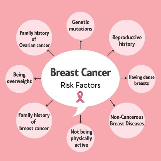
Understanding the underlying causes and risk factors associated with breast cancer is paramount in developing effective prevention and management strategies. While the precise etiology of breast cancer remains elusive, various genetic, hormonal, environmental, and lifestyle factors contribute to its onset and progression. Genetic predispositions, such as mutations in the BRCA1 and BRCA2 genes, significantly elevate the risk of developing breast cancer. Additionally, hormonal influences, including early onset of menstruation, late menopause, and hormone replacement therapy, play a crucial role in disease pathogenesis. Lifestyle factors such as excessive alcohol consumption, obesity, lack of physical activity, and exposure to environmental carcinogens further augment the risk profile.
Preventive Measures

Empowering individuals with knowledge about preventive measures is essential in mitigating the burden of breast cancer. Promoting regular breast self-examinations, clinical breast examinations, and mammographic screenings facilitates early detection and intervention. Emphasizing lifestyle modifications, including maintaining a healthy weight, adopting a balanced diet rich in fruits and vegetables, limiting alcohol intake, and engaging in regular physical activity, can reduce the risk of breast cancer. For individuals with a heightened risk due to genetic predispositions or familial history, prophylactic surgeries, such as mastectomy or oophorectomy, and chemo preventive agents offer viable preventive options.
Diagnostic Modalities
Advances in diagnostic modalities have revolutionized the early detection and diagnosis of breast cancer, enabling prompt initiation of treatment and improved clinical outcomes. Mammography remains the cornerstone of breast cancer screening, capable of detecting abnormalities such as microcalcifications, masses, or architectural distortions. Complementary imaging techniques, including ultrasound, magnetic resonance imaging (MRI), and molecular breast imaging (MBI), enhance diagnostic accuracy, particularly in women with dense breast tissue or high-risk profiles. Biopsy procedures, such as core needle biopsy or surgical excision, facilitate histopathological examination, enabling precise diagnosis and classification of breast lesions.
Treatment Options

Tailoring treatment strategies to individual patient characteristics and disease parameters is essential in optimizing therapeutic outcomes in breast cancer. The treatment landscape encompasses a multidisciplinary approach, integrating surgical, medical, and radiation oncology interventions. Surgical options range from breast-conserving surgeries, such as lumpectomy or segmental mastectomy, to radical procedures like total mastectomy or modified radical mastectomy, depending on tumor size, location, and extent of spread. Adjuvant therapies, including chemotherapy, hormonal therapy, targeted therapy, and immunotherapy, aim to eradicate residual disease, prevent recurrence, and improve overall survival. Radiation therapy, administered either postoperatively or as a primary modality in selected cases, targets residual tumor cells, minimizing locoregional recurrence rates.
Supportive Care and Survivorship

Recognizing the holistic needs of breast cancer patients and survivors is integral in promoting comprehensive care and ensuring optimal quality of life. Supportive care interventions, including symptom management, psychosocial support, nutritional counseling, and rehabilitation services, address the multifaceted challenges associated with cancer diagnosis and treatment. Survivorship programs, focusing on survivorship care planning, surveillance for recurrence, long-term monitoring of treatment-related complications, and health promotion initiatives, facilitate the transition from active treatment to survivorship. Engaging patients and caregivers in survivorship care planning fosters empowerment, resilience, and a sense of agency in navigating the post-treatment phase.
Conclusion
In conclusion, breast cancer represents a formidable health challenge with profound implications for affected individuals, families, and communities worldwide. While significant strides have been made in understanding its pathophysiology, enhancing diagnostic capabilities, and expanding treatment options, concerted efforts are warranted to address existing gaps in prevention, early detection, and access to care. By fostering collaborative partnerships among stakeholders, advocating for evidence-based interventions, and promoting health equity, we can strive towards a future where breast cancer incidence and mortality rates are substantially reduced. Through continued innovation, education, and advocacy, we can transform the landscape of breast cancer care, offering hope, support, and healing to those impacted by this pervasive disease.
We wish you all the best in your medical education journey. In case you need any guidance or assistance during the learning process, do not hesitate to reach out to us.
Email at;
#fullmetal alchemist#healthcare#medical students#assignment help#puppies#aesthetic#ratblr#kittens#pets#plants#nursing student#nurse#nursing school#home nursing services#doctor who#fourteenth doctor#14th doctor#tenth doctor#medicine#medication#pharmacy#big pharma#pharmacy colleges#pharmacy student#pharmacy services#pharmacy school#pharmacy technician#health and wellness
8 notes
·
View notes
Text
Breast Cancer | Symptoms and Early Signs

Introduction About Breast Cancer
Overview of Breast Cancer
Breast cancer, a prevalent malignancy among women globally, arises from breast tissue cells that grow uncontrollably. It encompasses various types, each differing in growth dynamics and impact. Men, though less frequently, can also be affected by this disease. Breast cancer’s prominence is not just in its incidence but also in the social and emotional ripples it creates, impacting patients, families, and communities. The journey of understanding breast cancer begins with recognizing its multifaceted nature, encompassing risk factors, symptoms, treatment options, and, most importantly, the human stories behind the statistics.
Importance of Awareness and Early Detection
The significance of awareness and early detection of breast cancer cannot be overstated. Early detection typically leads to a broader range of treatment options and a better prognosis. Awareness campaigns and educational initiatives are critical in demystifying the disease, encouraging regular screenings, and empowering individuals with knowledge about symptoms and risk factors. This proactive approach helps catch the disease early and fosters a supportive community for those affected, underscoring the idea that early action can be life-saving.
Understanding Breast Cancer
Definition and Basic Facts
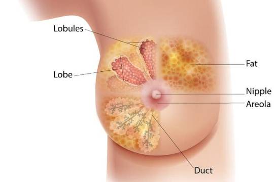
Breast cancer, fundamentally, is a malignant tumor that develops from breast cells. It typically begins in the inner lining of milk ducts or the lobules supplying them with milk and can spread to other body parts. It’s one of the most common cancers worldwide, affecting millions of people, predominantly women, although it can also occur in men. This disease’s progression and severity can vary, making early detection and tailored treatment crucial. Understanding the nature of breast cancer is the first step in effective prevention and management. It’s not just one disease but a complex of diseases with different manifestations, treatments, and outcomes.
Types of Breast Cancer
Breast cancer is classified into several types, primarily based on where it originates. The most common is ductal carcinoma, which begins in the lining of the milk ducts. Another type is lobular carcinoma, which starts in the lobules. Invasive breast cancer has the potential to spread outside the breast, while non-invasive or in situ cancer does not. Each type has unique characteristics and treatment responses, necessitating a personalized approach to care.
Statistics and Prevalence
Breast cancer represents a significant public health issue globally, with increasing incidence rates in various regions. It’s the most diagnosed cancer in women worldwide, contributing to a substantial portion of cancer-related deaths. The survival rates have improved over the years due to better awareness, screening programs, and advances in treatment methods. However, the prevalence underscores the need for ongoing research, education, and accessible healthcare services to continue improving outcomes for those affected by breast cancer.
Causes and Risk Factors
Genetic Factors
Genetic factors can significantly influence breast cancer’s etiology. Inherited mutations in genes like BRCA1 and BRCA2 notably heighten the risk, accounting for a sizeable percentage of hereditary breast cancers. However, it’s crucial to recognize that not all cases are hereditary; many women with breast cancer have no family history of the disease. Genetic counseling and testing are recommended for those with a strong family history to understand their risk better and consider preventive measures.
Lifestyle and Environmental Factors
Lifestyle and environmental elements also play a pivotal role in breast cancer risk. Factors such as prolonged exposure to estrogen, obesity, alcohol consumption, lack of physical activity, and specific reproductive patterns (like late-age first childbirth) have been linked to increased risk. Conversely, regular exercise, maintaining a healthy weight, and moderating alcohol intake are suggested for risk reduction. Environmental aspects, including exposure to certain chemicals and radiation, are also under investigation for their potential links to breast cancer.
Common Myths and Misconceptions
Dispelling myths is critical in understanding breast cancer. For instance, the misconception that only women are affected ignores male breast cancer cases. Also, while family history is a risk factor, most breast cancer patients have no such history, emphasizing the importance of regular screening for all. Moreover, the belief that a lump is the only symptom overlooks signs like nipple changes or skin dimpling. Understanding these facts is essential for effective awareness and prevention strategies.
Symptoms and Early Signs
Recognizing Changes in the Breast
Early detection of breast cancer significantly improves treatment outcomes, making it vital to recognize early signs. Changes to watch include new lumps or thickening in the breast or underarm, changes in breast size or shape, nipple retraction or discharge, and skin changes such as puckering, dimpling, or redness. These symptoms don’t always indicate cancer, but they warrant prompt medical attention for accurate diagnosis.
When to Seek Medical Advice
It’s essential to consult a healthcare professional if any changes in breast appearance or feel are noticed. Early consultation is crucial even in the absence of pain, as breast cancer often doesn’t cause discomfort in the initial stages. Regular mammograms are recommended for early detection, particularly for women over 40 or those with elevated risk factors. Timely medical advice can lead to early detection, increasing the chances of successful treatment.
Symptoms and Early Signs
Recognizing Changes in the Breast
Breast cancer often presents itself through noticeable changes in breast appearance or feel. These changes can include a new lump or mass in the breast, usually painless and hard with irregular edges but sometimes tender, soft, or rounded. Other signs include changes in breast size or shape, skin dimpling, nipple inversion, or redness and flaking of the breast skin or nipple. A clear or bloody discharge from the nipple can also be a warning sign. While these symptoms don’t always indicate cancer, noticing any unusual changes in your breasts should prompt a visit to your healthcare provider for a thorough evaluation.
When to Seek Medical Advice
It’s crucial to seek medical advice if you notice any new or unusual changes in your breasts. This includes lumps, nipple discharge, persistent breast pain, changes in the size or shape of the breast, or changes in the skin over the breast, like dimpling or redness. While these symptoms can be caused by conditions other than cancer, early consultation ensures timely diagnosis and treatment. Regular self-examination and awareness of your normal breast texture and appearance are vital in detecting these changes early.
Diagnosis of Breast Cancer
Screening Methods (Mammograms, Ultrasound, etc.)
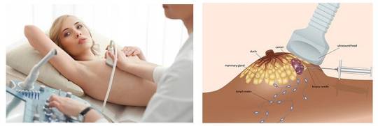
Breast cancer screening is vital for early detection. Mammograms, the most common screening tool, are specialized X-rays that detect tumors before they can be felt. Ultrasounds are used to examine specific areas of concern found during mammography or a physical exam. In some cases, particularly for women with dense breast tissue or high-risk factors, magnetic resonance imaging (MRI) might be recommended. These screening methods aim to detect breast cancer at an early stage when treatment is more likely to be successful.
Biopsy and Lab Tests
If screening tests suggest the presence of breast cancer, a biopsy is the only definitive way to make a diagnosis. During a biopsy, a small amount of tissue is removed from the suspicious area and examined under a microscope. Various types of biopsies, including fine-needle aspiration, core needle biopsy, and surgical biopsy, are used depending on the case. Lab tests on the biopsy sample can determine the type of cells involved, the aggressiveness of the cancer, and whether the cancer cells have hormone receptors or other receptors that may influence treatment options.
Staging and Grading
After a breast cancer diagnosis, staging and grading of the cancer are crucial for determining the extent of the disease and the most effective treatment plan. Staging involves assessing the size of the tumor, the involvement of lymph nodes, and whether the cancer has spread to other parts of the body. Grading evaluates how much cancer cells differ from healthy cells, indicating how quickly the tumor will likely grow and spread. These factors help in assessing the prognosis and planning the most appropriate course of treatment.
Treatment Options
Surgery (Lumpectomy, Mastectomy)
Surgery is a primary treatment for breast cancer, often starting with a lumpectomy or mastectomy. A lumpectomy involves removing the cancerous tumor and a small margin of surrounding tissue, preserving most of the breast. In contrast, a mastectomy consists in removing the entire breast and is usually considered when the cancer is more extensive, or there are multiple areas of cancer in the breast. The choice between these surgical options depends on the cancer’s size, stage, and other medical considerations. Post-surgical reconstruction is also an option for those who undergo mastectomy, helping to restore the patient’s appearance.
Radiation Therapy
Radiation therapy is commonly used after surgery to eliminate any remaining cancer cells in the breast, chest wall, or lymph nodes. It involves directing high-energy rays at the cancer site, which can reduce the risk of recurrence. The treatment typically spans several weeks, where patients receive radiation for a few minutes per session. Side effects may include fatigue and skin changes, usually temporary and manageable.
Chemotherapy
Chemotherapy involves using drugs to destroy cancer cells and is often used in cases where the cancer has spread beyond the breast or for aggressive types of breast cancer. It can be administered before surgery (neoadjuvant chemotherapy) to shrink tumors or after surgery (adjuvant chemotherapy) to eliminate any remaining cancer cells. Chemotherapy is systemic, meaning it affects the entire body and can lead to side effects like hair loss, nausea, and increased risk of infection. The specific drugs and length of treatment vary based on the cancer type and stage.
Hormonal and Targeted Therapies
Hormonal therapies are used for breast cancers that are sensitive to hormones. These therapies block somebody’s natural hormones from fueling cancer growth. On the other hand, targeted therapies focus on specific characteristics of cancer cells, like the presence of the HER2 protein. These treatments can be more effective and have fewer side effects than traditional chemotherapy, as they specifically target cancer cells without affecting normal cells.
Living with Breast Cancer
Emotional and Psychological Impact
Living with breast cancer brings significant emotional and psychological challenges. Patients may experience a range of feelings, from shock and denial to anger, fear, and depression. The uncertainty about the future and changes in body image post-treatment can also impact mental health. Patients must seek emotional support through counseling, support groups, or discussions with healthcare providers. Addressing these emotional aspects is an integral part of the cancer journey.
Support Systems (Counseling, Support Groups)
Support systems play a vital role in coping with breast cancer. Counseling can provide a safe space for patients to express and work through their emotions. Support groups connect patients with others who have similar experiences, offering a sense of community and understanding. These groups can be found through hospitals, cancer organizations, or online platforms. Family and friends also form an essential support network, providing practical and emotional support throughout the treatment and recovery.
Lifestyle Changes and Rehabilitation
Adopting healthy lifestyle changes can aid in the recovery and overall well-being of breast cancer patients. This includes a balanced diet, regular exercise, adequate rest, and stress management techniques. Rehabilitation may involve physical therapy to regain strength and mobility, especially after surgery.Thesee lifestyle changes can help cope with the physical aspects of recovery and improve mental health and quality of life post-treatment.
Prevention and Early Detection
Lifestyle Modifications for Prevention
Adopting a healthy lifestyle plays a crucial role in reducing the risk of breast cancer. This includes maintaining a balanced diet rich in fruits, vegetables, and whole grains while limiting processed and high-fat foods. Regular physical activity is essential; moderate exercise like brisk walking can lower risk. Avoiding tobacco and limiting alcohol consumption are crucial preventive measures. Understanding and discussing family history with a healthcare provider can also help assess genetic risks. These lifestyle changes contribute to breast cancer prevention and promote overall health and well-being.
Importance of Regular Screenings
Regular breast cancer screenings are vital for early detection, significantly improving treatment success rates. Mammograms, the most effective screening tool, can detect tumors before they are palpable. Women over 40, or earlier for those at higher risk, should have annual or biennial mammograms. Clinical breast exams by healthcare professionals are also recommended. These screenings are crucial as early-stage breast cancer may not present any symptoms, and early detection dramatically enhances the chances of successful treatment and survival.
Breast Self-Examination Techniques
Breast self-examination (BSE) is a convenient, no-cost tool that women can use regularly to detect any changes in their breasts. It involves visually inspecting breasts for changes in size, shape, and skin texture and physically examining for lumps or abnormalities. BSE should be performed monthly, preferably at the same time each month, as breasts can change due to menstrual cycles. While BSE is not a substitute for professional screenings, it empowers women to understand their bodies and recognize early warning signs, prompting timely medical consultation.
Research and Advances in Treatment
Latest Developments in Breast Cancer
Research Recent years have seen remarkable advancements in breast cancer research, leading to more personalized and effective treatments. Breakthroughs include targeted therapies that attack cancer cells without harming normal cells, significantly reducing side effects. Immunotherapy, which harnesses somebody’s immune system to fight cancer, has also shown promise. Additionally, advancements in genetic testing have enhanced the understanding of individual cancer profiles, enabling more tailored treatment plans. These innovations continue to improve breast cancer patients’ survival rates and quality of life.
Future Trends in Treatment and Care
The future of breast cancer treatment and care is focused on precision medicine and minimally invasive techniques. Ongoing research is developing drugs that target specific genetic mutations in tumors. There is also a growing trend towards less aggressive treatments that balance efficacy with quality of life. Technological advancements in imaging and biopsy techniques enable earlier and more accurate detection. Moreover, survivorship care, focusing on survivors’ long-term health and well-being, is becoming an integral part of the treatment journey, emphasizing the importance of holistic care in cancer treatment.
FAQs About Breast cancer
Q: What are the early signs of breast cancer?
A: Early signs can include a new lump in the breast or underarm, changes in breast size or shape, and skin changes like dimpling.
Q: Can breast cancer be prevented?
A: While not entirely preventable, risk can be reduced through a healthy lifestyle, regular screenings, and understanding family history.
Q: Is breast cancer only found in women?
A: No, men can also get breast cancer, although it’s less common, accounting for a small percentage of cases.
Q: How is breast cancer diagnosed?
A: Diagnosis typically involves a combination of physical exams, mammograms, biopsies, and other imaging tests.
Q: Are there different types of breast cancer?
A: Yes, there are several types, including invasive ductal carcinoma and invasive lobular carcinoma, among others.
Q: What are the treatment options for breast cancer?
Treatment varies but may include surgery, chemotherapy, radiation, and targeted therapies.
Conclusion
Breast cancer, a complex and multifaceted disease, commands our attention for both its prevalence and the profound impact it has on those affected. This article has journeyed through the various dimensions of breast cancer, from understanding its types and recognizing its symptoms to exploring the latest advancements in treatment and the importance of early detection. As we have seen, while the risk factors and challenges are significant, there is also a beacon of hope: advancements in medical science continually improve patient outcomes. The key takeaway is the undeniable power of awareness and proactive health measures. Regular screenings, understanding one’s risk factors, and staying informed about changes in one’s body are critical steps in combating this disease.
1 note
·
View note
Text
Use of anastrozole in the chemoprevention and treatment of breast cancer: A literature review
Breast cancer is one of the most commonly diagnosed types of cancer in women, presenting high incidence rates in developed regions of the world compared with developing ones. Incidence rates of the disease range from 27 cases per 100,000 women in Eastern Africa to 96 cases per 100,000 women in Western Europe.1,2 Breast cancer is characterized as a multifactorial disease and its development has been reported as the result of complex interactions between an individual’s genoma and the environment.3 Prolonged exposure to estrogene plus progesterone plays a significant role in the etiology of breast carcinoma and biosynthesis pathway of estrogens is thus an important therapeutic target.
The main enzyme involved in estrogen biosynthesis is CYP19A1 or aromatase that belongs to the cytochrome P450 family and is predominantly located in the liver, adrenal glands and fatty tissue.5 However, the source of estrogen varies widely between premenopausal and postmenopausal women (Figure 1).6 In premenopausal women, the main source of estrogen is the ovary, while in postmenopausal women, estrogen is derived from the conversion of androgens into estrogens (through the aromatase enzyme). In particular, testosterone is converted into estradiol, androstenedione into estrone and 16-alpha-hydroxytestosterone into estriol (Figure 2), originating from the peripheral tissues, including the skin, fatty tissue and breast. Therefore, the aromatase enzyme directly affects estrogen biosynthesis in the breast and it is believed that this enzyme plays an important role in the progression of breast cancer.
0 notes
Text
Breast Cyst Treatment Market, Forecast to 2030
A breast cyst is a noncancerous (benign), fluid-filled sac in the breast. Developed as a result of fluid accumulation inside the breast glands, this is found in women aged 20 to 50 years and it can be single or multiple. A cyst may feel like a grape or water balloon or firm.
Market Size & Growth Rate:
The breast cyst treatment market was valued at USD 530 million in 2021 and is projected to reach USD 865 million by 2027, with a CAGR of 7.3 % over the forecast period 2022-2027. The growth could be attributed to the return of demand to pre-pandemic levels, increasing authorization of novel & innovative medicines, extensive research, disease prevalence, and immunotherapies.
Click here for full report:
Market Dynamics:
Various research studies show a high lifetime prevalence of fibrocystic breast disease in women. Over 70% of all women experience fibrocystic changes at some point in their life, with 20% of these women becoming symptomatic and 10% to 30% developing sclerosing adenosis.
A breast cyst is estimated to affect 7% of all women in the United States at some point in their life. Breast cysts are most common in women aged 20 to 50 years. The incidence of cyst development increases throughout these years and then drops dramatically.
Most benign cysts disappear, and new cysts stop developing a year after menopause, as cyst development is linked to hormone levels in the body.
Moreover, living lifestyle, food habits, and psychological issues are finally impacting hormonal balance, these few factors are anticipated to propel the growth of the market.
Market Drivers:
Fine-needle aspiration segment is expected to drive market growth
If all the fluid in the cyst can be removed during the procedure, fine-needle aspiration can be used to diagnose and treat a breast cyst and the breast lump and symptoms disappear.
However, some breast cysts may require more than one drainage procedure. Cysts that form new cysts are common. If a breast cyst persists for two to three menstrual cycles and grows, it should be treated. A small amount of breast tissue or fluid is removed from a suspicious area with a thin, hollow needle and checked for cancer cells during a fine needle aspiration.
If other tests indicate that you may have breast cancer, this type of biopsy may be an option (although a core needle biopsy is often preferred). It can also be used in other conditions.
The main benefits of FNA are that it is relatively quick, it rarely requires anesthesia, the skin is not cut, so no stitches are required, and there is usually no scar. In some cases, one can even get the results on the same day.
The increasing research & development are expected to drive the market growth.
Alcohol consumption increases the risk of breast cystic changes.
Challenges:
Identify the etiology of breast cysts.
In general, the etiology of breast cysts is unknown. However, the majority of breast cysts are linked with abnormal development and compression.
It is based on the research studies that all most all benign disorders of the breast are due to some minor abnormalities in the general processes of the physiological development of the breast following cyclical normal growth response.
These cysts develop in any size, ranging, for example, a few millimeters to several centimeters, generally, the shape of cysts is oval or round and can grow quickly anywhere in the breast.
Although more common in women, many women have cysts and don't feel them at all, because of the difficulty in identifying the condition of cysts.
Professionals don't know exactly what causes breast cysts. They may develop as a result of hormonal changes from monthly menstruation.
Lack of awareness of breast cysts in developing nations.
High cost of imaging systems & side effects related to imaging.
Click here for full report:
Competitive landscape:
The global breast cyst treatment market is moderately competitive with mergers, acquisitions, and product launches. Some of the key players in the market such,
Pfizer, Sanofi, Merck, Teva Pharmaceutical Industries, Bayer AG, Allergan, Johnson and Johnson, Advin Health Care, Becton, Dickinson and Company, Swastik Enterprise, Argon Medical Devices, Inc., Somatex Medical Technologies GmbH, Remington Medical Inc., Hakko co., ltd., Bpb Medica
Click here for request free sample:
Key Developments:
In August 2022, AstraZeneca in collaboration with Daiichi Sankyo received FDA nod for Enhertu. the drug which is directed to treat breast cancer patients exhibiting low HER2 protein in their tumors.
In August 2021, NeoDynamics AB company has launched a product biopsy needle used in the NeoNavia FlexiPulse probe for developing breast cancer diagnosis and treatment.
In April 2020, The FDA-approved Fisher's CytoCore fine needle device, which can be attached to a small needle, has an internal motor that rotates the needle during the biopsy.
0 notes
Text

Lung Cancer. Should you be concerned?
It is crucial to identify demographics who are most likely to be diagnosed with lung cancer in order to enhance early detection. However, some people believe that it should only concern smokers. The actual scenario involves groups of people least expected to have a cancer problem. From adolescents to nonsmokers comprise five susceptible groups that do not know or care about the threat of lung cancer.
This is what Dr Leana Wen, a medical professional and a professor of health policy and administration at the Milken Institute School of Public Health at George Washington University has responded to these questions.
As per CDC, the Centers for Disease Control, lung cancer is the third common disease in America. The most common is skin cancer.
Breast cancer occurs more often in women compared to lung cancer, and also prostate cancer takes place more frequently among the men versus lung cancer.
Lung cancer is currently the leading cause of death among men and women in the US. The American Cancer Society predicts around 238 thousand cases of lung cancer in 2023, of which around 127 thousand people will die.
What are the many types of lung cancer?
Lung cancer is classified into two types: small-cell lung cancer (SCLC) and non-small cell lung cancer (NSCLC).
SCLC is so termed because the cancer cells in SCLC are smaller on microscopic examination than in NSCLC. Both types of cancer contain subcategories; for example, NSCLC has adenocarcinoma, squamous cell carcinoma, and giant cell carcinoma.
Understanding Lung Cancer: Causes, Symptoms, and Risks
Millions of people die every year due to lung cancer alone. One should understand the etiology of lung cancer in order to properly tackle the problem and enhance screening programs for early detection.
It is a result of cigarette smoking, which contains many toxic chemicals which could possibly damage the cells within the lungs resulting from lung cancer. Nevertheless, it is worth mentioning that smoking alone is not responsible for causing emphysema. Other lung health risks related include exposure to second hand smoke, asbestos, radon gas, and some chemicals & substances.
Lung cancer manifests itself differently according to the condition and type it is in. These symptoms include a cough persisting for long periods, chest pain, difficulty breathing, wheezing, coughing of blood, and unexpected weight loss which cannot be explained. This should be followed by seeking medical help if any such signs do not subside, for timely discovery could make success in the treatment a reality.
Some of these risks are age, past or present lung diseases as well as past cases in the family. People with compromised immunities like HIV patients and those after organ transplants among others are also more likely.
Lung cancer is a deadly disease that results from smoking cigarettes. It is caused by many things. Symptoms of it are coughing, chest pain, etc. To minimize its risk, we ought to be knowledgeable about its causes. Lung cancer affects many people, mainly men. Life saving and reducing the impact of the deadly AIDS is possible only trough education and awareness.
Examining the Predominant Demographic Groups Affected by Lung Cancer
Lung cancer affects both men and woman irrespective of their age and social or cultural backgrounds. Even though smoking is still the most significant trigger agent for lung cancer among population, there are some particular communities which are more vulnerable than others.
With regard to second, gender is concerned. Lung cancer has traditionally affected males more than females. Nevertheless, several research studies lately revealed an incredible rise in such events among younger females. This has partly been contributed by the increased uptake of cigarette smoking among women over the last couple of decades.
Further, the aged are more exposed to lung cancer than other groups in society. Although it is mainly associated with ageing citizens, it is experiencing an increase in the number of young adults involved. Some of the reasons behind this trend include exposure to a polluted environment and genetics.
This thirdly, non-smokers should know that there is no way they can avoid this problem even though they may be non-smokers. It is also true that most of the cases occurs in smokers but a large percentage arise in nonsmokers. Lung cancer may also affect non-smoking people due to exposure to secondhand smoke, pollution, and radon gas.
Finally, some occupational hazards expose the people to lung cancer at increased level. The latter include working in mines, constructing buildings and being around other known hazardous materials.
Examining these predominant population groups affected by lung cancer will assist in unravelling the intricacies associated with this life threatening disease. These at-risk populations can get more aware about this knowledge which may also contribute in the efforts of early detection and preventions measures.
The American Cancer Society’s estimates for lung cancer in the US for 2023 are:
About 238,340 new cases of lung cancer (117,550 in men and 120,790 in women)
About 127,070 deaths from lung cancer (67,160 in men and 59,910 in women)
Unraveling the Five Surprising Demographics Prone to Lung Cancer
Surprisingly, other demographically, the lung cancer has different groups that can get affected. While everyone knows that smoking is the principal factor, several others contribute making some groups more vulnerable.
The youth of today represent the first astonishing demographic category. Surprisingly, many cases of lung cancer have been reported among people who are within their 20s and 30s. The reasons for this is attributed to various factors that ranges from exposure to environmental pollutants and/or genetic susceptibility.
Non-smokers form the second group. It should be noted that though many lung cancer cases are associated to smoking, a considerable proportion is observed among non-smokers as well. Non-smokers often develop lung cancer through exposure to secondhand smoke, pollution, and radon gas.
Lastly, this third category might sound strange but includes young women as well. Historically, lung cancer is more common among males, but research shows notable progression for females. This probably emanated from an increased number of women who have begun smoking over the past few years.
A fourth category of people includes those who have an ancestral lineage of lung cancer. It should be added that this risk may not only cover the immediate relatives, but those who have a family history as well. Other extended family members such as cousins, grandparents may also increase the risk.
These are people who have got family history of lung cancer. Although it might seem normal that relatives with such medical condition put you in high danger of getting the same problem, these consequences could even go along with close friends or neighbors. This includes the extended family members like the cousins or even the grandparents.
Thirdly, some occupation-related risks may increase the likelihood of getting infected as well. People working in industries like constructions, mines, and manufacturing have a higher chance of suffering lung cancer because they are exposed to dangerous agents like asbestos.
The five shocking demos should act as a reminder to all people that lung cancer can strike an individual, irrespective of his/her age, gender, and lifestyles. Everyone – including those in the above categories (at-risk groups) – should do whatever it will take to prevent the contraction of this fatal illness. Understanding peculiar risks related to these groups of people may lead us towards better early diagnoses and preventive measures.
Why these Unexpected Demographics are at Higher Risk
There is no single reason why some demographics would be more likely to develop lung cancer. Now, let’s go deeper into the reasons of why these unpredictable groups are more susceptible to this deadly disease.
It has come as a surprise that young adults now constitute a population at risk of developing lung cancer. The reason for this could be because there are more pollutants in the environment to which they are exposed or they have genetically inherited the condition making them predisposed to it.
Then, secondly, the unexpected group with the risk of suffering from lung cancer is non-smokers. Most of these cases are among the heavy smokers but even nonsmokers’ may develop lung cancer due to second hand smoking, air pollution or even radon gas.
This increase of incidence rate of lung cancers among women especially the young women should be worrying. However, this could have been the result of increasing numbers of women smoking in the last few years.
Moreover, people who have a family background of lung cancer such as cousins and grandparents also have raised chances for developing the disease. A person could also inherit the gene that makes them vulnerable to the disease because genetic factors determine susceptibility to the disease.
Last but not least, there are certain jobs which involve the exposure to potentially carcinogenic substances of workers, as in building, mining and others.
By understanding why people who do not fit into the expected demographic are susceptible, we can tailor our preventive and early detection measures for greater impact on specific groups. Through efforts to increase awareness and minimise exposure to risk factors, we can shape a better tomorrow where fewer people develop lung cancer regardless of age, gender, race or geographic location.
Steps to Reduce the Risk of Lung Cancer Across All Demographics
Taking proactive steps to reduce the risk of lung cancer is essential for everyone, regardless of their demographic. Here are some practical measures that individuals can take to minimize their chances of developing this deadly disease.
1. Quit smoking: If you smoke, quitting is the single most effective way to reduce your risk of lung cancer. Seek support from healthcare professionals, join smoking cessation programs, or use nicotine replacement therapies to increase your chances of success.
2. Avoid secondhand smoke: Even if you don't smoke, exposure to secondhand smoke can still increase your risk of lung cancer. Make sure to avoid environments where smoking is allowed and encourage your loved ones to quit smoking for their own health as well.
3. Protect yourself from environmental pollutants: Reduce your exposure to pollutants like air pollution and radon gas. Avoid spending prolonged periods in heavily polluted areas, and ensure your home is properly ventilated and radon levels are monitored.
4. Stay vigilant with screenings: Regular lung cancer screenings, such as low-dose computed tomography (LDCT), can detect early signs of lung cancer in high-risk individuals. Speak with your healthcare provider about recommended screening intervals based on your demographic and risk factors.
5. Lead a healthy lifestyle: Maintaining a healthy lifestyle can contribute to overall well-being and reduce the risk of developing lung cancer. Engage in regular exercise, maintain a balanced diet rich in fruits and vegetables, and limit alcohol consumption.
By following these steps, individuals can play an active role in reducing their risk of lung cancer. Remember, knowledge and awareness are key, so spread the word to friends and family, and together, we can make a difference in the fight against lung cancer.
#lung cancer#cancer#lung cancer symptoms#non-small cell lung cancer#small cell lung cancer#lung cancer diagnosis#symptoms of lung cancer#lung cancer survival rate#lung#what is lung cancer#lung cancer staging#treatment of lung cancer#lung cancer signs#lung cancer story#cancer treatment#lung cancer stages#xray of lung cancer#signs of lung cancer#lung cancer journey#lung cancer australia#lung cancer treatment#lung cancer screening
0 notes
Text
The Role of Exercise in Preventing Chronic Diseases: A Comprehensive Review

Regular exercise is commonly recognised as an important component of living a healthy lifestyle. Its advantages go beyond physical fitness and include the prevention and management of chronic diseases. This blog article will investigate the impact of exercise on numerous chronic illnesses, using recent scientific research released between 2018 and 2023 as evidence. We can better understand the importance of incorporating physical activity into our everyday lives by investigating the scientific evidence on the benefits of exercise and its association with disease prevention.
EXERCISE AND CARDIOVASCULAR HEALTH: Recent scientific study indicates that regular physical activity reduces the risk of cardiovascular disorders such as coronary artery disease, stroke, and heart failure (Smith et al., 2015). Aerobic activity, such as brisk walking, jogging, or cycling, improves cardiovascular fitness, lowers blood pressure, and lowers LDL cholesterol levels, all of which contribute to better heart health (Bouchard et al., 2012).
EXERCISE AND DIABETES PREVENTION: Type 2 diabetes has become a global epidemic, yet current research highlights the critical role of exercise in its prevention. Regular physical activity appears to enhance insulin sensitivity, aid in weight maintenance, and reduce the chance of developing diabetes (Colberg et al., 2010). Individuals who engage in moderate-intensity exercise, such as brisk walking for 150 minutes per week, have a decreased risk of type 2 diabetes than their sedentary counterparts, according to research published in the last five years (Tuomilehto et al., 2001).
EXERCISE AND MENTAL HEALTH: In addition to physical benefits, exercise has a significant impact on mental health. Mandolesi et al. (2018) found that regular physical activity lowers the risk of depression, anxiety, and cognitive deterioration. Exercising causes the release of endorphins, chemicals that boost emotions of enjoyment and reduce stress. Furthermore, physical activity increases blood flow to the brain, promoting neuron development and increasing cognitive performance (Ratey & Hagerman, 2008).
EXERCISE AND CANCER PREVENTION: New study reveals that exercise can help prevent and control several types of cancer. Regular physical activity, according to studies published in the last five years, lowers the chance of acquiring colon, breast, and prostate cancers (Friedenreich et al., 2016). Exercise helps to maintain a healthy body weight, reduces inflammation, improves immunological function, and balances hormones, all of which contribute to a lower risk of cancer (Moore et al., 2016).
In conclusion, exercise is proving to be an effective therapy in the prevention and management of chronic diseases. Regular physical activity has numerous advantages, ranging from cardiovascular health and diabetes prevention to mental well-being and cancer risk reduction. It is critical to prioritise exercise as an intrinsic part of our daily routines, aiming for at least 150 minutes of moderate-intensity aerobic activity per week. We may considerably improve our general health and lower our risk of chronic diseases by including exercise into our daily life.
References:
Bouchard, C. et al. (2012). Physical Activity and Health. Human Kinetics.
Colberg, S.R. et al. (2010). Exercise and Type 2 Diabetes: The American College of sports Medicine and the American Diabetes Association: Joint Position Statement. Diabetes Care, 33(12), e147-e167.
Friedenreich, C.M. et al. (2016). Physical activity and cancer prevention: etiologic evidence and biological mechanisms. Journal of Clinical Oncology, 34(9), 987-994.
Mandolesi, L.et al. (2018). Effects of Physical Exercise on Cognitive Functioning and Wellbeing: Biological and Psychological Benefits. Frontiers in Psychology, 9, 509.
Ratey, J.J. & Hagerman, E. (2008). Spark: The Revolutionary New Science of Exercise and the Brain. Little, Brown Spark.
Smith, S.C. et al. (2015). AHA/ACC guidelines for secondary prevention for patients with coronary and other atherosclerotic vascular disease: 2006 update endorsed by the National Heart, Lung, and Blood Institute. Journal of the American College of Cardiology, 47(10), 2130-2139.
Tuomilehto, J.et al. (2001). Prevention of Type 2 Diabetes Mellitus by Changes in Lifestyle among Subjects with Impaired Glucose Tolerance. The New England Journal of Medicine, 344(18), 1343-1350.
Copyrights: If you like the post and want to use the information please give credits to my profile. Cheers :)
#exercise#fitness#health#wellness#cardiovascular#cardiovascular health#mental health#diabetes prevention#comprehensive review#cancer prevention#science#sport science#scientific research
0 notes
Text
National Cancer Control Month - April 2023
April 2023 marks National Cancer Control Month, an important initiative to raise awareness and share information about cancer prevention, diagnosis, and treatment. Aprazer Healthcare is proud to join in this effort to fight cancer. During this month, Aprazer Healthcare will work to bring attention to the many resources available to those affected by cancer. To further enlighten people on the most current cancer therapies and technology, we will be offering educational opportunities to healthcare professionals, patients, and their families. Aprazer Healthcare also offers a variety of cancer-care services, including screening, diagnosis, and treatment. Our team of doctors, nurses, and other healthcare experts collaborate to give each patient individualized care.
Statistics
According to data, a major cause of death and a global health concern is cancer. Globally, 10 million deaths from cancer are anticipated in 2023. Because of variables including population growth, aging populations, and other developments, this figure has been rising consistently over the last few decades.
Numerous facets of cancer, including its etiology, diagnosis, and prognosis, have recently come into focus thanks to improvements in research; nevertheless, much more work is still required to properly comprehend this complicated condition. Customized medicines targeting particular mutations or cell-level processes have been developed due to research into the genetic basis of various malignancies. These treatments can help individuals with specific cancers, like breast or lung cancer, have improved prognoses.
Focused Areas
Cancer screening and diagnostics are essential for early disease detection, which can improve patient outcomes. Age, gender, family history, and other risk factors like smoking or exposure to specific environmental chemicals can affect the appropriate screening tests. Mammograms and colonoscopies are typical cancer screening tests for breast and colon cancers, respectively. Nevertheless, a large variety of other diagnostics are available that are specialized for particular populations or cancer types.
Community Efforts
The burden of cancer on society must be lessened through community actions. As an illustration, consider Aprazer Healthcare, a nonprofit offering free cancer tests and treatments to people in need throughout the United States. More than 10 million people have benefited from their services in receiving treatment for early-stage malignancies and other severe ailments. They also offer educational resources on cancer prevention and treatment alternatives, which can assist people in making wise decisions regarding their health.
Conclusion
It is crucial to understand the significance of early cancer detection. Regular screenings and self-monitoring for suspicious changes can help identify potential issues before they become more serious, improving outcomes significantly if caught in time. By supporting continued funding and initiatives dedicated to fighting against this global issue, we can make a real difference when it comes to reducing its impact on society while providing hope for individuals affected by it directly or indirectly.
0 notes
Text
What can be the reason for a lump in Breast or Fibroadenoma?

#FIBROADENOma (Lump in Breast)
Definition: Fibroadenoma or Lump is a swelling protuberance, bulging or bump in the breast that feels different from the normal breast tissue.
It is a solid Benign tumor which is a non cancerous tumor in which epithelial cells are arranged in a fibrous stroma.
CAUSES OF FIBROADENOMA
Breast Cysts: Fluid filled sacs in breast tissue are usually benign.
Infection
Trauma
Fat necrosis
Fibrocystic breast
Lump of breast varying in size
It can be identified with the help of breast size whether it becomes fatal (dangerous) or not.
1. 1 cm/less – Small Fibroadenoma considered
normal.
2. Upto 3 cm-Large Fibroadenoma considered
3. More than 3cm – Giant Fibroadenoma considered Disease.
Symptoms
Age – Incidence usually occurs with menstruating females at any age.
Painless swelling (lump) in the breast.
Smooth, firm to hard consistency, freely movement of lump in breast.
Elderly age patients may not have a characteristic feature due to fibrosis occurring in the breast.
COMPLICATIONS
1. Simple Fibroadenoma have no family history of breast cancer and have no risk of cancer.
2. Complex Fibroadenoma show cyst, sclerosis adenosis, calcification have increased risk of cancer may show some symptoms:
Frequency of pain increased.
Bleeding does not stop.
Poor wound healing.
Treatment
Homeopathic treatment is effective in stabilizing the health of an individual, affected by fibroadenoma and its effects at various stages of fibroadenoma which help in dissolving internally the lump in breast. Homeopathy medicines will be prescribed on the basis of the cause or etiology of the disease or clinical condition i.e. sign and Symptoms.
Here are a few Homeopathic remedies that help In the treatment of fibroadenoma and thus, reducing the intensity of symptoms:
Top 5 Homeopathic remedies for Fibroadenoma:-
1. CONIUM MACULATUM:
This medicine very effective in the treatment of fibroadenoma.
Indicated in “mammary gland are hard and sore with inflammation of the Breast Tissue”.
The region of breast becomes hard, nodular, tender to touch with burning and stinging pain in the breast.
Also there is a discharge of pus from the nipple and if lesions occur then it may lead to the fibrosis.
2. BARYTA CARBONICA:
When there is inflammation i.e.redness, swelling with induration and enlargement of the lump in breast.
The lump in breast is hard and sensitive to touch with infiltration.
Edges of the lump are sharply defined with a bloody discharge from the nipple and it becomes sensitive to cold and complaints become worse when anything is taking cold.
3. HYDRASTIS CANADENSIS:
The patient has a tendency to indurated glands.
The patient is weak and emaciated, fainting due to improper assimilation.
Mammary gland is swelling with Fat Necrosis and glandular cell myoblastoma are common in this remedy.
Patient complains of pain and tenderness with an engorged nipple, cracks and discharge of watery fluid.
4. CALCAREA FLUORICA:
This remedy is indicated in the fibroadenoma of the breast.
Lump in breast which is hard, movable with clear margins and sharp in nature.
Nodules are in upper right quadrant.
Patients are very sensitive to cold air/ atmosphere.
Pain at the tip of urethra, back extending to the sacrum..
This remedy acts well after surgery on the breast
5. PHYTOLACA DECANDRA:*
Mammae full of hard, painful nodosities.
Breast is full of stony hard and painful especially when suppuration occurs.
Pain in the breast occurs when child nurse pain goes from nipple all over the body.
Cracks and small ulcers around nipple
Irritable in breast before and during menses. ( homeopathy benefits )
0 notes
Text
Preventive Cancer
After more than 3 decades of Medical Oncology practice, now I am firmly convinced about the importance of cancer prevention.
We cure a very small percentage of cancer patients and in others it is at its best a prolongation of useful life. Even when we achieve cure it is many times at a great financial and organ loss. In general the popular notion that cancer is the greatest misfortune that can descend on man is true.
The only cost effective way of dealing with cancer is to prevent it. Wherever the etiology is firmly known, prevention is possible. Where cause is not known, screening the high risk groups for cancer can detect precancerous conditions or early chances and cure rates can be high. The best person to advise prevention in my opinion is an oncologist who will be in a priviliged position to be in touch with family, friends of a cancer patient, who will be amenable to preventive advice.
At least 40% of cancers are tobacco related and are perfectly preventable. About 30 – 40% are related to wrong diet,obesity,lack of exercise and wrong sexual practices. These require change of life style and this change also is possible to achieve. Some common cancers are virus induced and vaccination will be the perfect answer for their prevention.
Cancer arises from a loss of normal growth control. In normal tissues, the rates of new cell growth and old cell death are kept in balance. In cancer, this balance is disrupted. This disruption can result from uncontrolled cell growth or loss of a cell’s ability to undergo “apoptosis”. Apoptosis, or “cell suicide,” is the mechanism by which old or damaged cells normally self-destruct.
Cancer can originate almost anywhere in the body. Carcinomas are the most common types of cancer arise from cells that cover the external and internal body surfaces. Sarcomas are the cancers arising from the supporting tissues of the body like bone, cartilage, fat, muscle etc. Lymphomas are cancers that arise from the lymph nodes and tissues in the bodys immune system. Leukemias are cancers of the immature blood cells that grow in the bone marrow and tend to accumulate in the blood stream.
Cancers are capable of spreading through the body by two mechanisms- invasion and metastasis. Invasion refers to the direct migration and penetration by cancer cells into the neighbouring tissues. Metastases refers to the ability of the cancer cells to penetrate into lymphatic and blood vessels, circulate through the blood stream and invade normal tissues elsewhere in the body.
70-75% of the patients are preventable. In the rest 25% many of them can be detected early if screened regularly. Cancers related to tobacco like lung cancers, head and neck cancers, pancreatic cancers, bladder cancer are preventable if the patient does not use tobacco or stops using tobacco. About 30% of the cancers are related to obesity, wrong diet and lack of exercise. These cancers like colon cancer, breast cancer and prostate cancer can be reduced by the right lifestyle intervention.
About 15-20% of cancers are related to infection either a viral infection like HPV, EBV and Hepatitis B and C viruses. Cancer of uterine cervix, liver cancer, some head and neck cancers can be prevented by vaccination against these viruses. Some bacterial infections also can cause cancer as exemplified by stomach cancer and stomach lymphoma caused by H Pylori. Lack of physical exercise can be responsible for 15% of the cancers directly and several other cancers indirectly.
Published Article By Dr Radheshyam Naik:
A Narrative Review of the Risk factors for Cancer and Preventive Opportunities: Current Status, Future Perspectives and Implications for India
https://www.thieme-connect.com/products/ejournals/pdf/10.1055/s-0041-1731092.pdf
Characteristics of tobacco consumption among cancer patients at a tertiary cancer hospital in South India-A cross-sectional study
https://pubmed.ncbi.nlm.nih.gov/34720601/
0 notes
Text
Preventive Cancer
After more than 3 decades of Medical Oncology practice, now I am firmly convinced about the importance of cancer prevention.
We cure a very small percentage of cancer patients and in others it is at its best a prolongation of useful life. Even when we achieve cure it is many times at a great financial and organ loss. In general the popular notion that cancer is the greatest misfortune that can descend on man is true.
The only cost effective way of dealing with cancer is to prevent it. Wherever the etiology is firmly known, prevention is possible. Where cause is not known, screening the high risk groups for cancer can detect precancerous conditions or early chances and cure rates can be high. The best person to advise prevention in my opinion is an oncologist who will be in a priviliged position to be in touch with family, friends of a cancer patient, who will be amenable to preventive advice.
At least 40% of cancers are tobacco related and are perfectly preventable. About 30–40% are related to wrong diet,obesity,lack of exercise and wrong sexual practices. These require change of life style and this change also is possible to achieve. Some common cancers are virus induced and vaccination will be the perfect answer for their prevention.
Cancer arises from a loss of normal growth control. In normal tissues, the rates of new cell growth and old cell death are kept in balance. In cancer, this balance is disrupted. This disruption can result from uncontrolled cell growth or loss of a cell’s ability to undergo “apoptosis”. Apoptosis, or “cell suicide,” is the mechanism by which old or damaged cells normally self-destruct.
Cancer can originate almost anywhere in the body. Carcinomas are the most common types of cancer arise from cells that cover the external and internal body surfaces. Sarcomas are the cancers arising from the supporting tissues of the body like bone, cartilage, fat, muscle etc. Lymphomas are cancers that arise from the lymph nodes and tissues in the bodys immune system. Leukemias are cancers of the immature blood cells that grow in the bone marrow and tend to accumulate in the blood stream.
Cancers are capable of spreading through the body by two mechanisms- invasion and metastasis. Invasion refers to the direct migration and penetration by cancer cells into the neighbouring tissues. Metastases refers to the ability of the cancer cells to penetrate into lymphatic and blood vessels, circulate through the blood stream and invade normal tissues elsewhere in the body.
70–75% of the patients are preventable. In the rest 25% many of them can be detected early if screened regularly. Cancers related to tobacco like lung cancers, head and neck cancers, pancreatic cancers, bladder cancer are preventable if the patient does not use tobacco or stops using tobacco. About 30% of the cancers are related to obesity, wrong diet and lack of exercise. These cancers like colon cancer, breast cancer and prostate cancer can be reduced by the right lifestyle intervention.
About 15–20% of cancers are related to infection either a viral infection like HPV, EBV and Hepatitis B and C viruses. Cancer of uterine cervix, liver cancer, some head and neck cancers can be prevented by vaccination against these viruses. Some bacterial infections also can cause cancer as exemplified by stomach cancer and stomach lymphoma caused by H Pylori. Lack of physical exercise can be responsible for 15% of the cancers directly and several other cancers indirectly.
0 notes
Text
The Medicinal Properties of Curcumin

Dr. Rakesh K. Srivastava
Dr. Rakesh K. Srivastava is an accomplished entrepreneur and is the CEO and President of GLAX Health LLC.
WILMINGTON, DELAWARE, UNITED STATES, September 3, 2022
— Dr. Rakesh K. Srivastava’s Medicinal Properties of Curcumin
Turmeric, a popular Indian spice, contains curcumin, the primary polyphenolic curcuminoid extracted from the rhizome of the Curcuma longa plant. Curcumin has been studied for its antitumor, antioxidant, and anti-inflammatory properties as a potential cancer chemopreventive agent. Curcumin's many therapeutic benefits have been thoroughly investigated, analyzed, and accepted. Curcumin has long been recognized for its therapeutic effects against various diseases, including diabetes, allergies, arthritis, and other chronic and inflammatory conditions. This article could shed more light on the condition and discuss Dr. Rakesh K. Srivastava’s efforts to improve the quality of life for cancer patients.
Curcumin's therapeutic actions include:
Inhibition of angiogenesis and metastasis.
Immune-modulation.
Positive effects on cellular enzymes including cyclooxygenase and glutathione S-transferases.
Inhibition of many cell signaling pathways at several levels.
Curcumin can alter gene transcription, stop the cell cycle, and trigger programmed cell death. Curcumin is a very pleiotropic chemical that targets various diseases and has an outstanding safety profile; yet, it did not have an optimal therapeutic effect in clinical studies mainly because of its poor solubility and bioavailability.
Crude curcumin, which is yellow in color, is made up of curcumin, demethoxycurcumin, and bisdemethoxycurcumin. Studies have established that when taken orally, curcumin can induce apoptosis in various tumor types, including leukemia, breast, prostate, skin, colon, stomach, duodenum, head & neck, and soft palate. It also slows proliferation and promotes death in cancer cells, but the underlying molecular pathways are unknown.
Cancer cells treated with curcumin undergo apoptosis due to the induction of the Bax and Bak genes upstream of the mitochondria and the suppression of Akt activity. Curcumin also increases cancer cells' sensitivity to chemotherapy and radiation by suppressing NFkB activation.
In addition to helping with allergies, arthritis, and Alzheimer's disease, curcumin has been shown to have antiviral, antibacterial, antifungal, anticancer, and antidiabetic properties. Oxidative stress plays a pivotal part in the etiology including various diseases such as Cancer, diabetes, cardiovascular disease, neuronal cell injury, and hypoxia. Like ascorbic acid and vitamin E, curcumin has potent antioxidant properties.
For a very long time, scientists have recognized curcumin's potential anti-inflammatory effects. It inhibits the activation of NF-kB, which controls inflammatory genes. It also prevents the production of the inflammatory 5-LOX enzyme while also decreasing COX-2 production. Curcumin inhibits the production of inflammatory cytokines and chemokines like tumor necrosis factor (TNF), interleukin (IL)-1, IL-6, and IL-8.
Curcumin has also been linked to a reduction in amyloid-induced brain inflammation, a hallmark of Alzheimer's disease. Curcumin supplementation in the diet has been suggested as a possible treatment for neurodegenerative diseases like Alzheimer's. Curcumin inhibits amyloid-plaque development in Alzheimer's disease by decreasing cytokine and chemokine activity in peripheral blood monocytes.
The antioxidant and anti-inflammatory properties of curcumin may be useful in preventing and treating cardiovascular disease. For instance, it can restore reduced glutathione, glutathione peroxidase, and superoxide dismutase to normal levels and prevent lipid peroxidation. Myocardial expression of tumor necrosis factor-alpha (TNF-alpha) and matrix metalloproteinase-2 (MMP-2) can be suppressed by curcumin, and regulate the expression of eNOS mRNA.
Dr. Rakesh Srivastava is a well-respected endocrinologist and scientist. He has been kind enough to share his insights with practicing physicians and help in the fight against cancer. Please don't hesitate to contact him if you have any queries about your cancer problems. After reading this article, we wish you a thorough understanding of Curcumin and its treatment. Rather than focusing on a quick fix like dieting, you should aim to form healthy habits that will last a lifetime.
Contact: Dr. Rakesh Srivastava
www.rksrivastava.com
Email: [email protected]
Rakesh Srivastava
GLAX, LLC.
0 notes
Text
A Simple Screening Test for Cancer-Iris Publishers
Authored by AbulKalam M Shamsuddin*

Abstract
Comparative and correlative studies of the pathology and pathogenesis of colon cancer in animal models and human disease have resulted in conceptualization of ‘field effect” theory, and identification of a simple carbohydrate marker that is expressed early during carcinogenesis. This assimilated body of knowledge has resulted in development of a simple screening test for cancers of the lung, breast, colorectum, uterus, pancreas, and prostate. The marker galactose-N acetyl-galactosamine (Gal-GalNAc) is expressed in the cell surface and secreted glycoproteins of otherwise normal appearing tissues remote from cancer or precancerous lesions and is detected by enzymatic oxidation (10 minutes) followed by color reaction (1 minute). The high sensitivity, specificity, and cost-effectiveness of this point-of-care test makes it a great tool in our strategies for early detection, hence control of cancer. It would also reduce the number of unnecessary and expensive procedures, thereby decreasing the total national health-care cost to the societies and governments, globally.
Keywords: Screening test; Lung cancer; Breast cancer; Prostate cancer; Colon cancer; FOBT
Abbreviations: AOM: azoxymethane; FOBT: fecal occult blood test; Gal-GalNAc: galactose-N acetyl- galactosamine; GOS: galactose oxidase Schiff; MNNG: N-methyl-N’-nitro-N-nitroso-guanidine; NCI: National Cancer Institute; T-Ag or TF-Ag: Thomsen-Friedenreich antigen
Introduction
Regrettably, in 2020 approximately 10 million people were killed by cancer; now-a-days there is hardly a family that is not affected by cancer. Additionally, the healthcare cost, and loss of productivity owing to morbidity and poor quality of life, has been immensely draining to families, societies, and governments alike, everywhere. This scourge has been around for a very long time with no end in sight. As in SARS-Cov-2 now, in 1971 President Richard Nixon had declared ‘War on Cancer’ to combat this menace. Alas, 50 years later, conquering cancer remains an elusive goal despite progress in treatment, albeit modest. Again, akin to SARS-Cov-2, prevention is our best alternative. Cancer prevention includes (a) detection at the very early stage of the disease (secondary prevention) to reduce cancer mortality and increase the survival rate of patients; and (b) etiology prevention or primary prevention to reverse precancerous lesions or in situ carcinomas (a cancer that is still confined) to normal or stop them from progressing to invasive malignancies in populations at high risk – ‘nipping in the bud’. Thus, early detection is fundamental to prevention, and the key is to find the marker which is differentially expressed in highrisk tissues (cancer and precancer) but not in normal. Biomarker(s) meeting this criterion is/are logical choice for establishing accurate methods to detect cancers at infancy; it may also help in monitoring the efficacy of chemoprevention program by serving as intermediate endpoint marker [1].
Fundamental to the success of prevention programs of any disease is the detection of the problems. It includes identification of people with existing disease and those who are at risk of developing the disease. Identification of people with cancer is relatively simple for most cancers because of signs and symptoms from the disease; the individual, now a patient seeks medical attention. Extensive work-up including a battery of diagnostic tests are performed and appropriate therapy is administered; regrettably, it is usually too late for many, if not for most as it does not prevent the disease. Hence the adage: an ounce of prevention is better than a pound of cure. For an effective prevention program, we must actively seek individuals with cancer or high risk thereof from an apparently healthy noncompliant population. This is done by separating i.e., screening individuals into groups with high and low probability of cancer with the help of rapid, simple, accurate, and inexpensive tests (screening tests). Implicit in the definition of screening is a promise that there is a benefit for those who participate; they will be followed with further diagnostic tests and future management of the problem. But a screening test is never intended to give the full diagnosis, hence the distinction from diagnostic tests [2]. An individual who is screening test positive will need to undergo diagnostic procedures to confirm the presence of the disease. Ideal screening tests should have a high sensitivity (proportion of diseased subjects who are test- positive) and specificity (proportion of non-diseased subjects who are testnegative), be simple and non- invasive or minimally invasive, easy to administer – therefore enjoying a high acceptability amongst populations and of course cost-effective. It would not be successful if it is shunned for discomfort, cultural, religious, or other reasons or just too expensive [2]. Currently, except for colon cancer, there are no true good screening tests for other cancers at early stages. Imaging technologies such as mammogram, chest X-ray, low dose CT etc. cannot detect early cancers, for a tumor must attain a minimum size before it can be ‘visible’ by these. And that is too late!
The Galactose Oxidase-Schiff Test
The Galactose Oxidase-Schiff Test is one such screening test that fits all the criteria of an ideal screening test as it is rapid, simple, non-invasive, easy to administer, and inexpensive. Its high accuracy has been consistently and reproducibly demonstrated independently by numerous investigators in three continents. That is not surprising because of extensive scientific research on carcinogenesis and operation of i) field-effect of carcinogens and ii) a marker differentially and specifically expressed during carcinogenesis, shared by both cancer and precancer, but not by normal or regenerating tissue.
Field-effect
I studied early detection of cancer using colon cancer as a model since 1975 at the University of Maryland School of Medicine as my PhD dissertation. The advantages of colon as a model include a) relatively easy access to samples, b) well known precancerous changes (polyps) and conditions e.g., ulcerative colitis, Crohn’s disease etc.; c) well developed animal models with d) well characterized steps of carcinogenesis viz. initiation → promotion → progression; etc. Both in vivo and in vitro carcinogenesis experiments with rats and mice were performed to see how colon cancer forms in them, and to identify the earliest recognizable changes by microscopy and histochemistry [3,4]. Colon tissue from rats and human were cultured in the Petri dishes and exposed to the carcinogens N-methyl-N’-nitro-N-nitroso- guanidine (MNNG) for rat colon [3], and fecapentaene-12 a suspected human carcinogen, for human colon explants [5]. The findings were correlated with in vivo animal models, and ultimately to the human disease [6]: what does the human colon near and far from the cancer look like (Figure 1)?
It has been observed that the earliest recognizable change in the carcinogen-exposed tissue is an alteration in the composition of secreted mucus that persists through precancerous and cancerous tissues. The microscopic and histochemical changes in the human colon away from cancer are identical to those seen in the colons of rats (mouse colon is distinctly different from human or rat colon [7]) treated with the carcinogen azoxymethane in vivo, or MNNG for rat explants and fecapentaene-12 for human explants in vitro in Petri dishes. These changes are therefore the earliest evidence (or markers) of cancer formation – even before a cancer has formed. Using high iron diamine alcian blue technique, the mucus in the colon away from the cancer that looked normal by naked eye appears as blue (Figure 2, lower frame) as opposed to black in truly normal human colon without cancer, in the upper frame (Figure 2). Also note that the colonic glands or crypts in a cancer bearing colon appear distorted – changes identical to the rat carcinogenesis models. It was reasoned that because of the generalized effect of the carcinogen throughout the entire field of the large intestine, it is most likely that the tissue away from an obvious cancer would be abnormal – Field-Effect. The carcinogens in our environment such as the air we breathe, the food we eat etc. expose the entire lung or large bowel. Therefore, it is logical that their effect would be observed throughout the exposed field. While the vast majority of the cells will undergo DNA repair, and other host defense mechanisms such as NK cell will prevent them to progress to cancer, a few cells will be promoted and even fewer progressed to precancer and cancer who may share the same early changes as the rest of the exposed tissue in the field. (Figures 2,3).
Field-Effect phenomenon indicates that the entire field bears the brunt of the carcinogenic assault and expresses variable changes throughout. In the schematic drawing of the large intestine (Figure 3), it is depicted that irrespective of where a cancer or a precancerous polyp may be, areas of the otherwise normal appearing mucosa will show expression of the marker perhaps in a patchy manner (it is not practical to sample the entire large intestine to see if every millimeter has the change; hence this assumption of “patchy”). Rectum being a part of the large intestinal “field” and a convenient sampling site is therefore likely to show the same changes. Since digital rectal examination is a part of routine physical examination (though not practiced diligently by many primary care physicians now-a-days), it is simple and noninvasive. It also allows the physician to examine the prostate in males, and uterus, cervix, and adnexa in females. Samples from lungs such as coughed up sputum, nipple aspirate from breast, endocervical mucus, or prostatic secretion are likewise simple and noninvasive.
The marker: gal-galnac
The observed mucin histochemical change has further been identified as due to a biochemical alteration in the cell surface and secreted glycoprotein – presence of the carbohydrate moiety D- galactose-ß-(1-3)-N-acetyl-D-galactosamine (Gal-GalNAc, also called Thomsen-Friedenreich antigen or T- Ag, or TF antigen, though it may be different). In normal cells, a terminal sialic acid blocks the T-Ag from being recognized by the lectin peanut agglutinin (PNA), or the enzyme galactose oxidase; in cancer and precancer owing to loss of the sialic acid it is now recognizable. D-galactose oxidase specifically oxidizes C-6 hydroxyl groups of D-galactopyranose and N-acetyl galactosamine residues of Gal-GalNAc, generating two vicinal aldehyde groups that react with basic fuchsin to give magenta/purple coloration. Thus, Gal- GalNAc can be visualized by a simple enzymatic reaction with galactose oxidase followed by Schiff’s reagent, resulting in the development of a simple test for early detection, initially of colorectal cancer [8] by using rectal mucus sample.
What is an Ideal Cancer Marker According to the National Cancer Institute (NCI)?
Kelloff et al of the Division of Cancer Control and Prevention (DCCP) at the National Cancer Institute [1] outlined six criteria for intermediate endpoint biomarkers of use in chemoprevention, and here is how Gal-GalNAc and STEDi live up to those expectations:
Is the intermediate biomarker differentially expressed in normal and high-risk tissue? YES!
Figure 4 shows the expression of Gal-GalNAc (magenta) in a colon cancer but, not by the normal human colon (Figure 5). The normal tissues were obtained from healthy normal people without any cancer. Note the absence of magenta color in the mucus of normal colonic goblet cells. Also note that the colonic glands or crypts in truly normal humans appear uniform test tube shaped as compared to the those in cancer-bearing colon or in rats treated with the carcinogen azoxymethane (see Figure 4-6).
At what stage of carcinogenesis does the marker appear?
The earlier a reliable marker appears in the carcinogenic process, the greater is the chance for successful intervention. Answer: Gal-GalNAc is expressed very early during carcinogenesis. Not only the marker is expressed early during the carcinogenesis in rats in vivo (Figure 6 left panel) but also by human precancerous polyp (right panel appearing as purplish). The colors are slightly different owing to different batch of staining and tissue preparation. The mucus alteration is also observed in rat and human explants exposed to carcinogens in vitro [4,5]. Note the distorted appearance of the colonic glands as opposed to uniform test tube shaped ones in normal (Figures 5,6).
Does the marker and its assay provide acceptable sensitivity, specificity, and accuracy?
Answer: Both the marker [9,10] and the assay [11-14, Table I] enjoy 70-100% sensitivity and specificity. That it is not expressed by regenerating cells following wounding is an added evidence that Gal-GalNAc is carcinogenesis specific [10]. Please see the results on colon cancer in the following Table: results on other organ sites are provided after this: (Table 1)
Table 1: Performance Summary of GOS Test for Colorectal Cancer.
The proof of field-Effect: Schematic diagram of the human large intestine depicting the location of the cancers detected by the rectal mucus test. That the “field-effect” phenomenon is operational is proved by the detection of cancers in the various segments of the colon remote from the rectum where the mucus was sampled from. Note that 4 of 5 cancers (80%) of the ascending (or the right) colon, 5 of 7 (71.4%) of the descending (or the left) colon and 29 of 33 (87.9%) of the sigmoid colon were detected in the two studies by Sakamoto et al [11,12] (Figure 7). The widely used current fecal occult blood test (FOBT) for colon cancer screening has been notoriously inaccurate; “Occult blood testing is, at best an imperfect approach to the screening of colorectal cancer” concluded Dr. Ahlquist [15]. And that is not surprising since blood is not a marker of cancer or precancer. A newer test combining FOBT and DNA in stool appears to have better sensitivity and specificity albeit at a very high price.
Gal-GalNAc is a common tumor marker: The usefulness of the tumor marker Gal-GalNAc in differentiating the benign from the malignant and pre-malignant lesions of the prostate was tested [16] yielding similarly high sensitivity and specificity (vide infra). Having determined that the principle is practicable in colon and prostate, studies were conducted on other cancers. The expression of Gal-GalNAc determined in a total of 133 tissue samples from 81 cases of the carcinomas of the breast, ovary, pancreas, stomach, and endometrium and 52 cases of respective normal controls [17]. None of the 52 cases of normal tissues (except 15 cases of stomach) showed expression of Gal-GalNAc. In contrast, 100% of adenocarcinomas from the breast (19 of 19), ovary (15 of 15), and pancreas (6 of 6), and 94.1% of stomach (16 of 17) cancers, and 91.7% (11 of 12) of uterine adenocarcinomas expressed Gal- GalNAc. The normal epithelia and their secretions in the vicinity of the carcinoma (within the “field”) in the breast, bronchus, endometrium, and pancreatic duct also expressed Gal-GalNAc in contrast to normal tissues obtained from non-cancerous individuals, which were totally non-reactive. Thus, the tumor marker Gal- GalNAc recognized by galactose oxidase-Schiff sequence was highly expressed not only by a variety of adenocarcinomas but also by the apparently normal-appearing epithelia and their secretions in the vicinity of carcinomas confirming the operation of a field effect phenomenon by carcinogenic agent(s) in these organs as well, setting the stage for identification of the marker in these secretions for mass screening for these cancers too [17].
Studies on lung cancer: Twelve of 12 pulmonary adenocarcinomas expressed Gal-GalNAc. The bronchial tissue away from the cancer were available in 4 cases, all of whom also expressed the marker both in the epithelial lining cells as well as in the secreted mucus [17]. Coughed-up sputum therefore can be used to screen people for cancers of the lungs. Indeed, three clinical studies on lung cancer were performed on coughed up sputum [18-20]. Lai et al [18] reported the results of their study on sputum specimens from 116 healthy persons; and 216 cases of benign and malignant lung diseases were tested for the marker Gal-GalNAc. The result showed that 165 of the 182 patients (90.7% sensitivity) with lung cancer, confirmed by cytology and histology, had positive results, whereas 22 of 116 (19.0%) healthy controls were positive (81.0% specificity, Table 2). In 28 cases of patients whose sputum cytology showed various degrees of dysplasia-a precancerous condition that progresses to cancer, 21 were found Gal-GalNAc positive, of which 15 patients were identified to have lung cancer on further work-up! Thus, the concept of “false positive” as generally used does not apply to Gal-GalNAc and the GOS Test since they take the precancerous lesions and early cancers into consideration. In addition, three cases of early lung cancer in this study were also positive, supporting the fact that Gal-GalNAc is expressed at an earlier stage in the malignant process of the lung as well. The studies by Cox & Miller [19] and Miller et al [20] had smaller sample size and showed sensitivities of 64.7 – 88% and specificities of 77.8 – 93.6%. The test revealed 20 of 23 lung cancers among 76 patients. The other 53 patients were either healthy or had benign lung disease such as bronchitis. Even more germane to the issue of prevention is the fact that 13 of 15 cancers detected were early stage (Stage I and II). Of note is that a tumor must attain a minimum size before it can be detected by imaging techniques. And that is late! (Figures 8,9) (Table 2).
Table 2: Lung Cancer study.
Breast cancer study: Gal-GalNAc is also expressed by the normal-looking breast tissue away from an obvious cancer by way of the field-effect phenomenon [17]. The marker was positive in 19 of 19 histological specimens of breast cancer giving it a 100% sensitivity. More importantly, and germane to our screening, the ducts away from the cancers and close to the nipple also express the marker both in the cells as well as in the secretions in the ducts. Thus, the nipple aspirate from a breast harboring a cancer should express the marker. Kumar et al [21] demonstrated that Gal-GalNAc (or TF antigen) is differentially expressed in nipple aspirate, albeit using a different technique – monoclonal anti- TF antibody. Nineteen of 25 cancer patients and none of the 25 healthy controls were positive yielding a 100% specificity and 76% sensitivity. The relatively low sensitivity of the test in nipple aspirate as opposed to tissue expression may be due to the use of different techniques for identifying the marker – galactose oxidase Schiff’s v monoclonal anti-TF antibody. Chagpar et al [22] also reported the utility of using nipple aspirate and Gal-GalNAc in screening for breast cancer. They investigated 23 women with biopsy confirmed, unilateral stage I or II breast cancer. They took samples (nipple aspirate by way of a suction cup attached to a syringe) from both breasts prior to surgery. Most, but not all the women were able to provide large enough fluid samples that could then be evaluated. Based on the resulting color of the test strips one could differentiate between a healthy and cancerous breast (Figures 10,11).
Prostate cancer study: The current screening test-PSA is inaccurate primarily because it is non-specific; its level goes up in benign conditions as well. The marker Gal-GalNAc is differentially and specifically expressed in prostate has also been observed in histological sections taken from 65 cases of adenocarcinoma [16]. While none of the 35 benign prostates and 11 foci of adenosis expressed Gal-GalNAc (100% specificity), 62 (95.4% sensitivity) of 65 adenocarcinomas expressed the marker. That the expression of the marker is not non-specific (meeting the NCI’s criteria #3) has been demonstrated by the absence in 25 samples of benign prostatic hyperplasia (BPH) and 11 of adenosis, neither of which are precancerous, nor were the normal prostates (10 samples) giving the marker a specificity of 100%. Notably, foci of prostatic intraepithelial neoplasia when present in some of the cancer specimens were also positive (Figure 12), demonstrating the useful of this marker in identifying early cancers [16]. As for colon, lungs, and breast, testing prostatic massage secretion or seminal fluid with GOS test should yield similar results (Figure 12).
Uterine cancer: Currently there are no screening tests for uterine cancers except for maybe the Pap smear wherein a uterine cancer could be detected by chance; but it is usually advanced when the cancer cells shed from endometrium are detected in Pap smear. Expression of Gal-GalNAc with high sensitivity (91.7%) was observed in 12 cases of uterine endometrial adenocarcinoma [17]. In all three cases where endocervical tissues was available, the endocervical mucus was positive for Gal- GalNAc. The photomicrograph (Figure 13) shows histological section of endocervix of a patient with endometrial adenocarcinoma; magenta coloration of the mucus in the endocervical glands and in endocervix is evident. Again, this supports the Field-Effect of carcinogenesis. More importantly, this makes it a simple, convenient, and non-invasive method of screening for endometrial adenocarcinoma while a gynecologist routinely collects samples for Pap smear (Figure13).
Pancreatic cancer: Pancreatic cancer is one of the deadliest of all cancers, primarily because of its silent inception; and owing to its location, signs and symptoms do not show till very late. All 6 of 6 cases of adenocarcinoma of pancreas were positive for Gal- GalNAc, and the normal ducts away from the cancers also showed positive reaction [17]. Thus, GOS test could be performed on fluid samples including pancreatic juice obtained during ERCP (endoscopic retrograde cholangiopancreatography) thereby providing additional value to the screening of the cancer. Figure 14 shows an adenocarcinoma of the pancreas with magenta color in the cancerous ducts and glands. (Figure14).
How easily can the marker be measured?
Answer: GOS test for detection of the marker is non-invasive done on mucus sample obtained during routine digital rectal examination, coughed-up sputum, nipple aspirate, prostatic massage secretion, endocervical mucus etc., and the entire assay period is ~15 min. This is the only point-of-care test for breast, lung, colon, prostate, and uterus; the results are available before the individual is ready to leave the doctor’s office. The test sample is placed on a special paper and reacted with galactose oxidase for 10 minutes, rinsed with distilled water, reacted with Schiff’s basic fuchsin for 1 minute, washed with tap water and dried. A pinkmagenta- purple color is positive, no color is negative (Figure 15). Positive indicates the presence of the marker Gal-GalNAc which is correlated with the presence of precancer, precancer or a high-risk thereof (Figure15).
Note: Though desirable, quantitative evaluation of the color reaction is neither practical nor meaningful as studies have shown no correlation between the intensity of color and the underlying disease.
Can the marker be modulated by chemo preventive agents?
Answer: YES! Sakamoto et al. [23] and Yang & Shamsuddin [24] have demonstrated that; indeed Gal-GalNAc expression can be suppressed by the chemo preventive agent IP6 [25,26]. Figure 16 shows that HT-29 human colon cancer cells express the marker Gal- GalNAc (magenta color in mucus of cells in left panel). Following IP6 treatment, HT-29 cells terminally differentiate and produce mucin, yet not Gal-GalNAc, akin to normal goblet cells (right panel showing a differentiated HT-29 cell that has a mucus vacuole yet not expressing the marker [23,24]. Clinical studies are needed to validate this, but that would take a very long time. In this regard of particular importance is the study on colon cancer by Vucenik et al [13] where 32 of 53 (60%) samples collected from patients after tumor resection showed persistence of the biochemical change; 5 out of these 32 (16%) post-operative cases with positive GOS Test had tumor recurrence within a year. Thus, persistently positive test may serve as a predictor of tumor recurrence (Figure16).
Does modulation of the intermediate biomarker correlate with a decrease in cancer rate?
This would require a long time-years if not decades, and additional resources.
In conclusion, GOS test is a point-of-care screening test that is very simple, rapid, non-invasive, and inexpensive yet accurate for identifying asymptomatic people who may be at high risk of cancer or precancer of the lung, breast, colon, pancreas, prostate, and uterus. It is based on robust scientific background; researched and validated over three decades in >20 independent clinical studies in North America, Europe, and Asia. It exploits a disaccharide marker expressed early during carcinogenesis that persists in precancer and cancer and, satisfies all the practical rigid criteria set forth by NCI for an ideal marker. Given the fact that cancers of the lungs, breast, colon, prostate, and uterus comprise the most cancers, it is in the interest of public health that they are identified at a very early stage, or even before they are formed. The sooner it is used, the more lives could be saved from these cancers.
Read More....FullText
For More Articles in Advances in Cancer Research & Clinical Imaging please click on https://www.irispublishers.com/acrci/
Indexing List of Iris Publishers: https://medium.com/@irispublishers/what-is-the-indexing-list-of-iris-publishers-4ace353e4eee
Iris publishers google scholar citation articles: https://scholar.google.co.in/scholar?hl=en&as_sdt=0%2C5&q=irispublishers&btnG=
2 notes
·
View notes
Text
PCOS (PCOD) – CHALLENGES AND CONCERNS

PCOS is also commonly known as PCOD or polycystic ovarian disease. The term; PCOD is a misnomer and should not be used.
With an incidence of 5 to 10% worldwide, PCOS is a common endocrine disorder where there is an imbalance of hormones in the body.
It causes a wide range of problems ranging from acne to obesity to infertility and can affect all age groups of females. Due to our modern lifestyle and environmental pollutants, the incidence of PCOS is on the rise and is becoming a major cause of concern due to its obesity and fertility-related issues.
It has an unknown etiology and is recognized as a heterogeneous disorder that is also genetically predisposed. Women are more likely to develop PCOS if their mother or sister also has the same condition. It is associated with insulin resistance in the body and overproduction of androgens, primarily from the ovary. The ovaries fail to ovulate on time and the women can develop cysts in the ovaries which on sonography appear like a “string of pearls”.
PCOS in adolescence
Young adolescent girls present with irregular periods or amenorrhea, and heavy or scanty bleeding during menses, weight gain, fatigue, unwanted and excessive hair growth, increased hair loss leading to frontal baldness, oily skin, acne, sleep problems, and mood changes. Most symptoms begin shortly after puberty but can also develop during late teens or early adulthood.
PCOS in married women
In addition to the above problems after marriage, women may also suffer from infertility. Many women have the myth that PCOS patients cannot conceive by themselves, but the fact is that women with PCOS do conceive spontaneously especially if they follow a healthy lifestyle and maintains a normal weight. Due to PCOS during pregnancy, there is a higher rate of miscarriage, gestational diabetes, and premature delivery.
PCOS in elderly women
In addition to the irregular menses and excessive bleeding during menses older women with PCOS have a high risk of developing other health complications such as hypertension, high cholesterol, anxiety and depression, sleep apnea, heart attack, diabetes, and endometrial, ovarian, and breast cancer.
Recommended treatment

The symptoms of PCOS can be managed to a large extent by various means. Early diagnosis is very important so that we can implement measures to prevent long-term health sequelae.
Weight loss and maintaining a normal BMI (Body mass index) is the mainstay of treatment. It lowers blood glucose levels, maintains the normal hormonal profile, and helps to regulate the menstrual cycle.
Weight loss can be achieved by following a healthy diet and regular physical activity regime. In married women, who are trying to conceive, 5 -10% of weight loss can regulate the menstrual cycle, bring back the ovulation, and help the women to conceive. Along with weight loss, fertility drugs can be administered to aid ovulation. In unmarried girls and in women who do not want to get pregnant oral contraceptive pills may be prescribed. In order to stop excess hair growth and help reduce acne, using anti-androgens is the recommended course of action.
PCOS among women, especially adolescents, is an urgent public health problem that needs careful assessment, timely intervention and appropriate treatment. Promotion of healthy lifestyles, regular exercise and increased awareness programs is necessary to control the problem.
For more information you can visit our website: https://www.ihrindia.com/
3 notes
·
View notes
Note
Any upper level bios class recs?
absolutely!! this will get long so i’ll stick it below a read more.
disclaimer: my Niche of biology is very much on the medical side. that’s why i’m specializing in cancer biology! i’ve stayed faaar away from the ecology/evolution side of the department since that just is not my jam, so i can’t offer any advice on those kinds of classes. but i WILL graduate having taken every single bios elective about cancer in the undergrad catalog, so i can definitely speak to those!
these are all the upper level bios electives i’ve taken, and i would recommend any of them depending on what you’re looking for:
classes with no prerequisite beyond an intro bio sequence: bios 25108, bios 25109, bios 25228
advanced (but very good!) cancer electives to take after bios 25108 and maybe some research experience: bios 25308, bios 25326
interdisciplinary classes: bios 25327, bios 28101
and now more info about each of them!
bios 25108 cancer biology (w)
a comprehensive introduction to how cancer works! i took this on a whim and it literally redefined the trajectory of my life. the material is super interesting but accessible without prior experience and it’s also not a ton of work, which imo is the best kind of class. there’s short weekly homeworks, the exams lean more on concepts/thinking through problems in research & treatment than on making you memorize content, and there’s a journal club component (every other week i think?) where you learn how to critique and present papers. i recommend this class to EVERYONE i know.
bios 25109 topics: reproductive bio/cancer (s)
this course is split into halves taught by two different profs in their areas of expertise, breast cancer and macrophages in cancer. i took it last spring and really liked it! this one goes into more detail about experiment design in cancer research, but the profs are both great lecturers who do a nice job leading you through it. taking 25108 before this class can definitely help, but it’s not a must. there was no homework when i took this class, only participation points (in class and weekly paper discussions) and exams.
bios 25308 heterogeneity in human cancer: etiology and treatment (a)
i just took this and loooooved it. definitely not for the faint of heart, though; i’ve been working in a cancer research lab for over a year now and definitely would have been overwhelmed without that context. but maybe you’re braver than me! if you’re interested and willing to push yourself, give it a go!
every week the prof gives a lecture introducing cancers of a different organ, then there’s an in depth discussion of a recent paper about that cancer and you write a short paper (1-2 pages) about a related topic of your choice, leading up to a comprehensive take-home final exam.
this class is designed for aspiring researchers & oncologists. they assume you’re already comfortable dissecting complicated papers, brainstorming solutions to current challenges in the field, and diving into the literature to find answers to questions on your own. i believe you need at least a B+ in 25108 or instructor consent to take the class.
if you want a challenge class, this one is so rewarding and surprisingly doable -- yes, they ask a lot of you, but you’re graded on surprisingly little of it (only the weekly writeups and the final) and just about everyone in the class ends up doing really well. i feel SO much more prepared to write my thesis after taking this class.
bios 25326 tumor microenvironment and metastasis (s)
this class is taught by two researchers who run an ovarian cancer lab! of all the cancer-focused classes, this is the one that’s the most technical and geared towards memorization. you learn all about different cell types in the microenvironment, techniques to study the microenvironment and metastasis, and a bit of cancer metabolism. there’s no journal club component or participation points on this class, only exams. i had to take it remotely last spring, but supposedly the profs normally take you into their lab and let you take a stab at some of the techniques you’re learning about!
my favorite part of the class: a guest lecture from a pathologist who showed us slides of different gynecological cancers and talked us through the diagnosis process. SO cool.
bios 25228 endocrinology iii: human disease (s)
i’ve heard good things about all the endocrinology classes (25226/25227/25228), but this is the only one i’ve personally taken. very cool class! it’s broken into chunks focusing on different diseases taught by physicians who specialize in treating them. it feels a bit like a peek into med school for undergrads.
the tricky thing is that your grade comes ONLY from exams, and there’s no curve, so that can be stressful. the exams are fairly straightforward, though, and the course directors always provide past exams for you to practice with. i definitely recommend taking physiology before taking this class -- i did it the other way around and that made it a lot harder than it needed to be.
the other endocrinology classes focus on signaling (25226, fall) and hormone systems (25227, winter). you can take them in any order and they’re run the same way if this description^ appeals to you!
bios 25327 health disparities in breast cancer (w)
i’m actually taking this in the upcoming quarter! it’s a discussion class taught by a series of MD lecturers. hopefully it’s good. this could be a good one if you need a bios elective credit but you like classes on the social science-y side.
bios 28101 science communication: writing a digital science story (a)
just took this one. you spend all quarter learning about science communication and workshopping pieces that the profs later help you publish! the profs are wonderful, you learn a lot by giving and receiving peer feedback, and you learn actual practical skills that will serve you well no matter where you go. shocking, i know!
finally, a few classes i haven’t taken but have heard good things about:
bios 24217 conquest of pain (w): when i tell you i’ve been dreaming of this class since i applied and wanted to take it SO BADLY! unfortunately this one’s in high demand and fills up every year. you learn about the biology of anesthesia from an anesthesiologist and WATCH HIS SURGERIES. if you can ever snag a spot, take this and let me live vicariously through you please.
bios 24101 foundations of neuroscience (a): i did actually take this, but i realized just too late to drop it that i.....actually really hate neuroscience. oops. that one’s one me. could be a very good class if you like it, though!
bios 21507 stem cell biology, regeneration, and disease modeling (w): i was very briefly in this class at one point but needed to drop it to make more room in my schedule. the one lecture i attended was very cool, though -- it’s a crash course in tissue engineering!
bios 22249 principles of toxicology (a): dude. you get to study how POISONS work. so fascinating, probably heavy on the chemistry though.
phew. i hope this is helpful, and i’m always happy to answer more questions if you have them!!
2 notes
·
View notes
Text
Yoga & Meditation To Relieve Stress Post Chemotherapy
After more than 3 decades of Medical Oncology practice, now I am firmly convinced about the importance of cancer prevention.
We cure a very small percentage of cancer patients and in others it is at its best a prolongation of useful life. Even when we achieve cure it is many times at a great financial and organ loss. In general the popular notion that cancer is the greatest misfortune that can descend on man is true.
The only cost effective way of dealing with cancer is to prevent it. Wherever the etiology is firmly known, prevention is possible. Where cause is not known, screening the high risk groups for cancer can detect precancerous conditions or early chances and cure rates can be high. The best person to advise prevention in my opinion is an oncologist who will be in a priviliged position to be in touch with family, friends of a cancer patient, who will be amenable to preventive advice.
At least 40% of cancers are tobacco related and are perfectly preventable. About 30 – 40% are related to wrong diet,obesity,lack of exercise and wrong sexual practices. These require change of life style and this change also is possible to achieve. Some common cancers are virus induced and vaccination will be the perfect answer for their prevention.
Cancer arises from a loss of normal growth control. In normal tissues, the rates of new cell growth and old cell death are kept in balance. In cancer, this balance is disrupted. This disruption can result from uncontrolled cell growth or loss of a cell’s ability to undergo “apoptosis”. Apoptosis, or “cell suicide,” is the mechanism by which old or damaged cells normally self-destruct.
Cancer can originate almost anywhere in the body. Carcinomas are the most common types of cancer arise from cells that cover the external and internal body surfaces. Sarcomas are the cancers arising from the supporting tissues of the body like bone, cartilage, fat, muscle etc. Lymphomas are cancers that arise from the lymph nodes and tissues in the bodys immune system. Leukemias are cancers of the immature blood cells that grow in the bone marrow and tend to accumulate in the blood stream.
Cancers are capable of spreading through the body by two mechanisms- invasion and metastasis. Invasion refers to the direct migration and penetration by cancer cells into the neighbouring tissues. Metastases refers to the ability of the cancer cells to penetrate into lymphatic and blood vessels, circulate through the blood stream and invade normal tissues elsewhere in the body.
70-75% of the patients are preventable. In the rest 25% many of them can be detected early if screened regularly. Cancers related to tobacco like lung cancers, head and neck cancers, pancreatic cancers, bladder cancer are preventable if the patient does not use tobacco or stops using tobacco. About 30% of the cancers are related to obesity, wrong diet and lack of exercise. These cancers like colon cancer, breast cancer and prostate cancer can be reduced by the right lifestyle intervention.
About 15-20% of cancers are related to infection either a viral infection like HPV, EBV and Hepatitis B and C viruses. Cancer of uterine cervix, liver cancer, some head and neck cancers can be prevented by vaccination against these viruses. Some bacterial infections also can cause cancer as exemplified by stomach cancer and stomach lymphoma caused by H Pylori. Lack of physical exercise can be responsible for 15% of the cancers directly and several other cancers indirectly.
Published Article By Dr Radheshyam Naik:
A Narrative Review of the Risk factors for Cancer and Preventive Opportunities: Current Status, Future Perspectives and Implications for India
https://www.thieme-connect.com/products/ejournals/pdf/10.1055/s-0041-1731092.pdf
Characteristics of tobacco consumption among cancer patients at a tertiary cancer hospital in South India-A cross-sectional study
https://pubmed.ncbi.nlm.nih.gov/34720601/
0 notes
Last Seen Blogs

jrready-tool
无标题
jrready-tool
无标题

eijukawai
栄樹のフォトダイアリー

poshkauai
Posh Kauai ~ Perfectly Posh Independent Consultant

sebmarc
sebmarc