#Pine Needles
Note
a spiky dragon made of pine needles :3
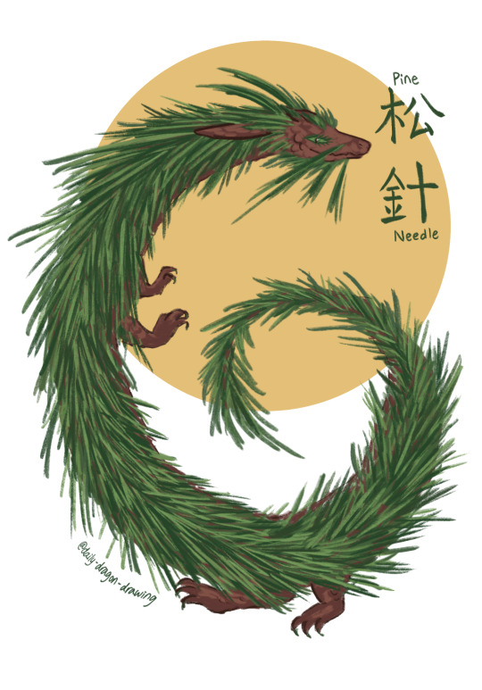
#91 - 松針 (sōngzhēn / pine needle) - Please refrain from putting ornaments on them...🌲🎄🪵
#daily dragon drawing#ask and you shall receive :3#art#art challenge#artists on tumblr#chinese artist#dragon#dragon a day#dragon art#dragon oc#dragons#daily drawing#daily dragon#chinese dragon#drawing challenge#drawing every day#drawing#illustration#year of the dragon#fantasy creature#creature design#zodiac#eastern dragon#dragon illustration#pine needles#pine dragon#pine needle dragon#pine trees#pine#plant dragon
1K notes
·
View notes
Text
Pine Needle Soda: Fermentation for Beginners
289 notes
·
View notes
Text
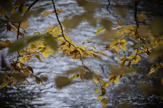

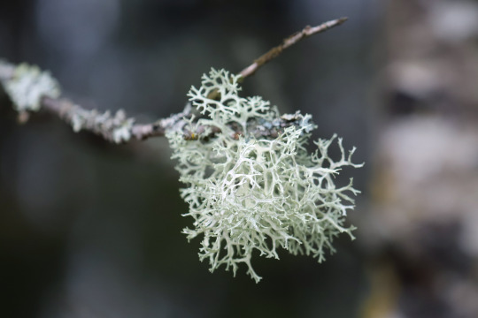
Autumnal Collections
(c) riverwindphotography, October 2023
359 notes
·
View notes
Text

by Elizabeth Johnson-Wold
122 notes
·
View notes
Photo

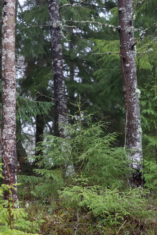


Värmland, Sweden (February 23, 2019).
450 notes
·
View notes
Text

Fiery Hot Cocoa (Vegan)
#vegan#drinks#hot cocoa#hot chocolate#cacao#plant milk#pine needles#rosemary#chocolate#vanilla#cornstarch#whipped cream#coconut sugar#non alcoholic rum
32 notes
·
View notes
Text

Scented Carpet
The scent of pine, a symphony sweet
Awakens our senses, sets our hearts to beat
With every breath, we’re filled with hope
Ready to conquer, to climb life's slope
Have a great day. 💋💋
#pine needles#pine tree forest#pine forest#pine trees#naturephotooftheday#naturephotography#nature#dress#purple dress#pretty smile#beautiful smile#smile
19 notes
·
View notes
Photo


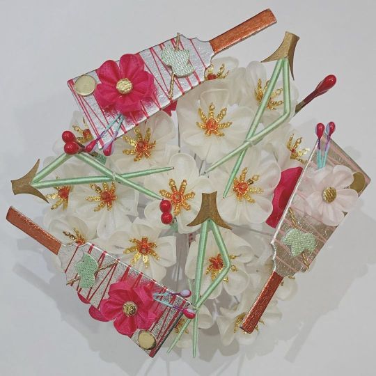


Spotlight: Alternative January Kanzashi
While the yearly kanzashi are always in the spotlight, it’s important not to forget the kanzashi that the maiko of Kamishichiken and Gion Higashi wear too. Often featuring the same design elements as the other kagai, they’re unique in that they don’t need to feature kangiku as a base. They still use the same motifs that the other kagai pull from, but they feature them in unique combinations. The kanzashi above feature the following motifs:
Kanzashi #1
Cranes (鶴), Pine (松), and Plum Blossoms (梅)
Kanzashi #2
Folding Fans (扇子) and Pine (松)
Kanzashi #3
Battledores (羽子板), Pine Needles (松葉), and Plum Blossoms (梅)
Kanzashi #4
Bobbins (糸巻), Plum Blossoms (梅), and Pine (松)
Kanzashi #5
Plum Blossoms (梅), Pine Needles (松葉), and Pine (松)
Kanzashi #6
Cranes (鶴) and Pine (松)
All images are courtesy of Gion Dteel Product [1] [2].
#maiko#geiko#geisha#kyoto#kanzashi#january#crane#pine#plum blossom#pine needles#bobbins#battledore#舞妓#芸妓#芸者#京都#かんざし#簪#一月
151 notes
·
View notes
Photo


Sleek modern goth styling of an antique kurotomesode by Dali (the hat is truly the star of this outfit ^^). The kimono has sasa (bamboo) with circles of seasonal flowers, paired with a lovely obi with matsuba (pine needles).
305 notes
·
View notes
Text

20 notes
·
View notes
Text
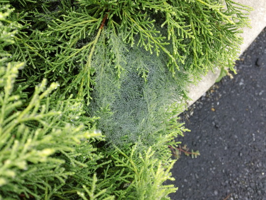
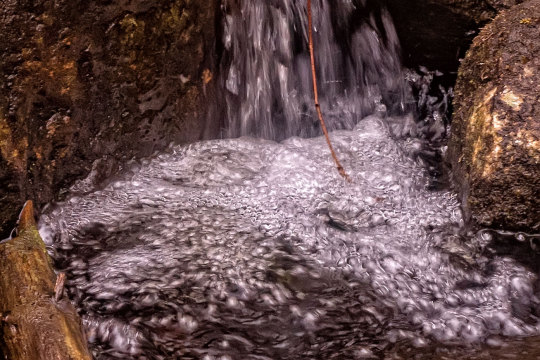
#Photography#Sept. 2018#Outdoors#Close-Up#Wet Spiderwebs#Wet Cobwebs#Raindrops#Pine Needles#Pine Bushes#Pine Branches#Curb#Gravel#Stones#Rocks#Nature#Pavement#Concrete#Spiderwebs#Cobwebs#Webs#Rain#Needles#Pine#Bushes#Branches#My Snaps#My Photos#My Photography
8 notes
·
View notes
Text

Molepelt
#Molepelt#Medicine Cat#Shadowclan#Cloudstar's Journey#Firestar's Quest#Yellowfang's Secret#Pine needles
14 notes
·
View notes
Text
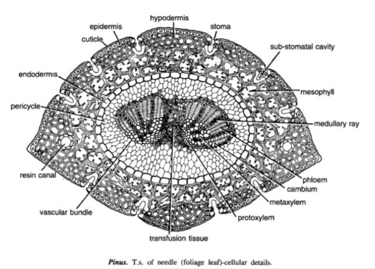
Mod 3: Gymnosperms
Pinus Needle T.S.
It is circular in outline in P. monophylla, semicircular in P. sylvestris and triangular in P. longifolia, P. roxburghii, etc.
Outermost layer is epidermis, which consists of thick-walled cells. It is covered by a very strong cuticle.
Many sunken stomata are present on the epidermis.
Each stomata opens internally into a substomatal cavity and externally into a respiratory cavity or vestibule.
Below the epidermis are present a few layers of thick-walled sclerenchymatous hypodermis. It is well developed at ridges
In between the hypodermis and endodermis is present the mesophyll tissue.
Cells of the mesophyll are polygonal and filled with chloroplasts. Many peg-like infoldings of cellulose also arise from the inner side of the wall of mesophyll cells.
Few resin canals are present in the mesophyll, adjoining the hypodermis. Their number is variable but generally they are two in number.
Endodermis is single-layered with barrel-shaped cells and clear casparian strips.
Pericycle is multilayered and consists of mainly parenchymatous cells and some sclerenchymatous cells forming T-shaped girder, which separates two vascular bundles. Transfusion tissue consists of tracheidial cells.
Two conjoint and collateral vascular bundles are present in the center. These are closed but cambium may also be present in the sections passing through the base of the needle.
Xylem lies towards the angular side and the phloem towards the convex side of the needle.
Idk where to find xeric nature info
Williamsoniaceae
Occurrence of Williamsonia
Williamsonia belongs to family Williamsoniaceae of Bennettiales.
It has been reported from Upper Triassic period but was more abundant in Jurassic.
This was earlier discovered under the name Zamia gigas by Willamson in 1870 but has now been named as Williamsonia.
Professor Birbal Sanhi (1932) described W. sewardiana from Rajmahal Hills of Bihar (India).
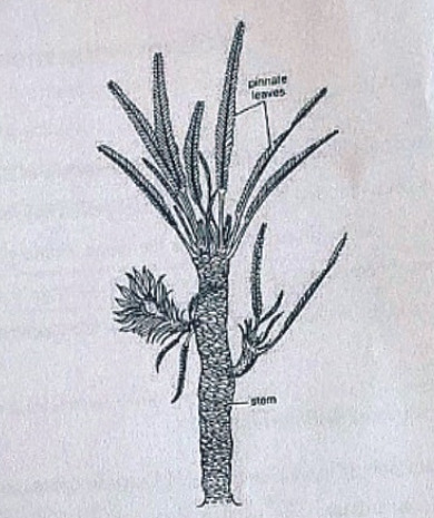
External Features of Williamsonia
Williamsonia resembled Cycas in appearance, but its best-knows species is W. sewardiana. The plant had an upright, branched, and stout stem covered by persistent leaf bases.
A terminal crown of pinnately compound leaves was present. For the stem genus Bucklandia, Sharma (1991) opined that features of leaf bases such as their shape, size and arrangement pattern are of taxonomic significance.
He observed that leaves in Williamsoniaceae show syndetocheilic stomata with rachis possessing collateral endarch vascular bundles.
Reproduction in Williamsonia
The fructifications of Williamsonia were large and attained a diameter of about 12 cm.
They were borne on a peduncle.
Many spirally arranged bracts were present around the base of the floral axis.
In W. gigas the cones were present among the crown of leaf bases while in W. sewardinia they were present on the short lateral branches.
Williamsonia plants were unisexual.
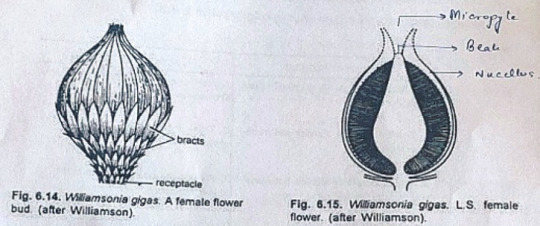
Female Flower
The female 'cones' of W. gigas and W. sewardiana have been investigated in detail. Instead of 'strobili' or 'cones', Sporne (1965) proposed to use the term 'flower'.
The conical receptacle was surrounded by many perianth-like bracts. The ovules were stalked.
The apex of the receptacle was naked and sterile. The nucellus was surrounded by a single vascularize integument, which was fused with the nucellus. The nucellus had a well-marked beak and a pollen chamber. In young ovules the micropylar canal was long and narrow.
In mature ovules, the canal widened because of the formation of nucellar plug and disappearance of interlocking cells. In the apical part of the endosperm, Sharma (1979) observed 2 or more archegonia.
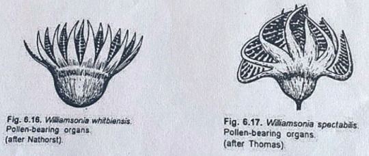
Male Flower
Male flowers consisted of a whorl of microsporophyll's which were united to form a more or less cuplike structure. In majority of the investigates species the sporophylls were un-branched but in some species they were also pinnately branched.
Sitholey and Bose discovered W. santalensis from Upper Gondwana, and observed that microsporophyll's in the species were bifid.
One of the branches of microsporophyll was fertile while the other was sterile. The fertile part has finger-like structures called synangia. Each synangium had two rows of chambers enclosing microsporangia.
The fertile branch of the bifid sporophyll possessed many purse-like capsules, in each of which there were present many monocolpate pollen grains.
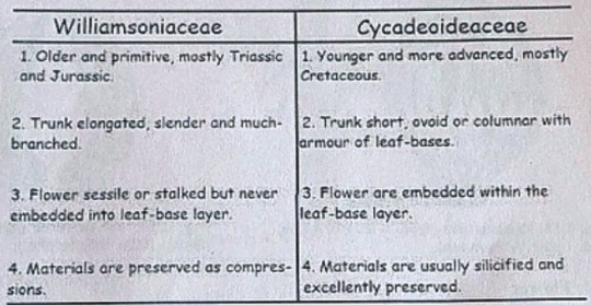
Cycadeoideaceae
Classification:
Division - Cycadeoidophyta
Order - Cycadeoideales
Family - Cycadeoideaceae
Genus - Cycadeoidea
Introduction:
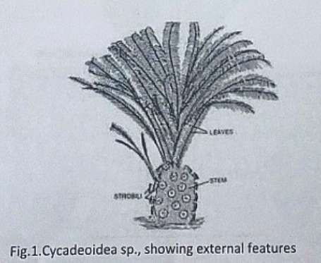
Cycadeoidea is the only genus of family Cycadeoidaceae, represented by thirty species. They are entirely extinct and resemble cycads in the outward stumpy appearance of trunk and an apical crown of pinnate compound leaves. This fossil group of plants flourished during the Triassic to Cretaceous periods of the Mesozoic era. They are reported from various places in the world, in India the Cycadeoidales are found in Rajmahal Hills in Bihar. The petrified trunks of C. entrusca are the oldest fossil ever collected by man.
External Features:
The genus Cycadeoidea had a short, branched, or unbranched spherical, conical, or irregular trunk. The diameter of the trunk is 50cm and the highest rarely reached a meter except in C. jenneyana, it attended the height of several meters. These trunks are covered by rhomboidal leaf bases having multicellular hairs in between. Crown of 10ft long pinnate compound leaves are present at the top.
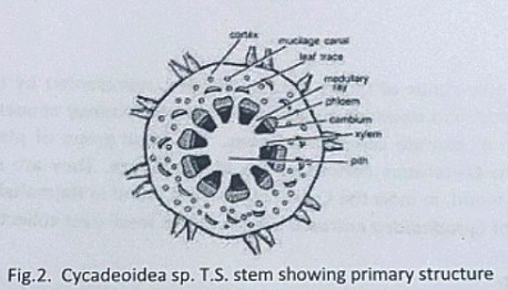
Anatomy of Stem:
The transverse section of the stem shows roughly a circular outline. The epidermis is not very distinct due to the presence of heavy armor of leaf bases. The cortex is parenchymatous and traversed by mucilage canals and numerous leaf traces. The primary vascular structure consists of a ring of endarch, collateral, conjoint, and open vascular bundles encircling the pith. Pith is wide and parenchymatous. A ray-like extension passes between the vascular bundles that make their appearance discrete.
There is a cambium ring with a thin zone of secondary wood. The secondary wood encircles the primary xylem and consists of tracheids with scalariform and bordered pits. The secondary medullary rays traverse the secondary xylem and secondary phloem.
The C-shaped leaf traces arise singly from the primary vascular strand and entering the cortex divided into several masarch strands and enters straight into the leaf.
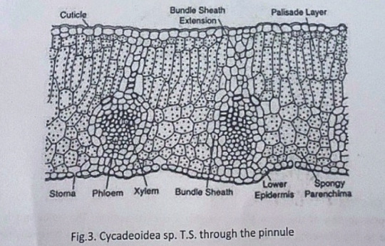
Anatomy of Leaf:
The pinnules show xerophilous structure. The upper and lower epidermis is heavily cutinized and thick walled. The mesophyll cells are distinguished into palisade and spongy parenchyma. The vascular bundles are mesarch and surrounded by bundle sheath.
Reproduction:
The reproductive structure is represented by flowers. In most of the species, the flowers are bisexual and arise in the axil of each leaf.
Structure of Flower:
The flowers are bisporangiate, stalked, and partially sunken in the leaf base armor. Rach such mature flower is 5-10cm in diameter and 10cm long. From the base of such flowers about 100 to 150 hairy bracts arise in close spiral little below the apex. These bracts formed a perianth like structure and protect the megasporangiate and microsporangiate parts of a flower. The microsporophyll or androecium forms a whorl united at the base into a sheath. The megasporophyll or gynaecium consists of numerous stalked ovules born around a central receptacle. Between the ovules, interseminal scales with expanded tips are present. These expanded tips fused to form a continuous surface with pores, through which the micropyle of ovules extended. The vascular supply of flowers consists of many branches from leaf traces.
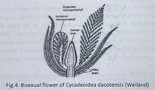
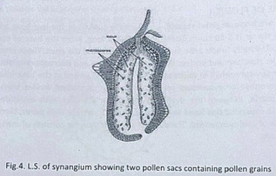
Microsporophyll or Androecium:
The microsporophyll is 10-12cm long, consists of a central rachis bearing numerous pinnae. The pinnae bear two rows of bean-shaped shortly stalked pollen capsules or synangia. These pollen capsules are born on the trabeculae within the fertile region of microsporophyll. A line of dehiscence is also visible at the base of each microsporophyll. This suggest that the entire microsporophyll might have been shed as a unit. The pollen capsule or synangia measures about 3.5x2.5mm and its wall is several layers thick, the outer layer made up of palisade like cells, and the inner layer is made up of thin-walled cells followed by a tapetum. The tapetum was not demarcated. A ring of microsporangia arranged around the periphery of each synangium. The microsporangia dehisce longitudinally and release the microspores into the synangial cavity. At maturity, the synangia liberate these microspores outside by an apical opening that splits into two valves. The liberated microspores or pollens are oval, measures up to 68µ that represents the male gametophytes. Pollen grains of Cycadeoidea are multicellular.
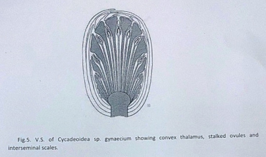
Megasporophyll or Gynoecium
The gynoecium consists of a spherical or conical receptacle that bears numerous stalked orthotropous ovules and interseminal scales. Each ovule is about 1mm long and consists of the single integument that fused with the nucellus except at its apex.
According to Lignier, in C. morieri, nucellus is free from the integument. Each ovule has a pollen chamber and a nucellar beak. This nucellar beak is the extension of the integument. The ovules also have long micropyle, extended from the flat surface of interseminal scales. The fused tips of interseminal scales form an external protective covering or pericarp surrounding the seeds.
Crepet and Delevoryas discovered many of bisporangiate cones from the Cretaceous of black hills. They studied the structure of these ovules in detail. These ovules are urn-shaped and resemble with the ovules of C. wellsii. According to them the micropyle of these ovules are funnel-shaped due to the constriction below the flaring. The inner wall of the micropyle is lined with large cells, considered to be epidermal cells. The integument has three distinct layers. The outer fleshy layer of radially elongated cells, the middle stony layer made up of thick-walled cells, and the inner layer is fleshy.
The young nucellus is made up of thin-walled cells. The cells at the micropylar end are much elongated (80µ long) in comparison to the cells of the chalazal end. The cell at the nucellar tip is pointed up tp whereas cells on either side are bend outward to give the nucellus a distinct shape.
Crepet and Delevoryas reported a linear tetrad or row of three cells in the center of the nucellus.
The seeds are somewhat elongated or oval and possessed two cotyledons.
#exam season#send help#biology#notes#science#botany#gymnosperms#pines#pine trees#pine needles#plants#plant science#plant biology#nature#long post
8 notes
·
View notes
Photo
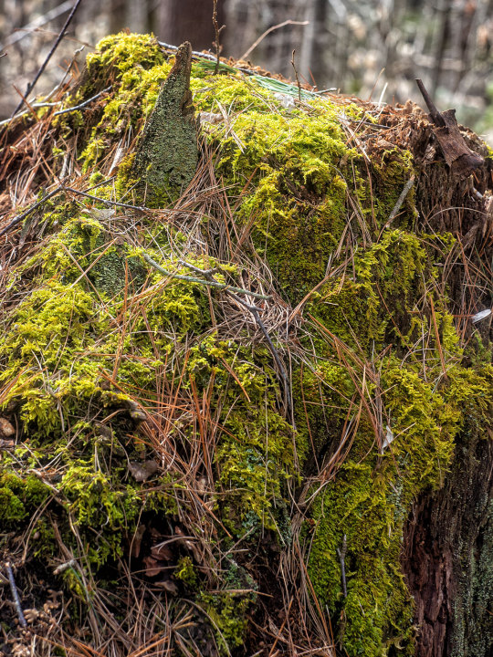

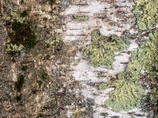
Forest Textures
Helios-44 58mm f/2.0
Sony A7
#forest textures#nature#original photography#photographers on tumblr#original photographers#forest#woods#stump#moss#twigs#pine needles#lichens#tree stump#old#weathered#verdent#regeneration#microcosom#recycling#hiking#hiking adventured#nature photography#close up#ecology#new england#vintage lens#vintage lens photography#helios#sony alpha#Guy Biechele
55 notes
·
View notes
Text
31 notes
·
View notes
Text
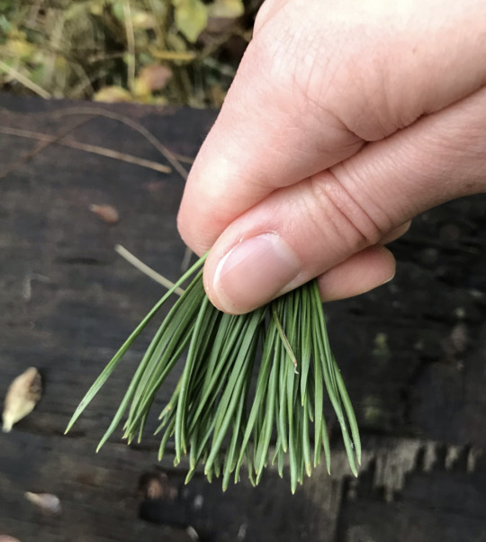


In the winter in southern England, evergreen trees like the Scots Pine (Pinus sylvestris) stand out. Ancient people saw the pine tree as a symbol of eternal life, which gave rise to our modern custom of Christmas trees.
The Scots Pine is the only native pine species in the British Isles. This species ranges from Western Europe to Eastern Siberia. The Scots Pine is only naturally found in Scotland in modern times, but in ancient times pine forests grew all over the British Isles. These forests died out due to climate change and human activity, but Scots Pine have been subsequently reintroduced as ornamentals and for forestry.
In these photos, we can see the two types of reproductive structures found on Scots Pine and other conifers. The classic pinecones are the female reproductive structures, known as ovulate cones, seed cones, or megastrobilus. These cones contain the ovules which will develop into seeds once fertilised by pollen. These cones open up their scales to allow wind blown pollen into the cones, after which the cones close up to allow the fertilised ovules to mature into seeds. After 6-8 months the cone opens again to let the winged seeds fly away on the wind to colonise new habitats. The second photo shows the male cones (microstrobilus or pollen cones) which contain pollen sacs under their scales. Scots Pine need to exchange pollen with another individual tree in order to reproduce.
Pine needles are also a delicious and healthy ingredient for herbal tea! Native Americans and East Asians have used pine needles from species in the Pinus genus for centuries as a healthy winter tonic. Pine needle tea is made by crushing and cutting the pine needles into small pieces and steeping them in boiling water. Medical research indicates pine needles are rich in antioxidants, vitamin A, and vitamin C. The taste is not like "pine scented air freshener" and more of a mellow citrusy flavour that combines exquisitely with cinnamon and gunpowder green tea. According to what I've read, all needles from Pinus genus species are safe for brewing unless you're pregnant.
#katia plant scientist#botany#plant biology#plants#plant science#pine trees#pine tree forest#tree#trees#scots pine#british nature#nature#foraging#herbal tea#pine needles#forestcore#naturecore#conifers#evergreens#english countryside#pine cones#branches#forest#woods#winter#wintercore#winter aesthetic
11 notes
·
View notes
Last Seen Blogs

padstowpm-blog
PPM

fallenparty
FallenParty

sp00kyjew
Life as a Pizza Bagel

skyclan-funny-name-squad
Firestar's Quest Is Good Actually

umarovaeva
⚜️