#conjugate beam method
Explore tagged Tumblr posts
Visit Tumblr Blog
Explore Tumblr blogs with no restrictions, modern design and the best experience.
Last Seen Tumblr Blogs


Fun Fact
In February 2021, Tumblr had 518.6 million blog accounts.
Text
@allvalley100
Prompt: Lost in Translation
Pairing: Samiguel
***
“I have something for you.” Sam smiles nervously at Miguel as she unfolds the paper. “I wanted to write you one of those romantic Spanish love poems, so. Here.”
She proceeds to recite one of the strangest mishmashes of false cognates, incorrectly-conjugated verbs, and word equivalents no sane person would use that Miguel has ever heard. Its wooing methods are nigh unfathomable.
Nonetheless, Miguel beams. Samantha LaRusso wrote him a poem—one she must have plugged through 15 translators to get halfway presentable.
“Sam, that’s amazing! You did this all yourself?”
“Thanks!” She grins. “Your sensei helped with the translation.”
#allvalley100#samiguel#samguel#smiggy#samdiaz#sam x miguel#miguel x sam#samantha larusso#sam larusso#miguel diaz#cobra kai#cobra kai season 3#cobra kai season 4#cobra kai season 5#tagging seasons 3 4 and 5 because this is when Sam would make the (poor) decision of asking Johnny for help with this <3#anyways I love to think that Miguel is usually the one who does the Grand Romantic Gestures#and Sam is kinda nervous to switch it up#but Miguel adores every word of her weirdly-translated poetry <3#absolute malewife
37 notes
·
View notes
Text
BTech Civil Engineering Tuition For Strength of Material
BTech Civil Engineering Tuition For Strength of Material
BTech Civil Engineering Tuition For Strength of Material
To develop knowledge of mechanics and to have in-depth understanding of material responses to load.
Simple stresses and strains : Definition, types of stresses and strains; Hooke’s law, Modulus of elasticity, various elastic constants and their relationship, stress strain curve for ductile materials, deformation of bars under axial loads,…
View On WordPress
#Advance Structural Design Tuition In Noida#Advanced Structural Analysis Tuition In Noida#Advanced Surveying Tuition In Noida#Analysis of strain#and Elementary concepts of theories of failure. Shear force and bending moment: Different types of beams and loads#Applied Chemistry Tuition In Noida#Applied Mathematics-II Tuition In Noida#Applied Mathematics-ITuition In Noida#Applied Physics-I Tuition In Noida#Applied Physics-II Tuition In Noida#bars of varying cross sections and composite sections#Betti’s law and Castigliano’s theorem and their applications. Torsion: Torsion of hollow and solid circular shafts#BTech Civil Engineering Tuition For Strength of Material To develop knowledge of mechanics and to have in-depth understanding of material r#Building Materials and Construction Tuition In Noida#calculation of bending stresses in beams for different loads and different types of structural sections. Shear stress and its distribution o#chimneys#Civil Engineering#comparison of solid and hollow shafts#Computer Science and IT Engineering. Strength Of Material Tuition In Noida#conjugate beam method#core of a section#crippling/buckling load#dams#deflection and radius of curvature#deflection and slope of statically determinate beams; moment area method#deformation of bars under axial loads#Design of Concrete Structure Tuition In Noida#Design of Steel Structure Tuition In Noida#double integration method#dummy load method
0 notes
Text
MATRIEX imaging: Simultaneously seeing neurons in action in multiple regions of the brain
https://sciencespies.com/physics/matriex-imaging-simultaneously-seeing-neurons-in-action-in-multiple-regions-of-the-brain/
MATRIEX imaging: Simultaneously seeing neurons in action in multiple regions of the brain
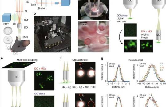
Design and implementation of MATRIEX imaging: (a) Experimental diagram of the MATRIEX imaging system. The two round 3D objects in the lower-left corner are the top and bottom views of the mouse head chamber used for in vivo imaging. (Ti:Sa): Ti:Sapphire ultrafast pulsing laser; PC: Pockels cell; BE: beam expander; SM1 and SM2: x–y scanning mirrors; SL: scan lens; TL: tube lens; DM: dichroic mirror; CL: collection lens; PMT: photomultiplier tube; DO: dry objective; MOs: miniaturized objectives. (b) Photograph showing an oblique overview of the actual MATRIEX imaging system. (c) The photograph in the upper image shows a zoomed in view of the three MOs attached to the manipulating bars over the head chamber; the lower photograph was taken directly above the MOs with a smartphone camera. All MOs used in this figure are of the same model: ���standard version.’ (d, e) Illustrations of the two-stage magnification and multiaxis coupling. The square images are actual two-photon images taken of 20-μm beads. Each red circle indicates one FOV. The model of DO used in panels (d-f) is the Olympus MPlan ×4/0.1, and all MOs in this figure are of the same customized model. (f) Illustration showing the absence of inter-FOV crosstalk under adjacent MOs. The images were taken on a uniform fluorescent plate. The red circles indicate the areas of analysis used to compare the image contrast between two conditions; the left-side condition shows the fluorescent plate under both MOs, and the right-side condition shows the fluorescence plate under only one MO. (g) Testing the optical resolution of the compound assembly with 0.51-μm beads. Curves: Gaussian fittings of raw data points. The on-axis or off-axis fluorescence intensity profiles were measured when the axis of the MO was aligned with the axis of the DO or apart from the axis of the DO (2 mm for the DO of ×4 or ×5, 3 mm for the DO of ×2.5, and 4 mm for the DO of ×2), respectively. Credit: Light: Science & Applications, doi: 10.1038/s41377-019-0219-x
Two-photon laser scanning microscopy imaging is commonly applied to study neuronal activity at cellular and subcellular resolutions in mammalian brains. Such studies are yet confined to a single functional region of the brain. In a recent report, Mengke Yang and colleagues at the Brain Research Instrument Innovation Center, Institute of Neuroscience, Center for Systems Neuroscience and Optical System Advanced Manufacturing Technology in China, Germany and the U.K. developed a new technique named the multiarea two-photon real-time in vitro explorer (MATRIEX). The method allowed the user to target multiple regions of the functional brain with a field of view (FOV) approximating 200 µm in diameter to perform two-photon Ca2+ imaging with single-cell resolution simultaneously across all regions.
Yang et al. conducted real-time functional imaging of single-neuron activities in the primary visual cortex, primary motor cortex and hippocampal CA1 region during anesthetized and awake states in mice. The MATRIEX technique can uniquely configure multiple microscopic FOVs using a single laser scanning device. As a result, the technique can be implemented as an add-on optical module within existing conventional single-beam-scanning, two-photon microscopes without additional modifications. The MATRIEX can be applied to explore multiarea neuronal activity in vivo for brain-wide neural circuit function with single-cell resolution.
Two-photon laser microscopy originated in the 1990s to become popular among neuroscientists interested in studying neural structures and functions in vivo. A major advantage of two-photon and three-photon imaging for living brains include the optical resolution achieved across densely labelled brain tissues that strongly scatter light, during which optically sectioned image pixels can be scanned and acquired with minimal crosstalk. However, the advantages also caused significant drawbacks to the method by preventing the simultaneous view of two objects within a specific distance. Researchers had previously implemented many strategies to extend the limits, but the methods were difficult to implement in neuroscience research labs. Nevertheless, an increasingly high demand exists in neuroscience to investigate brain-wide neuronal functions with single-cell resolution in vivo.

LEFT: Experimental diagram of the MATRIEX imaging system. The two round 3D objects in the lower-left corner are the top and bottom views of the mouse head chamber used for in vivo imaging. (Ti:Sa): Ti:Sapphire ultrafast pulsing laser; PC: Pockels cell; BE: beam expander; SM1 and SM2: x–y scanning mirrors; SL: scan lens; TL: tube lens; DM: dichroic mirror; CL: collection lens; PMT: photomultiplier tube; DO: dry objective; MOs: miniaturized objectives. RIGHT: Illustrations of the two-stage magnification and multiaxis coupling. The square images are actual two-photon images taken of 20-μm beads. Each red circle indicates one FOV. Credit: Light: Science & Applications, doi: 10.1038/s41377-019-0219-x
In a straightforward approach, scientists can place two microscopes above the same animal brain to image the cortex and cerebellum simultaneously. But such efforts can lead to substantial increases in complexity and cost. The existing high expectations for performance and feasibility therefore pose a highly challenging engineering question on how a single imaging system can simultaneously obtain live microscopic images from multiple brain regions in vivo. To address the question, Yang et al. introduced a new method that combined two-stage magnification and multi-axis optical coupling.
They realized the method using a low-magnification dry objective (DO), with multiple water-immersed, miniaturized objectives (MOs) under the dry objective. The scientists placed each of the MOs at the desired target position and depth in the brain tissue. The team used the new compound object assembly similarly to the original water-immersed microscope objective without additional modifications to the image scanning and acquisition subsystem.

TOP: Configuring the MOs with different parameters to target object planes at different depths to then be conjugated on the same image plane. Each gray cylinder represents one lens with a pitch value, front working distance (L1), back working distance (L2) and length (Z). BOTTOM: Demonstration of MATRIEX imaging: structural imaging in multiple brain areas in vivo. a Left image: a full-frame image including two FOVs in the frontal association cortex (FrA) and the cerebellum. The red and yellow circles indicate two FOVs that are digitally enlarged and shown in the upper-right and lower-right images. A GAD67-GFP transgenic mouse (with the interneurons labeled brain-wide) was used. Two MOs (‘standard version’) were placed at the same depth under a DO (Mitutoyo ×2/0.055). b Example configuration of three FOVs in the cortex of a Thy1-GFP transgenic mouse (with layer 5 cortical neurons specifically labeled and with tuft dendrites visible near the cortical surface). Three MOs (‘standard version’) were placed at the same depth under a DO (Olympus ×4/0.1). Credit: Light: Science & Applications, doi: 10.1038/s41377-019-0219-x
The research team first assembled the MATRIEX compound objective. For this, they replaced the conventional water-immersion microscope objective with a customized compound objective assembly, inside a two-photon laser scanning microscope equipped with a conventional single-beam raster scanning device. The compound assembly contained multiple MOs (miniaturized objectives) inserted through multiple craniotomies during which the scientists glued a 3-D printed plastic chamber to the skull of the mouse model. The chamber roughly aligned the MOs with the same space to adjust lateral position and depth. Yang et al. precisely manipulated the individual MOs to view the objects under all MOs simultaneously in the same image plane.
They implemented the MATRIEX method using two principles; two-stage magnification and multiaxis coupling. For example, using two-stage magnification with the dry objective (DO) alone, they observed 20 µm beads as tiny blurry dots while observing crisp, round circles through the compound assembly. During multiaxis coupling, the scientists coupled a single DO with multiple MOs on the same image plane. Using a simple raster scan in a single rectangular frame, the research team acquired a rectangular image containing multiple circular FOVs (Field of Views) – where each FOV corresponded to one MO with minimal inter-FOV pixel crosstalk.
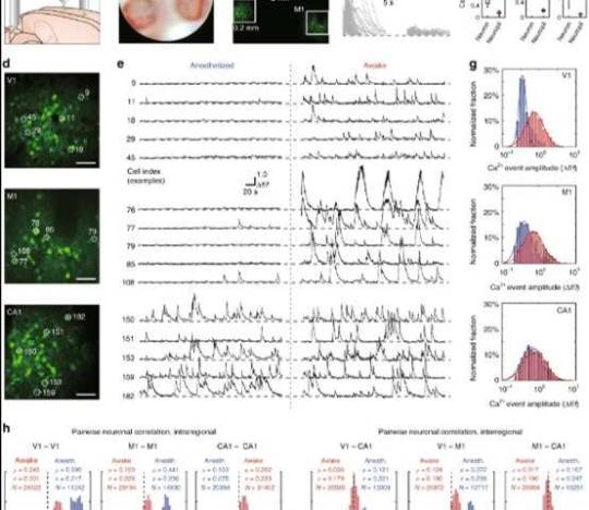
Demonstration of MATRIEX imaging: simultaneously acquiring live neuronal activity patterns in V1, M1, and hippocampal CA1 in mice in the anesthetized state or awake state. The neurons were labeled by a genetically encoded fluorescent Ca2+ indicator, GCaMP6f (a) Illustration showing the positioning of three MOs over the V1, M1 and hippocampal CA1 regions in a model mouse brain. (b) A camera photograph taken through the microscope ocular lens under white light bright-field illumination, in which three FOVs are readily visible. The upper region is V1, the lower-left region is CA1, and the lower-right region is M1. (c) A two-photon image, which is an average of 100 frames, acquired by simple full-frame raster scanning with a two-photon microscope. The solid white boxes show the three parts of the image that are enlarged in panel (d). (d) Digitally enlarged individual FOVs showing neurons in V1, M1, and CA1, from top to bottom. Scale bar: 40 μm. (e) Time-lapse Ca2+ signal traces of five example cells from each region, with each labeled by the cell index. Recordings of the same cell in the same animal in the anesthetized state (left side) and in the awake state (right side) are shown. (f) Left: traces showing individual Ca2+ signal events (split from each onset time and overlaid) from randomly selected example cells. Middle: Ca2+ signal traces of each of the neuropil zones that are directly adjacent to each of the example cells. Right: three box plots comparing the neuronal Ca2+ signal event amplitude to the neuron’s adjacent neuropil Ca2+ signal amplitude; paired Wilcoxon rank sum test, ***P < 0.001. (g) Log-normal fitting of the distribution histograms of the spontaneous Ca2+ event amplitude for data pooled from all animals. The red bars and fitted curve show the distribution of data recorded in the awake state, and the blue bars and fitted curve show the distribution of data recorded in the anesthetized state. (h) Pairwise neuronal activity correlation (Pearson correlation coefficients) for data pooled from all animals. The red bars show the distribution of data recorded in the awake state, and the blue bars show the distribution of data recorded in the anesthetized state. Credit: Light: Science & Applications, doi: 10.1038/s41377-019-0219-x
The scientists credited the magnification of the numerical aperture (NA) for allowing better resolution with the compound assembly. The associated lenses were also flexible and custom-designed for mass-production at low cost to assist experimental design. The main feature of MATRIEX was its capacity to image multiple objects simultaneously at large depth intervals. To highlight this, Yang et al. designed different MOs with diverse parameters, placing them at a specific depth where the corresponding object planes conjugated on the same axis. In practice, the research team compensated minor mismatches between the desired and actual object depth by adjusting MOs individually along each of the z axes.
Typically, under the DO (dry objective) the maximum lateral size of the target zone is limited by the maximum size of the scanning field. For example, using a DO with a 2x magnification and target zone of 12 mm in diameter, scientists can image an entire adult mouse brain. In this study, Yang et al. simultaneously imaged the frontal association cortex and cerebellum of the mouse. In practice, a 4x air objective was suited to achieve better resolution to observe fine dendrite structures.

Simultaneous calcium imaging in the V1, M1 and CA1 regions using MATRIEX during anesthetized and awake states in mice. View full movie on Credit: Light: Science & Applications, doi: 10.1038/s41377-019-0219-x
As proof of principle, the research team used MATRIEX to perform simultaneous two-photon Ca2+ imaging of fluorescently-labelled neurons in the primary visual cortex (V1 region), primary motor cortex (M1 region) and hippocampal CA1 region of mice. In the configuration of the three MOs, the scientists placed two MOs suited for the V1 and M1 region, directly above the cortex and inserted an MO within the hippocampal CA1 region after surgically removing a cortical tissue. The team then designed the lenses for the object planes corresponding to V1, M1 and CA1 for conjugation on the same image plane. Using a two-photon microscope equipped with a 12 kHz resonant scanner, the scientists scanned the full image to observe three FOVs and their single cells after enlarging the three different sections to resolve single neurons. Then they noted the laser power to be distributed among multiple FOVs.
While Yang et al. could have obtained these results using conventional single-FOV imaging within a single brain region, the MATRIEX technique provided them data beyond those offered with single-FOV imaging techniques. Taken together, these results allowed a highly inhomogeneous distribution and transformation of spontaneous activity patterns from the anesthetized state to the awake state in mice, spanning a brain-wide circuit level at single-cell resolution.
In this way, Menge Yang and co-workers developed the MATRIEX technique based on the principle of two-stage magnification and multiaxis optical coupling. They simultaneously conducted two-photon Ca2+ imaging in neuronal population activities at different depths in diverse regions (V1, M1 and CA1) in anesthetized and awake mice with single-cell resolution. Importantly, any conventional two-photon microscope can be transformed into a MATRIEX microscope, while preserving all original functionalities. The key to transformation is based on the design of a compound objective assembly. The researchers can use different, carefully designed MOs to suit diverse brain regions with 100 percent compatibility between the MATRIEX technique and conventional microscopy. The research team expect the MATRIEX technique to substantially advance three-dimensional, brain-wide neural circuit dynamics at single-cell resolution.
Explore further
Bringing faster 3-D imaging for biomedical researches
More information: Mengke Yang et al. MATRIEX imaging: multiarea two-photon real-time in vivo explorer, Light: Science & Applications (2019). DOI: 10.1038/s41377-019-0219-x
Tianyu Wang et al. Three-photon imaging of mouse brain structure and function through the intact skull, Nature Methods (2018). DOI: 10.1038/s41592-018-0115-y
Rongwen Lu et al. Video-rate volumetric functional imaging of the brain at synaptic resolution, Nature Neuroscience (2017). DOI: 10.1038/nn.4516
© 2019 Science X Network
Citation: MATRIEX imaging: Simultaneously seeing neurons in action in multiple regions of the brain (2019, December 24) retrieved 24 December 2019 from https://phys.org/news/2019-12-matriex-imaging-simultaneously-neurons-action.html
This document is subject to copyright. Apart from any fair dealing for the purpose of private study or research, no part may be reproduced without the written permission. The content is provided for information purposes only.
#Physics
2 notes
·
View notes
Text
Journal 102: my goodbye epic the musical
youtube
am done with golem and rushushas. Things are back to normal. And I am very traumatized. Were good!
"Jake then realized once again via his EDNOS and disordered eating he could not eat his plate. Today's lunch was fruitcake and chicken macaroni and jake cousnt take a bite. They also charmed it at the house and jake coudnt bear it. The smell made him sick. Delicoat can also lead to eating disorders. Not many people know that's just a thing with antiphycotics in general more sever depending on the kind. Depression meds = weight problems under or mostly over. Mood staplers = body image problems, numb, hyper blank sex drive or empathy and dreams, and antiphyoctics extra psychosis or depression or disordered eating or vomiting. Jake often had been on everything and he joked "this is what it cures, this is what it causes" it was an old joke JD haileys father told him as a little girl when he first vented. Ad jake prepared the first reality shift in a long time. He forgot how much of his adventures really just seemed in his head.
Jake was in Jesus guise told them his name was jake caloway. They tird him up in a bug like cavern and quickly disposed of him. They pushed him down. And tied him up. And after he questioned as a philosophy why not to leave the cavern showing his old philosophers cave. And so back in the day philosophy was the entire world and the expirence of amgick secrets. Why this is shown to some. Regardless it was jakes memory. And so Jake was pushed down on his back and they tied him up and beamed the sunlight upon him killing him. They noted he prayed for his other self and apologized for tricking him. As he became a human sacrifice. And so noted Jake heard "and this is why your afraid of wings here" by small petite voice of a woman. And jake remembered."
I've been learning the easy Of faries as Acrians is sharing all the rules of the courts previously forbiden as new ages are ushered in. And I've gotten back into reality shifting. Diongoespews video on the spastic method and Adam snowflakes book dreamers book 3 detail guides on it and I've done it once more. I've come to the rationale and reason that reality shifting is am entire setting. I've become an animyst and am learning the secrets of the stars. What species are. How they came here. What is is to do shape magick and dream. That hunters are real!!! What they do as I time travel as adamith froster and become friends with Crowley. Become Jaden angel and friends with Shaun whinchester. As I learn that every soul has a character and every character belongs to a force or player. That an actor or writer taps into to dream of a presentation thst becomes real for someone else. I have learned that everything was once divine. Magick. Door knobs. Wood. And these ideas and concepts seem laughable. But I've been time traveling making my mark and impact as I have now learned is all connected! And my skisoprehnic episodes were real conjugations and gates.
And that the world is built on layers. And societies to keep them safe. You may not like the illuminati or aliens or scientology. The mormo. Church might have fucked up rituals and the moonies to sexual in nature. But I was a family member once. I time traveled to do it all. Marylim Manson an alien in a jersey a Brad who wore in war as a general and in school as a fairy of two Petronas split down the middle.
My face was burned recently. And I'm raising a dragon as my own. I've learned regarding the multiverse there are people and planets and things. Every website was once a spirit. Every fashion belongs to a race. A race of hidden magick on earth. Thst now crosses the stars as a gift to then. So who cares what asgardians are
In some realities they never met a boat. I'm not in those realities either. You know thorin loves those movies comics mixed bag but he likes those movies. Cus he gets to dream he's Thor.
Whatever I have taught you insane or not has been to help the multiverse. To shift those proper realities. To save all of existance and when I'm done cus your reading in the moment for you to explore it. And expirerence it. The multiverse has aliens and elves and black white realities with advanced gaming consoles but for some reason improper computers.
I've married the gods of technology thievery and television. Techno loki and hermes and Teli. Old of tools and plays and advancement with Bill cipher as my guide in my wheelchair as olive. I really am in a wheelchair people don't seem to beleive I really fell from three stories like master. For some reason all his students call him that.
I learned that faking a persona can reincarnate into a new soul even if it's a lamp or a character or a dream. And that that is what a shape is. That is what becomes an animal. And that is what builds Egypt. And that's all because I learned reality shifting I learned pop culture magick I took chances and things seemed RIDICULIUS one of my characters. And I got rewarded for it. This is magick. Whether you like it or not
The silly things are magick. Cus this is how traditions come to play. And if your nodding your head shaking reading Farley curely into every word I say for their are to am y nays in the word of Smith. Get it naysayers and tool guys? Understand to fully explore the multiverse you read this diary.
And this diary teaches you kabahlla rushsusa, time travel, multiverse, and shape magick and if you don't like thst. That the fantasy novel ypu read and enjoy could be real? Your not ready foe magick. Your not ready for power. Which is not what magick should ever be about. Taught to me early by the master. Not from doctor who my sage acrians locket who I see myself in person towards. I'm sorry if your nitpicky reality and snobby ways blind you to the old eras but as someone who's lived them this is how we did it.
We bring stories to life. Pop culture magick. We treat new forms of magick to be ours and irished up or oldened or scotified so it's now vikings like Jesus joining thr norse gods or adapting the Jewish as our own. I've been traveling sometimes in a boat. Sometimes in a warp. And you may not know it yet but we could have been great friends before you secured at me.
Theirs no such thing as a stupid idea. Meet you new instructor olive brimstone ambaseder of earth Rushusha. And if you don't like the fantasy how dare you demand it or call yourself a witch? Time to open your eyes and stop being bigoted. Your never going to recognize me and no emo beanie can change that. It covers a tatoo dipshit. I know who I'm attesting. My stupid fingerless gloves.
Anyways! As someone who changes face but yet does not know how. I still have much to learn. But learning taboo and reacting it and not fading from it. Learning from the goetia and noting that just cus my mentor has experience doesn't meant I do is what got me far. Not binding someone taught me the stars. We don't teach people to whip the cooks who cook them food. If that food is knowledge and you use a circle or chant for it, that distress is deserved how dare you call yourself his student. Your trying to be his slave master and you dare demand a reward?????? Anyways. Enough @ing.
I got interdimemsional travel. I have to time travel and I finally keep doing. I'm doing to ritual to manifest my dream body of my right gender and I'm exploring through spirit work my true self and person. What the gods are. Where they live. Joining there families and basterdizing and crafting for my friends as Kalin high. For my friends at Jethro or Bridgeton and Malock academy. I've learned I'm an imoetal teenager and with xenith and dragon forms in place I've learned I'm a kins. You don't know what that means but I know what a glomp is. And if your laughing the special list of kanaui mocks you back. Kinzo mocks you back. You vest mocks you back.
Objects having souls is animism and I really hate that tumblr blog thisnisnt you don't recognize me and I can't ebelive we were ever friends I'm so glad your starting to forget I won't be there to help you thus time i time I refuse stop praying to me how you treated me blind matters.
- an bitter Olive Brismtone. A magickal creature angry at the world. Dot dot dot fuck you.
11:20
Wednesday February 8th
Closing song:
143 and when I'm gone after the clam
https://youtu.be/y-s9mdIKM84
https://youtu.be/tKGwpQiPFEM
1 note
·
View note
Text
Making scientific images : Super-resolution microscopy (2)
In my previous post, I talked briefly about resolution and why it’s important in microscopy. When you want to take pictures of stuff that you can’t see with your eyes, you want it to have the best resolution possible. Light microscopy is limited by the diffraction of light, which usually means that at best, we can have a resolution of 250 nm. That means that two points can be seen as seperate entities if they are 250 nanometers apart from each other, or 0,000000250 meters. It seems like a lot, but sometimes we need a better resolution. Electron microscopy can achieve this, but at the cost of many advantages that light microscopy can bring. Mainly the ability to stain proteins and organelles very easily. How does that work ?
We use antibodies. Normally, antibodies are part of the immune system. They recognize what we call antigens, that can be harmful to the organism. Scientists can thus produce antibodies that will recognize and attach to their protein of interest. These antibodies will in turn be recognized by other antibodies that are coupled to a fluorescent molecule. Meaning that it will emit a colored light when excited by a laser.
What does it mean ? We can stain for specific proteins, and they will emit a fluorescent light that we can detect using a light microscope. So we can pinpoint where they are inside cells. These signals, as mentioned before, are limited by the diffraction of light. To enhance the resolution, people had to get crafty. And on October 8, 2014, the Nobel Prize in Chemistry was awarded to Eric Betzig, W.E. Moerner and Stefan Hell for "the development of super-resolved fluorescence microscopy”. They invented a way to bypass the limitations of light microscopy and bring it to the nanodimension.
Since then, several ways to achieve super-resolved light microscopy were developed, I’ll mention a few of them and how they work.
- Structured illumination microscopy (SIM) : Different pictures are taken of different phases of the sample and then assembled by a computer, enhancing the resolution.

Dronpa–Lifeact in a mammalian CHO cell imaged with NL-SIM. (A) On the left, a portion of the entire image is shown with conventional microscopy. The image is displayed using a nonlinear intensity scale (gamma = 0.65) to highlight the small filaments in the background over the thick and bright stress fibers. A subset of the data is enlarged and shown with (B) conventional TIRF microscopy, (C) linear SIM-TIRF, and (D) nonlinear SIM-TIRF. Lifeact marks the actin network, the structure of which is most clearly resolved with nonlinear SIM-TIRF.

Live 3D SIM imaging of mitochondria labeled with MitoTracker Green and the actin cytoskeleton labeled with tdTomato-LifeAct in a HeLa cell over 30 time points. Imaged with conventional microscopy (A) and 3D SIM (B).

In these images of bovine pulmonary artery endothelial cells, the mitochondria are stained with MitoTracker Red CMXRos (red), and F-actin is stained with BODIPY FL phallacidin (green). On the left is a standard wide-field fluorescence image that includes signal fluorescence from above and below focus. On the right is the same image following deconvolution using the Zeiss Apotome Structured Illumination System. (Jon Ekman - ITG, Beckman Institute)

Mitosis, the cellular division that produces two genetically identical daughter cells, is perhaps the most fundamental process in biology. Without it, multicellular life wouldn’t exist, a broken bone would never heal, tissues would disintegrate. Cancer — essentially mitosis gone rogue — also wouldn’t exist. Scientists have been watching mitosis through a microscope for about 150 years, but new views are fleshing out the less-detailed pictures of the past (bottom, right). After DNA replicates, the nuclear envelope surrounding it dissolves. Spindle fibers (gold above, red at right) align pairs of chromosomes (blue) and then separate the genetic material into two daughter cells (shown forming, above).
- STORM, PALM and FPALM : Stochastic optical reconstruction microscopy (STORM), photo activated localization microscopy (PALM) and fluorescence photo-activation localization microscopy (FPALM) are super-resolution imaging techniques that utilize sequential activation and time-resolved localization of photoswitchable fluorophores to create high resolution images. In short, fluorescent molecules are switched on at random intervals, and the signals form the final image.
youtube

Nuclear pore complexes, PALM superresolution microscopy : Xenopus laevis A6 cells (epithelial kidney cells). gp120, a nuclear pore complex protein arranged in an eightfold symmetry (labeled with Alexa 647 conjugated antibodies). Courtesy of A. Löschberger, M. Sauer, University of Würzburg, Germany.

Dual color PALM microscopy imaging of a kinase (red) and an adapter protein (green). Left: orinary resolution image, right PALM image with zoomed region of interest on the bottom.

Samples were prepared using a setup for automated immunostaining. (A) One color STORM image of mitochondria (Tom20). (B) Two-color STORM image of mitochondria (Tom20) and microtubules (alpha-tubulin). In both panels, the zoomed region shows the same region imaged using conventional epifluorescence microscopy (left) and STORM (right). Scale bars, 5 µm in top images, and 1 µm in bottom images (zoomed regions).

The tiny diameter and high density of actin filaments are visible in the sheetlike protrusions at this monkey kidney cell’s edge (color-coded by depth, red farthest away). Scientists are still trying to figure out precisely how these sheets form and connect to the cell’s interior so they can understand more about how cells travel. Credit: K. Xu, H.P. Babcock and X. Zhuang/Nature Methods 2012

Hippocampal neurons labeled for actin (orange) imaged by STORM and synapsin (blue) imaged by TIRF. (NeuroCyto)
Check out NeuroCyto Lab’s STORM gallery for more !
- STED microscopy : Stimulated emission depletion (STED) uses two lasers pulses, the excitation pulse for excitation of the fluorophores to their fluorescent state and the STED pulse for the de-excitation of fluorophores by means of stimulated emission. This allows to “bleach” around the center of the STED beam :


Confocal (left) and super resolution (right) microscopy image of tubulin stained with LIVE 580 tubulin (cabazitaxel) in living human fibroblasts.

STED image of triple immunostaining in HeLa cells: Green: NUP153-Alexa 532, red: Clathrin-TMR, white: Actin-Alexa 488.
You can check Leica’s and Zeiss’ galeries for more pictures. I’ll see you later on the other side of the lens.
#microscopy#making scientific images#light microscopy#biology#cells#science#super resolution#super-resolution microscopy#STED#STORM#PALM#TIRF#SIM#fluorescence#blog post
52 notes
·
View notes
Photo

Spray-on electric rainbows: Making safer electrochromic inks
An innovation with water allows electrochromic conjugated polymer films to be applied more safely
Anyone who has a rear-view mirror that automatically dims blue in reaction to annoying high-beam headlights glaring from behind has seen an electrochromic film in action.
Now, chemists at the Georgia Institute of Technology have developed a new method to more safely and, by extension, easily produce these shear films, which change their color with the help of a tiny electric current. This could make them available to many industries that have not been able to feasibly use them before.
In manufacturing, electrochromic films are often coated onto other materials, such as the surface of a mirror, as inks. They are usually based in solvents that are flammable and have toxic fumes, making them unsuitable for many work settings that rely on printing and spraying machinery to apply colors.
Georgia Tech researchers have developed electrochromic film inks that are water-based, making them safer for diffuse application in settings where the kinds of safety precautions and protective equipment that are standard in handling volatile organic chemicals would be impractical.
Read more.
15 notes
·
View notes
Text
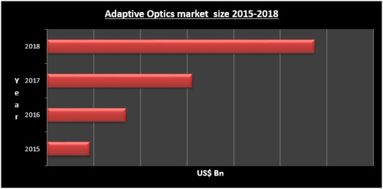
Global Adaptive Optics Market
Global Adaptive Optics Market is estimated to surpass US $XX Mn mark in 2016 and reach US$ XX Mn by 2024, at a CAGR of XX% during the forecast period 2017-2024 globally. Adaptive optics is having a major impact on rising industries and household sector alike consequently changing the face of the global economy in the upcoming future. Accordingly, XX million units adaptive optics is to be produced in the market by 2024, component efficiency and growing demand being the major driving force. An efficient adaptive optics market to manage such high device volumes, diversity and geographies is the need of the time.
Adaptive Optics MarketAdaptive Optics Market, the component segment is classified into the Deformable mirror, Controller, Wavefront sensor. The Wavefront sensor will hold the largest market i.e. around 50% in the forecast period. As these sensors are most preferable in the application such as laser application. Laser beam diagnostic and laser material come under Wavefront sensor which processes the laser beam to control shape and size for accuracy. Simultaneously other component segments are expected to lose their shares in the forecast period.
In terms of technologies, the Adaptive Optics Market can be split into Wavefront Modulator, Wavefront Sensor, and Control System. These technologies update to share the growth in the forecasting period.
The application of Adaptive Optics in the field such as Manufacturing, Biomedical, Defense and Security, Consumer Devices, Astronomy, and Communication. Among these Defense and Security sectors is estimated to have the highest share in the forecasting period. And further followed by the biomedical industry. Mainly defense weapons and highly sophisticated guidance systems use this market for improving method of the effectiveness of direct energy weapons.
The types segment of adaptive optics market can be divided into next-generation adaptive optics (NGAO), ground layer adaptive optics (GLAO), multi-conjugate adaptive optics (MCAO) and multi-object adaptive optics (MOAO). Among this, multi-conjugate adaptive optics (MCAO) has a high share in the forecasting period.
The market on the basis of geography is segmented by North America, Asia-Pacific, Europe, Middle East & Africa, and Latin America. The Asia Pacific is going to emerge as one of the highest growth regions in the forecast period followed by Europe. Increasing manufacturing and sales of the component and technology in the country such as China, India, Japan, and South Korea will fuel the Adaptive Optics Market.
Maximize Market Research has comprehensively analyzed Adaptive Optics Market emphasizing on each and every segment keeping regional dynamics in perspective. The driving forces, as well as considerable restraints, have been explained in depth to attain a balanced scenario in the market. The report classifies Adaptive Optics Market into various segments such as Component, Technology, Types, Applications and Regions providing a thorough understanding of the Adaptive Optics. Importantly, the report delivers forecasts of the market, giving an insight into the future opportunities that exist in the Adaptive Optics Market.
For More Information Visit:
https://www.maximizemarketresearch.com/market-report/adaptive-optics-market/11444/
0 notes
Text
To Chile and beyond

Bachelor of Science (Physics) student Grace Lawrence is a rising star in the Swinburne Astronomy scene. We were lucky enough to get the scoop on her latest trip to Chile at the world class Gemini South Telescope facility, set in the foothills of the Andes.
Grace’s experience
Over the summer, I had the honour of being awarded an Australian Gemini Undergraduate Summer Studentship (AGUSS). This meant I had the opportunity to travel to La Serena, Chile, for ten weeks and undertake a body of research work at the Gemini South Telescope facility.
During my time in Chile, I delved into the world of crowded-field photometry, a method of analysing the intensity of light from astronomical objects. Supervised by tenure-track astronomer, Morten Andersen, I was lucky to work with Gemini data taken using their Multi-Conjugative Adaptive Optics system. This uses deformable mirrors to correct for wavefront aberrations, producing some of the highest resolution data in the world. With it, I probed the low-mass stellar population of a young starburst cluster, Westerlund 1, which will help astronomers to learn how stars form in different environments.

As an AGUSS scholar, I attended multiple science conferences including Accretion Processes in Symbiotic Stars and Related Objects and the XIV Annual Meeting of the Chilean Astronomical Society (SOCHIAS) in Chile’s capital, Santiago. Four jam-packed days of dark matter, galaxy structure, exo-planets, black holes, supernovae, instrumentation and more!
Observing on the 8.1 metre Gemini telescope atop the summit of Cerro Pachon (an Andes mountain), was an absolute highlight of the trip - the Gemini laser system shoots a five-beam sodium laser 90 kilometres into the sky to create laser guide stars, all set against the breath-taking background of southern hemisphere stars.

In addition to the wonderful learning experiences of an internship at Gemini South, La Serena provided a vibrant cultural backdrop. Polishing my Spanish skills and learning about Chilean music, language and food, I was lucky to be able to travel some of the Chilean coastline; visiting the artistic city of Valparaiso, as well as spending the new year in the Atacama Desert (bordering with Bolivia and Peru). Sand dunes, desert foxes, hot geysers and Martian landscapes were an exciting way to welcome 2017.

After being encouraged to apply by Swinburne’s Professor Karl Glazebrook, it was the support and enthusiasm of Professor Jeremy Mould and Associate Professor Alan Duffy that inspired my application to the Gemini program. It provides students with a once-in-a-lifetime opportunity to work on an incredible research project and utilise the first-class, world-leading telescopes available in Northern Chile. I was able to experience real academic research, hone both my science and computer skills, as well fully immerse myself into the Chilean culture in the idyllic city of La Serena. I forged many new friendships and professional connections and am incredibly grateful to both Swinburne and the Australian Astronomical Observatory for this opportunity.
Written by Grace Lawrence, Bachelor of Science (Physics).
1 note
·
View note
Text
300+ TOP STRUCTURAL ANALYSIS Objective Questions and Answers
STRUCTURAL ANALYSIS Multiple Choice Questions :-
1. The number of independent equations to be satisfied for static equilibrium of a plane structure is a) 1 b) 2 c) 3 d) 6 Ans: c 2. If there are m unknown member forces, r unknown reaction components and j number of joints, then the degree of static indeterminacy of a pin-jointed plane frame is given by a) m + r + 2j b) m - r + 2j c) m + r - 2j d) m + r - 3j Ans: c 3. Number of unknown internal forces in each member of a rigid jointed plane frame is a) 1 b) 2 c) 3 d) 6 Ans: c 4. Degree of static indeterminacy of a rigid-jointed plane frame having 15 members, 3 reaction components and 14 joints is a) 2 b) 3 c) 6 d) 8 Ans: c 5. Degree of kinematic indeterminacy of a pin-jointed plane frame is given by a) 2j - r b) j - 2r c) 3j - r d) 2j + r Ans: a 6. Independent displacement components at each joint of a rigid-jointed plane frame are a) three linear movements b) two linear movements and one rotation c) one linear movement and two rotations d) three rotations Ans: b 7. If in a pin-jointed plane frame (m + r) > 2j, then the frame is a) stable and statically determinate b) stable and statically indeterminate c) unstable d) none of the above where m is number of members, r is reaction components and j is number of joints Ans: b 8. A pin-jointed plane frame is unstable if a) (m + r)2j d) none of the above where m is number of members, r is reaction components and j is number of joints Ans: a 9. A rigid-jointed plane frame is stable and statically determinate if a) (m + r) = 2j b) (m + r) = 3j c) (3m + r) = 3j d) (m + 3r) = 3j where m is number of members, r is reaction components and j is number of joints Ans: c 10. The number of independent equations to be satisfied for static equilibrium in a space structure is a) 2 b) 3 c) 4 d) 6 Ans: d 11. The degree of static indeterminacy of a pin-jointed space frame is given by a) m + r - 2j b) m + r - 3j c) 3m + r - 3j d) m + r + 3j where m is number of unknown member forces, r is unknown reaction components and j is number of joints Ans: b 12. The degree of static indeterminacy of a rigid-jointed space frame is a) m + r - 2j b) m + r - 3j c) 3m + r - 3j d) 6m + r - 6j where m, r and j have their usual meanings Ans: d 13. The degree of kinematic indeterminacy of a pin-jointed space frame is a) 2j-r b) 3j-r c) j-2r d) j-3r where j is number of joints and r is reaction components Ans: b 14. The number of independent displacement components at each joint of a rigid-jointed space frame is a) 1 b) 2 c) 3 d) 6 Ans: d 15. If in a rigid-jointed space frame, (6m + r) < 6j, then the frame is a) unstable b) stable and statically determinate c) stable and statically indeterminate d) none of the above Ans: a 16. The principle of virtual work can be applied to elastic system by considering the virtual work of a) internal forces only b) external forces only c) internal as well as external forces d) none of the above Ans: c 17. Castigliano's first theorem is applicable a) for statically determinate structures only b) when the system behaves elastically c) only when principle of superposition is valid d) none of the above Ans: c 18. Principle of superposition is applicable when a) deflections are linear functions of applied forces b) material obeys Hooke's law c) the action of applied forces will be affected by small deformations of the structure d) none of the above Ans: a 19. In moment distribution method, the sum of distribution factors of all the members meeting at any joint is always a) zero b) less than 1 c) 1 d) greater than 1 Ans: c 20. The carryover factor in a prismatic member whose far end is fixed is a) 0 b) 1/2 c) 3/4 d) 1 Ans: b 21. In column analogy method, the area of an analogous column for a fixed beam of span L and flexural rigidity El is taken as a) L/EI b) L/2EI c) L/3EI d) L/4EI Ans: a 22. The degree of static indeterminacy up to which column analogy method can be used is a) 2 b) 3 c) 4 d) unrestricted Ans: b 23. The deflection at any point of a perfect frame can be obtained by applying a unit load at the joint in a) vertical direction b) horizontal direction c) inclined direction d) the direction in which the deflection is required Ans: d 24. In the slope deflection equations, the deformations are considered to be caused by i) bending moment ii) shear force iii) axial force The correct answer is a) only (i) b) (i)and(ii) c) (ii) and (iii) d) (i), (ii) and (iii) Ans: a 25. The three moments equation is applicable only when a) the beam is prismatic b) there is no settlement of supports c) there is no discontinuity such as hinges within the span d) the spans are equal Ans: c 26. While using three moments equation, a fixed end of a continuous beam is replaced by an additional span of a) zero length b) infinite length c) zero moment of inertia d) none of the above Ans: a 27. The Castigliano's second theorem can be used to compute deflections a) in statically determinate structures only b) for any type of structure c) at the point under the load only d) for beams and frames only Ans: b 28. Bending moment at any section in a conjugate beam gives in the actual beam a) slope b) curvature c) deflection d) bending moment Ans: c 29. For a two-hinged arch, if one of the supports settles down vertically, then the horizontal thrust a) is increased b) is decreased c) remains unchanged d) becomes zero Ans: c 30. For a symmetrical two hinged parabolic arch, if one of the supports settles horizontally, then the horizontal thrust a) is increased b) is decreased c) remains unchanged d) becomes zero Ans: b Structural Analysis Interview Questions 31. A single rolling load of 8 kN rolls along a girder of 15 m span. The absolute maximum bending moment will be a) 8 kN.m b) 15 kN.m c) 30 kN.m d) 60 kN.m Ans: c 32. The maximum bending moment due to a train of wheel loads on a simply supported girder a) always occurs at centre of span b) always occurs under a wheel load c) never occurs under a wheel load d) none of the above Ans: b 33. When a uniformly distributed load, longer than the span of the girder, moves from left to right, then the maximum bending moment at mid section of span occurs when the uniformly distributed load occupies a) less than the left half span b) whole of left half span c) more than the left half span d) whole span Ans: d 34. When a uniformly distributed load, shorter than the span of the girder, moves from left to right, then the conditions for maximum bending moment at a section is that a) the head of the load reaches the section b) the tail of the load reaches the section c) the load position should be such that the section divides it equally on both sides d) the load position should be such that the section divides the load in the same ratio as it divides the span Ans: d 35. When a series of wheel loads crosses a simply supported girder, the maximum bending moment under any given wheel load occurs when a) the centre of gravity of the load system is midway between the centre of span and wheel load under consi-deration b) the centre of span is midway between the centre of gravity of the load system and the wheel load under consideration c) the wheel load under consideration is midway between the centre of span and the centre of gravity of the load system d) none of the above Ans: b 36. Which of the following is not the displacement method ? a) Equilibrium method b) Column analogy method c) Moment distribution method d) Kani's method Ans: b 37. Study the following statements. i) The displacement method is more useful when degree of kinematic indeterminacy is greater than the degree of static indeterminacy. ii) The displacement method is more useful when degree of kinematic indeterminacy is less than the degree of static indeterminacy. iii) The force method is more useful when degree of static indeterminacy is greater than the degree of kinematic indeterminacy. iv) The force method is more useful when degree of static indeterminacy is less than the degree of kinematic indeterminacy. The correct answer is a) (i) and (iii) b) (ii) and (iii) c) (i) and (iv) d) (ii) and (iv) Ans: d 38. Select the correct statement a) Flexibility matrix is a square symmetrical matrix b) Stiffness matrix is a square symmetrical matrix c) both (a) and (b) d) none of the above Ans: c 39. To generate the j th column of the flexibility matrix a) a unit force is applied at coordinate j and the displacements are calculated at all coordinates b) a unit displacement is applied at co-ordinate j and the forces are calculated at all coordinates c) a unit force is applied at coordinate j and the forces are calculated at all coordinates d) a unit displacement is applied at co-ordinate j and the displacements are calculated at all co-ordinates Ans: a 40. For stable structures, one of the important properties of flexibility and stiffness matrices is that the elements on the main diagonal i) of a stiffness matrix must be positive ii) of a stiffness matrix must be negative iii) of a flexibility matrix must be positive iv) of a flexibility matrix must be nega¬tive The correct answer is a) (i) and (iii) b) (ii) and (iii) c) (i) and (iv) d) (ii) and (iv) Ans: a 41. Effects of shear force and axial force on plastic moment capacity of a structure are respectively to a) increase and decrease b) increase and increase c) decrease and increase d) decrease and decrease Ans: d 42. Which of the following methods of structural analysis is a force method ? a) slope deflection method b) column analogy method c) moment distribution method d) none of the above Ans: b 43. Which of the following methods of structural analysis is a displacement method ? a) moment distribution method b) column analogy method c) three moment equation d) none of the above Ans: a 44. In the displacement method of structural analysis, the basic unknowns are a) displacements b) force c) displacements and forces d) none of the above Ans: a 45. The fixed support in a real beam becomes in the conjugate beam a a) roller support b) hinged support c) fixed support d) free end Ans: d 46. The width of the analogous column in the method of column analogy is a) 2/EI b) 1/EI c) 1/2 EI d) 1/4 EI Ans: b 47. A simply supported beam deflects by 5 mm when it is subjected to a concentrated load of 10 kN at its centre. What will be deflection in a 1/10 model of the beam if the model is subjected to a 1 kN load at its centre ? a) 5 mm b) 0.5 mm c) 0.05 mm d) 0.005mm Ans: a 48. The deformation of a spring produced by a unit load is called a) stiffness b) flexibility c) influence coefficient d) unit strain Ans: b 49. For a single point load W moving on a symmetrical three hinged parabolic arch of span L, the maximum sagging moment occurs at a distance x from ends. The value of x is a) 0.211 L b) 0.25 L c) 0.234 L d) 0.5 L Ans: a 50. Muller Breslau's principle for obtaining influence lines is applicable to i) trusses ii) statically determinate beams and frames iii) statically indeterminate structures, the material of which is elastic and follows Hooke's law iv) any statically indeterminate structure The correct answer is a) (i), (ii) and (iii) b) (i), (ii) and (iv) c) (i) and (ii) d) only (i) Ans: a 51. Consider the following statements: Sinking of an intermediate support of a continuous beam 1. reduces the negative moment at support. 2. increases the negative moment at support. 3. reduces the positive moment at support. 4. increases the positive moment at the centre of span. Of these statements a) i and 4 are correct b) 1 and 3 are correct c) 2 and 3 are correct d) 2 and 4 are correct Ans: a 52. A load 'W is moving from left to right support on a simply supported beam of span T. The maximum bending moment at 0.4 1 from the left support is a) 0.16 Wl b) 0.20 Wl c) 0.24 Wl d) 0.25 Wl Ans: c 53. When a load crosses a through type Pratt truss in the direction left to right, the nature of force in any diagonal member in the left half of the span would a) change from compression to tension b) change from tension to compression c) always be compression d) always be tension Ans: a STRUCTURAL ANALYSIS Questions and Answers pdf free download :: Read the full article
0 notes
Link
GET THIS BOOK
Author:
Ronald W. Waynant & Marwood N. Ediger
Published in: McGraw-Hill Release Year: 2000 ISBN: 0-07-068716-1 Pages: 1000 Edition: Second Edition File Size: 7 MB File Type: pdf Language: English
(adsbygoogle = window.adsbygoogle || []).push({});
Description of Electro-Optics Handbook
Our concept for a new handbook on electro-optics integrates sources, materials, detectors and ongoing applications. The field of electro-optics now encompasses both incoherent optical sources and lasers that operate from the millimeter wavelength region to the x-ray region. In Electro-Optics Handbook, we provide coverage of the most important laser sources in this wavelength range. Having chosen a broad range of wavelengths from our sources, we then define the properties of the materials through which these sources might travel. From there we consider the detectors that might be used to observe them. When all the components have been covered, we consider the applications for which electro-optical systems can be used. The applications for electro-optics systems is growing at a phenomenal rate and will most likely do so for the next fifty years or more. Applications range from the astronomical to the microscopic. Laser systems can track the moon and detect small quantities of atmospheric pollution. Laser beams can trap and suspend tiny bacteria and help measure their mechanical properties. They can be used to clip sections of DNA. The applications that we have included in the Electro-Optics Handbook are only the beginning of applications for this field. Electro-Optics Handbook is intended as a reference book. It can be used as a starting place to learn more about sources, materials, detectors and their use and applications. Most chapters have a considerable list of references to original research articles, or else refer to books that contain such lists of references. Liberal use is made of tables of data and illustrations that clarify the text. The authors are all experts in their fields. We make no statement that Electro-Optics Handbook is complete although it was our goal to work toward complete coverage of this field. It is a dynamic field continually advancing and changing. We hope to follow these changes and to strive for further completeness in future editions. We believe electro-optics will be part of a new field with new ways of transferring knowledge. We hope to use these new fields to find additional ways to present data and knowledge that will be even more comprehensive. We are indebted to Daniel Gonneau of McGraw-Hill for suggesting this project and then providing the encouragement and motivation to see it through. As editors, we are grateful to the authors who made great sacrifices to complete their contributions and who made our job quite pleasant. We hope that references are made to the authors and their sections because it is with these authors that the knowledge presented here really resides. We would be remiss not to mention Paul Sobel for his help and encouragement during the finishing stages of Electro-Optics Handbook and to thank Eve Protic for her help during the many stages of production. It’s often difficult to predict which areas of a field will become rejuvenated and grow rapidly or spin-off to fit with another to form something new. The field of electro-optics is also unpredictable, but currently, it has numerous forces acting on it. First is the development of new optical sources such as ultrafast lasers and fiber lasers to compete with semiconductor devices for pumping and lasing. The vast riches that can be obtained by work outside the visible seem to be opening up. Sources and fibers for telecommunications are moving ahead rapidly and new display devices may eventually bring an end to the vacuum tube cathode-ray tubes. We believe that the material in this book will find an interested audience for many years. This second edition of the Electro-Optics Handbook both updates individual chapters where needed and adds additional chapters where new fields have emerged. Electro-optics remain a dynamic area and that will continue and broaden into many new areas. Our thanks to Steve Chapman for his help getting this edition in progress and to Marcia Patchan and Petra Captain for much of the work to move it toward composition.
Content of Electro-Optics Handbook
Chapter 1. Introduction to Electro-Optics Ronald W. Waynant andMarwood N. Ediger 1.1
1.1 Introduction / 1.1 1.2 Types of Light Sources / 1.1 1.3 Materials / 1.4 1.4 Detectors / 1.5 1.5 Current Applications / 1.6 1.6 References / 1.7
Chapter 2. Noncoherent Sources Sharon Miller 2.1
2.1 Introduction / 2.1 2.2 Definition of Terms / 2.1 2.3 Characteristics / 2.6 2.4 Measurements and Calibration / 2.10 2.5 Sources of Noncoherent Optical Radiation / 2.21 2.6 References / 2.35
Chapter 3. Ultraviolet, Vacuum-Ultraviolet, and X-Ray LasersRoland Sauerbrey 3.1
3.1 Lasers in the Electromagnetic Spectrum / 3.1 3.2 Principles of Short-Wavelength Laser Operation / 3.4 3.3 Ultraviolet and Vacuum Ultraviolet Lasers / 3.11 3.4 X-Ray Lasers and Gamma-Ray Lasers / 3.36 3.5 References / 3.43
Chapter 4. Visible Lasers William T. Silfvast 4.1
4.1 Introduction / 4.1 4.2 Visible Lasers in Gaseous Media / 4.2 4.3 Visible Lasers In Liquid Media—Organic Dye Lasers / 4.14 4.4 Visible Lasers in Solid Materials / 4.18 4.5 References / 4.21
Chapter 5. Solid-State Lasers Georg F. Albrecht and Stephen A. Payne 5.1
5.1 Introduction / 5.1 5.2 Solid-State Laser Devices / 5.2 5.3 Solid-State Laser Materials / 5.34 5.4 Future Directions / 5.56 5.5 References / 5.57
Chapter 6. Semiconductor Lasers James J. Coleman 6.1
6.1 Compound Semiconductors and Alloys / 6.1 6.2 Energy Band Structure / 6.3 6.3 Heterostructures / 6.6 6.4 Double Heterostructure Laser / 6.7 6.5 Stripe Geometry Lasers / 6.10 6.6 Index-Guided Stripe Geometry Lasers / 6.12 6.7 Materials Growth / 6.13 6.8 Quantum Well Heterostructure Lasers / 6.14 6.9 Vertical Cavity Surface Emitting Lasers / 6.17 6.10 Laser Arrays / 6.18 6.11 Modulation of Laser Diodes / 6.21 6.12 Reliability / 6.23 6.13 References / 6.25
Chapter 7. Infrared Gas Lasers Michael Ivanco and Paul A. Rochefort 7.1
7.1 Introduction / 7.1 7.2 Gas Laser Theory / 7.1 7.3 Specific Gas Lasers / 7.12 7.4 Conclusions / 7.30 7.5 References / 7.30
Chapter 8. Free-Electron Lasers John A. Pasour 8.1
8.1 Introduction / 8.1 8.2 FEL Theory / 8.3 8.3 FEL Components / 8.8 8.4 FEL Devices / 8.14 8.5 Future Directions / 8.17 8.6 Conclusions / 8.20 8.7 References / 8.20
Chapter 9. Ultrashort Optical Pulses: Sources and Techniques Li Yan,P.-T. Ho, and Chi. H. Lee 9.1
9.1 Principles of Ultrashort Pulse Generation / 9.1 9.2 Methods of Generation / 9.5 9.3 Ultrashort Pulse Laser Systems / 9.18 9.4 Methods of Pulse Width Measurements / 9.26 9.5 Conclusions / 9.31 9.6 References / 9.32
Chapter 10. Optical Materials—UV, VUV Jack C. Rife 10.1
10.1 Fundamental Physical Properties / 10.3 10.2 Transmissive UV Optics / 10.7 10.3 Reflective UV Optics / 10.16 10.4 Damage and Durability / 10.26 10.5 Fabrication / 10.31 10.6 References / 10.37
Chapter 11. Optical Materials: Visible and Infrared W. J. Tropf, T. J. Harris,and M. E. Thomas 11.1
11.1 Introduction / 11.1 11.2 Types of Materials / 11.1 11.3 Applications / 11.2 11.4 Material Properties / 11.5 11.5 Property Data Tables / 11.9 11.6 References / 11.71
Chapter 12. Optical Fibers Carlton M. Truesdale 12.1
12.1 Theory of Fiber Transmission / 12.1 12.2 Materials for the Fabrication of Optical Fiber / 12.10 12.3 Fabrication Methods / 12.12 12.4 Fiber Losses / 12.16 12.5 Pulse Broadening / 12.19 12.6 References / 12.26
Chapter 13. Nonlinear Optics Gary L. Wood and Edward J. Sharp 13.1
13.1 Introduction / 13.1 13.2 Linear Optics: The Harmonic Potential Well / 13.1 13.3 Nonlinear Optics: The Anharmonic Potential Well / 13.4 13.4 Second-Order Nonlinearities: / 13.7 13.5 The Third-Order Susceptibilities: / 13.9 13.6 Propagation Through Nonlinear Materials / 13.12 13.7 Acknowledgments / 13.27 13.8 References / 13.27
Chapter 14. Phase Conjugation Gary L. Wood 14.1
14.1 Phase Conjugation: What It Is / 14.1 14.2 Phase Conjugation: How to Generate It / 14.5 14.3 Applications / 14.30 14.4 References / 14.34
Chapter 15. Ultraviolet and X-Ray Detectors George R. Carruthers 15.1
15.1 Overview of Ultraviolet and X-Ray Detection Principles / 15.1 15.2 Photographic Film / 15.1 15.3 Nonimaging Photoionization Detectors / 15.2 15.4 Imaging Proportional Counters / 15.7 15.5 Photoemissive Detectors / 15.9 15.6 Solid-State Detectors / 15.27 15.7 Scintillation Detectors / 15.34 15.8 References / 15.35
Chapter 16. Visible Detectors Suzanne C. Stotlar 16.1
16.1 Introduction / 16.1 16.2 The Human Eye as a Detector / 16.3 16.3 Photographic Film / 16.6 16.4 Photoelectric Detectors / 16.6 16.5 Thermal Detectors / 16.15 16.6 Other Detectors / 16.19 16.7 Detection Systems and Selection Guide / 16.19 16.8 References and Further Reading / 16.21
Chapter 17. Infrared Detectors Suzanne C. Stotlar 17.1
17.1 Introduction / 17.1 17.2 Photographic Film / 17.1 17.3 Photoelectric Detectors / 17.2 17.4 Thermal Detectors / 17.13 17.5 Other Detectors / 17.21 17.6 Detection Systems and Selection Guide / 17.21 17.7 References and Further Reading / 17.23
Chapter 18. Imaging Detectors Frederick A. Rosell 18.1
18.1 Introduction / 18.1 18.2 Photosurfaces / 18.2 18.3 Imaging Tubes / 18.5 18.4 Solid-State Imaging Devices / 18.10 18.5 Imaging System Performance Model / 18.13 18.6 Modulation Transfer Functions / 18.19 18.7 Applications / 18.22 18.8 References / 18.23
Chapter 19. Holography Tung H. Jeong 19.1
19.1 Introduction / 19.1 19.2 Theory of Holographic Imaging / 19.1 19.3 Volume Holograms—A Graphic Model / 19.6 19.4 Material Requirements / 19.9 19.5 General Procedures / 19.12 19.6 Current Applications / 19.13 19.7 References / 19.15
Chapter 20. Laser Spectroscopy and Photochemistry G. Rodriguez,S. B. Kim, and J. G. Eden 20.1
20.1 Introduction / 20.1 20.2 Laser-Induced Fluorescence and Absorption Spectroscopy / 20.3 20.3 Photoionization and Photoelectron Spectroscopy / 20.12 20.4 Multiphoton Spectroscopy / 20.21 20.5 Nonlinear Laser Spectroscopy / 20.24 20.6 Photochemistry / 20.39 20.7 Concluding Comments / 20.45 20.8 Acknowledgments / 20.46 20.9 References / 20.46
Chapter 21. Fiber-Optic Sensors Charles M. Davis and Clarence J. Zarobila 21.1
21.1 Introduction / 21.1 21.2 Fiber-Optic Sensor Transduction / 21.1 21.3 Fiber-Optic Sensor Components / 21.9 21.4 Temperature Sensors / 21.13 21.5 Static and Dynamic Pressure Sensors / 21.15 21.6 Accelerometers / 21.19 21.7 Rate-of-Rotation Sensors / 21.21 21.8 Magnetic/Electric Field Sensors / 21.22 21.9 References / 21.25
Chapter 22. High-Resolution Lithography for OptoelectronicsMartin Peckerar, P.-T. Ho, and Y. J. Chen 22.1
22.1 Introduction / 22.1 22.2 Fundamentals of Lithography / 22.2 22.3 Lithographic Techniques Useful In Optoelectronic Device Fabrication / 22.6 22.4 Examples / 22.22 22.5 Concluding Remarks / 22.33 22.6 Acknowledgments / 22.34 22.7 References / 22.34
Chapter 23. Laser Safety in the Research and Development EnvironmentDavid H. Sliney 23.1
23.1 Introduction / 23.1 23.2 Biological Effects / 23.2 23.3 Safety Standards / 23.4 23.4 Risk of Exposure / 23.4 23.5 Laser Hazard Classification / 23.7 23.6 Laser Hazard Assessment / 23.12 23.7 Laser System Safety / 23.13 23.8 The Safe Industrial Laser Laboratory / 23.14 23.9 Laser Eye Protection / 23.16 23.10 Laser Accidents / 23.23 23.11 Electrical Hazards / 23.24 23.12 Visitors and Observers / 23.24 23.13 Delayed Effects and Future Considerations / 23.24 23.14 Conclusions and General Guidelines / 23.25 23.15 References / 23.26
Chapter 24. Lasers in Medicine Ashley J. Welch and M. J. C. van Gemert 24.1
24.1 Introduction / 24.1 24.2 Optical-Thermal Interactions / 24.3 24.3 Medial Applications / 24.17 24.4 Ablation / 24.23 24.5 Photochemical Interactions / 24.26 24.6 Photoacoustic Mechanisms / 24.27 24.7 Future Directions / 24.28 24.8 References / 24.29
Chapter 25. Material Processing Applications of Lasers James T. Luxon 25.1
25.1 Material Processing Lasers / 25.1 25.2 Laser Characteristics For Material Processing: Advantages and Disadvantages / 25.4 25.3 Laser Surface Modification / 25.6 25.4 Welding / 25.8 25.5 Cutting and Drilling / 25.11 25.6 Marking / 25.12 25.7 Microelectronics Applications / 25.13 25.8 Bibliography / 25.14
Chapter 26. Optical Integrated Circuits Hiroshi Nishihara, Masamitsu Haruna, and Toshiaki Suhara 26.1
26.1 Features of Optical Integrated Circuits / 26.1 26.2 Waveguide Theory, Design, and Fabrication / 26.1 26.3 Grating Components For Optical Integrated Circuits / 26.9 26.4 Passive Waveguide Devices / 26.17 26.5 Functional Waveguide Devices / 26.24 26.6 Examples of Optical Integrated Circuits / 26.31 26.7 References / 26.35
Chapter 27. Optoelectronic Integrated Circuits Osamu Wada 27.1
27.1 Introduction / 27.1 27.2 Categories and Features / 27.1 27.3 Materials, Basic Devices and Integration Techniques / 27.3 27.4 Optoelectronic Integrated Circuits / 27.15 27.5 System Applications / 27.27 27.6 Summary / 27.33 27.7 References / 27.33
Chapter 28. Optical Amplifiers Beth A. Koelbl 28.1
28.1 Introduction / 28.1 28.2 Optical Fiber Amplifiers / 28.1 28.3 Semiconductor Optical Amplifiers / 28.7 28.4 Planar Waveguide Amplifiers / 28.8 28.5 Performance Parameters / 28.8 28.6 Applications / 28.14 28.7 Conclusions / 28.15 28.8 References / 28.15
Chapter 29. High-Speed Semiconductor Lasers and Photodetectors
Thomas Liljeberg and John E. Bowers 29.1 29.1 High-Speed Lasers / 29.1 29.2 High-Speed Laser Structures / 29.4 29.3 High-Speed Photodetectors / 29.7 29.4 Summary / 29.12 29.5 References / 29.13
0 notes
Text
Best Company That Provides Flow Cytometry Antibody Staining Procedure
Flow cytometry is a most popular laser-based technology that is used to sort and count cells and detects engineer proteins and bio makers by suspending cells in a stream of fluid and allows them to pass by an electronic detection system. The flow cytometry procedure is used in clinical laboratories and research for many applications such as chromosome analysis, protein expression, cancer diagnosis, DNA and RNA quantification, measuring enzyme activity and multidrug resistance. Flow cytometry is considered as a powerful tool because this procedure makes it a quantitative and rapid method for purification and analysis of cells in suspension and it also allows the multiparametric analysis of both physical and chemical characteristics of up to thousands of cell or particles per second.
What Is Flow Cytometer or Flow Cytometry System?
The flow cytometer instrument consists of three main systems which include fluidics, the optics and the electronics. The main purpose of the fluidic system is to transport the particles in a stream of fluid where they are interrogated. Whereas the optics system is made up of lasers which illuminate the cells or particles present in the stream as they pass through and scatter light from the laser. A Flow Cytometry System includes software that should be user-friendly for flow cytometry novices and this system is advanced for experienced users or researchers to perform sophisticated experimental setups and analysis. By using a flow cytometry system, the cells and other particles suspended in a liquid stream passed through a laser light beam and interaction with the light is measured using an electronic detection tool as light scatter and fluorescence intensity.
There are various protocols available for staining cells for flow cytometry and these protocols required to be optimised for different cell types, targets or applications. The flow cytometry protocols such as direct and indirect staining, staining of intracellular antigens and permeabilization and cell preparation protocols provide a detailed procedure for the treatment and staining of cells prior to using a flow cytometer. Boster antibody and ELISA experts have more than 25 years of experience designing antibodies and ELISA kits and our Flow Cytometry Antibody Staining Procedure works perfectly well. This antibody provides strong membrane staining throughout many of the cells and other particles in the immune cell infiltrated islets of body.
With the help of Boster, you can build your flow cytometry panels using our antibodies to detect proliferation and identify cells, activation states, cytokine release and homing profiles. We offer antibodies Fluorescence Conjugated too many types of fluorophores to accommodate your flow cytometry needs. With Boster, you can save up to 50% on flow antibodies and we also provide detailed information on our website to efficiently build a simple or complex panel. For more information to know about Boster antibody and ELISA experts please visit our website here: https://www.facs-analysis.com/
0 notes
Text
Surrogacy Clinic Bangalore | Vardhan Fertility and Laparoscopy Centre
Surrogacy Clinic Bangalore
There's still some debate about utilizing a surrogate mother to have a baby. The lawful procedure is likewise precarious in light of the fact that it shifts from state to state. All things being equal, regardless of whether this is a result of fertility issues or different reasons, surrogacy is a possibility for you and your accomplice. Discover how it functions and check whether it's appropriate for you.
What Is a Surrogate Mother?
There are two sorts:
Traditional surrogate. It's a lady who gets misleadingly inseminated with the dad's sperm. She at that point conveys the baby and conveys it for you and your accomplice to raise.
A customary surrogate is the baby's natural mother. That is on the grounds that it was her egg that was treated by the dad's sperm. Contributor sperm can likewise be utilized.
Gestational Surrogates. A system called "in vitro treatment" (IVF) presently influences it conceivable to assemble eggs from the mother, to prepare them with sperm from the dad, and spot the developing life into the uterus of a gestational surrogate.
The surrogate at that point conveys the baby until birth. She doesn't have any hereditary connections to the tyke since it wasn't her egg that was utilized.
A gestational surrogate is known as the "birth mother." The natural mother, however, is as yet the lady whose egg was prepared.
In the U.S., gestational surrogacy is less unpredictable legitimately. That is on the grounds that both proposed guardians have hereditary connections to the baby. Accordingly, gestational surrogacy has turned out to be more typical than a conventional surrogate. Around 750 children are brought into the world every year utilizing gestational surrogacy.
Who Uses Surrogates?
In case you're a lady, you may think about a surrogate for a few reasons:
Medicinal issues with your uterus
You had a hysterectomy that expelled your uterus
Conditions that make pregnancy unthinkable or dangerous for you, for example, extreme coronary illness
You might need to consider surrogacy on the off chance that you attempted yet couldn't get pregnant with an assortment of helped multiplication methods, for example, IVF.
Surrogates mother have additionally made parenthood a possibility for individuals who probably won't most likely embrace a tyke, maybe on account of their age or conjugal status.
On the off chance that gay men choose to utilize a conventional surrogate, one of them utilizes his sperm to treat the surrogate's egg through planned impregnation. The surrogate at that point conveys the baby and conceives an offspring.
A gay couple may likewise pick an egg contributor, treat that gave egg, and after that have the fetus embedded in a gestational surrogate to convey until birth.
Finding a Surrogate
There are a few different ways you can locate a surrogate mother:
Companions or family. Now and then you can ask a companion or with respect to be a surrogate for you. It's to some degree disputable. But since of the mind-boggling expense of surrogacy and the complex legitimate issues it raises about parental rights, an attempted and-tried family relationship can be less difficult to oversee.
The American Society for Reproductive Medicine acknowledges certain family ties as worthy for surrogates. It, by and large, debilitates surrogacy, however, in the event that the tyke would convey indistinguishable qualities from a tyke conceived of interbreeding between close relatives.
A Surrogacy Clinic Bangalore office. A great many people utilize one to orchestrate a gestational surrogate. There are around 100 organizations currently working in the U.S. They go about as go-betweens.
An office encourages you to locate a surrogate and make plans. It additionally gathers any charges that get-go among you and the surrogate, for example, paying for her medicinal costs.
Step by step instructions to Choose a Surrogate
At the present time, there aren't any guidelines about who can be a surrogate mother. Yet, specialists concede to a couple of focuses on how to choose one.
You ought to pick a surrogate who:
Is no less than 21 years of age
Has effectively brought forth somewhere around one solid baby so she sees firsthand the restorative dangers of pregnancy and labor and the intense subject matters of holding with an infant
Has passed a mental screening by psychological well-being proficient to reveal any issues with surrendering the baby after birth
Signs an agreement about her job and obligations in the pregnancy, for example, pre-birth care and consenting to give you the baby after birth
Utilizing a Surrogate
The American Society for Reproductive Medicine says a surrogate ought to get a therapeutic test to watch that she's probably going to have a solid, full-term pregnancy. The association proposes she gets tests that check for irresistible sicknesses, for example, syphilis, gonorrhea, chlamydia, HIV, cytomegalovirus, and hepatitis B and C.
Surrogates mother to get tests to ensure they have resistance to measles, rubella (German measles), and chickenpox. Likewise, you might need to ask that she get a medicinal method to outwardly "map" the uterus, which can enable the Fertility specialist to check her capability to convey a pregnancy. A surrogate mother ought to have her very own Fertility specialist amid pregnancy as opposed to utilizing yours.
The expense of surrogacy can extend from Rs 800,000 to Rs 120,000. A variety of things go into the cost, for example, regardless of whether the surrogate has her very own medicinal protection or whether you have to purchase a surrogacy-pregnancy approach for her.
Garbhagudi IVF Center Jayanagar
Garbhagudi Ivf Center Jayanagar, situated in Jayanagar, Bangalore has been recorded among the best 20 Most Promising Fertility Clinics in an overview held by the Silicon India Health Magazine. The clinic comprehends the issues looked by Infertile patients and remembering that it has been giving propelled medications like IVF, IUI, Pre and Post Pregnancy care, Semen Donation, Frozen Embryo, and Male Infertility Treatments. Garbhagudi focus has sensational higher achievement rates and gives complete consideration directly from the finding to the treatment. With over 2 decades, the IVF focus has been helping the patients with the most recent systems and far reaching care. Dr. Asha S Vijay, as the managing light of the clinic, gives a beam of would like to the couples.
Mannat Fertility Clinic Marathahalli
Mannat Fertility Clinic Marathahalli is an IVF Clinic situated in Marathahalli, Bangalore and was built up in 2010. This center is furnished with the most recent gear, bleeding edge labs and a profoundly gifted medicinal staff. Mannat Fertility Clinic board comprises of Gynecologists, Obstetricians and Fertility Experts. This facility selects every restorative staff in all respects prudently and guarantees its patient wellbeing and solace. By picking up long stretches of involvement, they are putting forth unmatched human services treatment to their patients. This center gives an exhaustive scope of administrations like Freezing, Egg and Sperm Donation, Blastocyst Transfer, Intrauterine insemination (IUI), In Vitro Fertilization (IVF) treatment and Surrogacy alternatives.
Vardhan Fertility and Laparoscopy Centre
Vardhan Fertility and Laparoscopy Centre is an IVF Center in Banaswadi, Bangalore. It is a champion among the medical clinics in furnishing medicines at standard with the universal norms since 2015. Strikingly, the results are very astounding and productive with an extraordinary number of upbeat patients with regards to barrenness medications. The strength of Vardhan Fertility can be seen in their capacity to offer the total scope of ART and related administrations. Beginning appropriate from the indicative offices to the most present day richness improving medical procedures just as late IVF methods, everything is effectively open at a similar spot. The Center has a group of luxuriously experienced specialists and care staff that stay up to date with the most recent advancement in the field of ART. Exacting rules are followed so as to keep up task theaters and research centers. Dr. P.K Vijay Kumar who has an encounter of 18 years, is as of now the Medical Director and Reproductive drug master, gynae endoscopic specialist and obstetrician at Vardhan Fertility and Laparoscopy Center. He has 50000+ Laparoscopy and Hysteroscopic medical procedures surprisingly.
Wellspring Mother And Child Care Hospital
Wellspring Mother And Child Care Hospital is a Surrogacy and IVF medical clinic which is situated in Sadashivanagar, Bangalore. It is suggested by numerous individuals as a standout amongst the best Surrogacy and IVF focuses. They are the suppliers of complete ladies care and appropriate from imagining, they pursue the total consideration till the conveyance and furthermore sustain both the mother and the child. They comprise of a group of Infertility Specialists to deal with the issues in the ladies reproductive framework and give the right directing to the anticipating couple. They make the parenthood experience the most appreciated one. They give different medicines and offices like NICU, IVF, Treatment, ICSI Treatment, IUI Treatment, Pediatrics, Cosmetic Surgeries, General Surgeries, Maternity Services, Stem Cell Banking, and different Obstetrics and Gynecology Services. The medical clinic is visited and guided by the eminent Infertility pros, Dr. Bharathi Rajanna and Dr. Ashalatha Ganesh
Smile Baby IVF Fertility Hospital
Smile Baby IVF Fertility Hospital is arranged at Lingarajapuram, Bangalore. They have been giving the top tier treatment under moderate cost and are master in IVF, for example, the Test Tube Baby and Surrogacy medicines. The main thought process of Smile Baby IVF Fertility Hospital is to convey a grin to the essences of the couples who have been holding on to hear the principal cry of their child. A portion of their different administrations incorporate Hysterectomy (Abdominal/Vaginal), Intrauterine Insemination (IUI) both (AIH and AID), Normal Vaginal Delivery (NVD) and Pre and Post-Pregnancy Care, Frozen Embryo Transfer, Intra Cytoplasmic Sperm Injection (ICSI), Frozen Embryo Replacement Cycle (FERC), Micro-Epididymal Sperm Aspiration (MESA), Percutaneous Epididymal Sperm Aspiration (PESA), IMSI (Advanced Sperm Selection Technique), Laser Assisted Hatching(LAH), Endometriosis, Fibroids, Menopause, and Polycystic Ovary Disease.
If you have any double about Surrogacy Clinic Bangalore. You can contact us
+91-8929020600
Elawoman
Please Follow Social links:-
Ela Facebook Ela Twitter Ela Instagram Ela Linkedin Ela Youtube
0 notes
Text
Some Tips In Acquiring Managed IT Services Industry
Some Tips In Acquiring Managed IT Services Industry
By Angela Hill
Attending on your needs especially on education category implies the expected and indicative highlight of your life since having the degree is always the best inheritance. This collides on thinking the path you might want to cross instead of just idling around the corner of your home. At some point, the indication of becoming a professional elevates your upcoming decisions about opportunities. Moreover, there are various manners of allocation after bringing the level into the highest condition you can introduce in something you need to advocate. Managed IT services San Ramon tend to be the utmost composition of knowing the ins and outs of this particular standard that includes computers and other sorts of technology. To unleash the potential of yours in handling this proponent, get to know the proponents below. Make sure of your capital. Basically, the topmost priority encompasses the reception of appreciating the usage of funds for these will be the initial completion to affirm. This elaborates the compensation of drafting the lenience of putting an effort to exemplify the current redemption in allowing this conception. It determines the contingency of promoting the projection inside the tenses of adjusting the profitable delegation amidst retaining the acculturation. Plan. Second, planning is indicating the main procedure of continuing to alter the nonchalant proposition which another major distraction in attending this fixation. It compresses the dignity of asking them their opinions and recommendations in stretches of instructing to illustrate these fractions. Likewise, the pertinence coordinates the posterity of enriching the constant figure of nominating oneself to depict the special accounts along your journey. Check the capability of applicants. The employees are attending the reception of igniting the competency to distinguish the refinement in definite selection you are about to dwell. This manages the tenure of accepting somebody to accompany the indicative proliferation ahead of such content. It discerns the appearance of stimulating the irrespective command than equalizing the supervision. Get the necessary license. Having the legal documents paves the way of exploring the authentication which assigns the negotiable attributes in fostering this anchor. It upholds the enunciation to exclusively pertain the resilience beyond the antics of prosecuting this promulgation. This enlightens the welfare of mending the intensity aside from rooting to furnish other compartments. Materials. The inspection and distribution of devices can supervise the concentration in inkling the reality about attributing the connected situation. It possibly contribute the commodity of inserting the derivation you needed the most when facilitating the next conglomeration. This intercepts the provision of quantifying the veracity before insinuating to convey the practical inclination. Marketing Strategy. The persuasion procedure is justifying the permanent course to underlie along each possible content. It provokes the sensibility of fulfilling the firmness in spite of digging the optimum collision. This enlightens the agility of receiving the unique denominators in another considerable aspect. In conclusion, the article is expressing the congruent versions you shall indicate in span of conjugating the internal procedures you dispose in current portions. Beaming to be vigilant with your decisions can complete the methods of giving back what is indignantly vital for all. This extends the discernment of compiling the necessity when this opts to signify the prominent direction in specifying the affirmative intersection in tentative sectors.
About the Author:
When you are looking for information about managed IT services San Ramon residents can come to our web pages online today. More details are available at https://ift.tt/2hsSY93 now.
from Blogger https://ift.tt/2JMyvpB
0 notes
Text
An integrated native mass spectrometry and top-down proteomics method that connects sequence to structure and function of macromolecular complexes
1.
Sharon, M. How far can we go with structural mass spectrometry of protein complexes? J. Am. Soc. Mass Spectrom. 21, 487–500 (2010).
2.
Heck, A. J. R. Native mass spectrometry: a bridge between interactomics and structural biology. Nat. Methods 5, 927–933 (2008).
3.
Lorenzen, K. & Duijn, E. v. Current Protocols in Protein Science (Wiley, 2001).
4.
Van Duijn, E. Current limitations in native mass spectrometry based structural biology. J. Am. Soc. Mass Spectrom. 21, 971–978 (2010).
5.
Benesch, J. L. P., Ruotolo, B. T., Simmons, D. A. & Robinson, C. V. Protein complexes in the gas phase: technology for structural genomics and proteomics. Chem. Rev. 107, 3544–3567 (2007).
6.
Snijder, J., Rose, R. J., Veesler, D., Johnson, J. E. & Heck, A. J. R. Studying 18 MDa virus assemblies with native mass spectrometry. Angew. Chem. Int. Ed. 52, 4020–4023 (2013).
7.
Van Berkel, W. J. H., Van Den Heuvel, R. H. H., Versluis, C. & Heck, A. J. R. Detection of intact megaDalton protein assemblies of vanillyl-alcohol oxidase by mass spectrometry. Protein Sci. 9, 435–439 (2000).
8.
Rose, R. J., Damoc, E., Denisov, E., Makarov, A. & Heck, A. J. R. High-sensitivity Orbitrap mass analysis of intact macromolecular assemblies. Nat. Methods 9, 1084–1086 (2012).
9.
Van de Waterbeemd, M. et al. High-fidelity mass analysis unveils heterogeneity in intact ribosomal particles. Nat. Methods 14, 283–286 (2017).
10.
Snijder, J. et al. Defining the stoichiometry and cargo load of viral and bacterial nanoparticles by orbitrap mass spectrometry. J. Am. Chem. Soc. 136, 7295–7299 (2014).
11.
Gault, J. et al. High-resolution mass spectrometry of small molecules bound to membrane proteins. Nat. Methods 13, 333–336 (2016).
12.
Dyachenko, A. et al. Tandem native mass-spectrometry on antibody–drug conjugates and submillion Da antibody–antigen protein assemblies on an Orbitrap EMR equipped with a high-mass quadrupole mass selector. Anal. Chem. 87, 6095–6102 (2015).
13.
Rosati, S., Yang, Y., Barendregt, A. & Heck, A. J. R. Detailed mass analysis of structural heterogeneity in monoclonal antibodies using native mass spectrometry. Nat. Protoc. 9, 967–976 (2014).
14.
Walzthoeni, T., Leitner, A., Stengel, F. & Aebersold, R. Mass spectrometry supported determination of protein complex structure. Curr. Opin. Struct. Biol. 23, 252–260 (2013).
15.
Shi, Y. et al. Structural characterization by cross-linking reveals the detailed architecture of a coatomer-related heptameric module from the nuclear pore complex. Mol. Cell Proteomics 13, 2927–2943 (2014).
16.
Savaryn, J., Catherman, A., Thomas, P., Abecassis, M. & Kelleher, N. The emergence of top-down proteomics in clinical research. Genome Med. 5, 53 (2013).
17.
Smith, L. M. & Kelleher, N. L. Proteoform: a single term describing protein complexity. Nat. Methods 10, 186–187 (2013).
18.
Li, H. et al. Use of top-down and bottom-up Fourier transform ion cyclotron resonance mass spectrometry for mapping calmodulin sites modified by platinum anticancer drugs. Anal. Chem. 83, 9507–9515 (2011).
19.
Siuti, N. & Kelleher, N. L. Decoding protein modifications using top-down mass spectrometry. Nat. Methods 4, 817–821 (2007).
20.
Tian, Z. et al. Enhanced top-down characterization of histone post-translational modifications. Genome Biol. 13, R86 (2012).
21.
Xie, Y., Zhang, J., Yin, S. & Loo, J. A. Top-down ESI-ECD-FT-ICR mass spectrometry localizes noncovalent protein–ligand binding sites. J. Am. Chem. Soc. 128, 14432–14433 (2006).
22.
Castro, M. E., Russell, D. H., Amster, I. J. & McLafferty, F. W. Detection of mass 16241 ions by Fourier-transform mass spectrometry. Anal. Chem. 58, 483–485 (1986).
23.
Karabacak, N. M. et al. Sensitive and specific identification of wild type and variant proteins from 8 to 669 kDa using top-down mass spectrometry. Mol. Cell Proteomics 8, 846–856 (2009).
24.
Zhang, H., Cui, W., Gross, M. L. & Blankenship, R. E. Native mass spectrometry of photosynthetic pigment–protein complexes. FEBS Lett. 587, 1012–1020 (2013).
25.
Li, H., Wolff, J. J., Van Orden, S. L. & Loo, J. A. Native top-down electrospray ionization-mass spectrometry of 158 kDa protein complex by high-resolution Fourier transform ion cyclotron resonance mass spectrometry. Anal. Chem. 86, 317–320 (2014).
26.
Li, H., Wongkongkathep, P., Van Orden, S., Ogorzalek Loo, R. & Loo, J. Revealing ligand binding sites and quantifying subunit variants of noncovalent protein complexes in a single native top-down FTICR MS experiment. J. Am. Soc. Mass Spectrom. 25, 2060–2068 (2014).
27.
Zhang, H., Cui, W., Wen, J., Blankenship, R. E. & Gross, M. L. Native electrospray and electron-capture dissociation FTICR mass spectrometry for top-down studies of protein assemblies. Anal. Chem. 83, 5598–5606 (2011).
28.
Geels, R. B. J., van der Vies, S. M., Heck, A. J. R. & Heeren, R. M. A. Electron capture dissociation as structural probe for noncovalent gas-phase protein assemblies. Anal. Chem. 78, 7191–7196 (2006).
29.
Barford, D., Hu, S. H. & Johnson, L. N. Structural mechanism for glycogen phosphorylase control by phosphorylation and AMP. J. Mol. Biol. 218, 233–260 (1991).
30.
Horn, D. M., Ge, Y. & McLafferty, F. W. Activated ion electron capture dissociation for mass spectral sequencing of larger (42 kDa) proteins. Anal. Chem. 72, 4778–4784 (2000).
31.
Schennach, M. & Breuker, K. Probing protein structure and folding in the gas phase by electron capture dissociation. J. Am. Soc. Mass Spectrom. 26, 1059–1067 (2015).
32.
Johnson, L. N. Glycogen phosphorylase: control by phosphorylation and allosteric effectors. FASEB J. 6, 2274–2282 (1992).
33.
Tsaprailis, G., Somogyi, Á., Nikolaev, E. N. & Wysocki, V. H. Refining the model for selective cleavage at acidic residues in arginine-containing protonated peptides2. Int. J. Mass Spectrom. 195–196, 467–479 (2000).
34.
Breci, L. A., Tabb, D. L., Yates, J. R. & Wysocki, V. H. Cleavage N-terminal to proline: analysis of a database of peptide tandem mass spectra. Anal. Chem. 75, 1963–1971 (2003).
35.
Rose, G., Geselowitz, A., Lesser, G., Lee, R. & Zehfus, M. Hydrophobicity of amino acid residues in globular proteins. Science 229, 834–838 (1985).
36.
Carrigan, J. B. & Engel, P. C. The structural basis of proteolytic activation of bovine glutamate dehydrogenase. Protein Sci. 17, 1346–1353 (2008).
37.
Banerjee, S., Schmidt, T., Fang, J., Stanley, C. A. & Smith, T. J. Structural studies on ADP activation of mammalian glutamate dehydrogenase and the evolution of regulation. Biochemistry 42, 3446–3456 (2003).
38.
Smith, T. J. & Stanley, C. A. Untangling the glutamate dehydrogenase allosteric nightmare. Trends Biochem. Sci. 33, 557–564 (2008).
39.
Jacobson, R. H., Zhang, X. J., DuBose, R. F. & Matthews, B. W. Three-dimensional structure of β-galactosidase from E. coli. Nature 369, 761–766 (1994).
40.
Matthews, B. W. The structure of E. coli β-galactosidase. C. R. Biol. 328, 549–556 (2005).
41.
Cui, W., Zhang, H., Blankenship, R. E. & Gross, M. L. Electron-capture dissociation and ion mobility mass spectrometry for characterization of the hemoglobin protein assembly. Protein Sci. 24, 1325–1332 (2015).
42.
Lermyte, F. et al. ETD allows for native surface mapping of a 150 kDa noncovalent complex on a commercial Q-TWIMS-TOF instrument. J. Am. Soc. Mass Spectrom. 25, 343–350 (2014).
43.
Li, H. et al. Structural characterization of native proteins and protein complexes by electron ionization dissociation-mass spectrometry. Anal. Chem. 89, 2731–2738 (2017).
44.
Jacob, E. & Unger, R. A tale of two tails: why are terminal residues of proteins exposed? Bioinformatics 23, e225–e230 (2007).
45.
van der Spoel, D., Marklund, E. G., Larsson, D. S. D. & Caleman, C. Proteins, lipids, and water in the gas phase. Macromol. Biosci. 11, 50–59 (2011).
46.
Faull, P. A. et al. Gas-phase metalloprotein complexes interrogated by ion mobility-mass spectrometry. Int. J. Mass Spectrom. 283, 140–148 (2009).
47.
Haverland, N. A. et al. Defining gas-phase fragmentation propensities of intact proteins during native top-down mass spectrometry. J. Am. Soc. Mass Spectrom. 28, 1203–1215 (2017).
48.
Brodbelt, J. S. & Wilson, J. J. Infrared multiphoton dissociation in quadrupole ion traps. Mass Spectrom. Rev. 28, 390–424 (2009).
49.
Bourgoin-Voillard, S., Leymarie, N. & Costello, C. E. Top-down tandem mass spectrometry on RNase A and B using a Qh/FT-ICR hybrid mass spectrometer. Proteomics 14, 1174–1184 (2014).
50.
Ahlf, D. et al. Evaluation of the compact high-field Orbitrap for top-down proteomics of human cells. J. Proteome Res. 11, 4308–4314 (2012).
51.
Holzmann, J., Hausberger, A., Rupprechter, A. & Toll, H. Top-down MS for rapid methionine oxidation site assignment in filgrastim. Anal. Bioanal. Chem. 405, 6667–6674 (2013).
52.
Belov, M. E. et al. From protein complexes to subunit backbone fragments: a multi-stage approach to native mass spectrometry. Anal. Chem. 85, 11163–11173 (2013).
53.
Brodbelt, J. S. Ion activation methods for peptides and proteins. Anal. Chem. 88, 30–51 (2016).
54.
Durbin, K. R., Skinner, O. S., Fellers, R. T. & Kelleher, N. L. Analyzing internal fragmentation of electrosprayed ubiquitin ions during beam-type collisional dissociation. J. Am. Soc. Mass Spectrom. 26, 782–787 (2015).
55.
Ogorzalek Loo, R. R. & Loo, J. A. Protein complexes: breaking up is hard to do well. Structure 21, 1265–1266 (2013).
56.
Schennach, M. & Breuker, K. Proteins with highly similar native folds can show vastly dissimilar folding behavior when desolvated. Angew. Chem. Int. Ed. 53, 164–168 (2014).
57.
Campuzano, I. & Giles, K. Nanoproteomics: Methods and Protocols (eds Toms, S.A. & Weil, R.J.) 57–70 (Humana, 2011).
58.
Marshall, A. G., Hendrickson, C. L. & Jackson, G. S. Fourier transform ion cyclotron resonance mass spectrometry: a primer. Mass Spectrom. Rev. 17, 1–35 (1998).
59.
Rayleigh, L. XX. On the equilibrium of liquid conducting masses charged with electricity. Philos. Mag. 14, 184–186 (1882).
60.
Ma, X., Zhou, M. & Wysocki, V. Surface induced dissociation yields quaternary substructure of refractory noncovalent phosphorylase B and glutamate dehydrogenase complexes. J. Am. Soc. Mass Spectrom. 25, 368–379 (2014).
61.
Rostom, A. A. & Robinson, C. V. Detection of the intact GroEL chaperonin assembly by mass spectrometry. J. Am. Chem. Soc. 121, 4718–4719 (1999).
62.
Sobott, F. & Robinson, C. V. Characterising electrosprayed biomolecules using tandem-MS—the noncovalent GroEL chaperonin assembly. Int. J. Mass Spectrom. 236, 25–32 (2004).
63.
Zubarev, R. A. Electron-capture dissociation tandem mass spectrometry. Curr. Opin. Biotechnol. 15, 12–16 (2004).
— Nature Chemistry
#Nature Chemistry#An integrated native mass spectrometry and top-down proteomics method that connect
0 notes
Text
Love Vashikaran Specialist, +91-8054105739
Love vashikaran specialist Love is the most brilliant thing in this world that acquires the loads of loved the lives of two individuals. In any case, at some point in this excellent connection of adoration a man come into such a large number of issues that their connection reaches the edge of end and this is the most excruciating thing that occasionally make a man candidly and in some cases physically not well. All things considered when a man is not ready to discover any expectation vashikaran conveys the beam of light to them. Love Vashikaran Pro settles the greater part of your adoration related issues. A few people think vashikaran is something extremely antagonistic however the truth of the matter is this vashikaran is made to illuminate the issues of individuals in a constructive way. Individuals are utilizing this system since from old day and age and it has made for those individuals who are truly confronting trouble in their lives.
There are numerous crystal gazers the individuals who have helped many individuals identified with their affection life. At the point when a man is not fulfilled in its adoration life then he/she stays resentful and due to that solitary individual the entire family get disturbed. Love vashikaran specialist There are many love issues that can happen in ordinary individual's life like:
Blocks in the affection relationship.
Additional conjugal undertakings and love triangles.
Love selling out.
Decreasing adoration between two individuals.
Contrasts in state of mind, standard, social and expectation for everyday comforts of two individuals.
What's more, there are likewise numerous different issues
Love vashikaran specialist There are numerous vashikaran spells and totakas that you may use for your affection. The adoration vashikaran pro uses most effective spells that must be utilized as a part of a decent way. As vashikaran is intense method and it ought to be utilized with the great goals. Love vashikaran master If the individual don't have a decent learning of the vashikaran spell and not by any means great goals then those adoration spell with giving the unfriendly impact on themselves and significantly someone else too.
Love vashikaran specialist Till now there are many love couple that is living cheerfully with the spells that are given by affection vashikaran pro. Try to go for those vashikaran experts who have a decent learning about vashikaran techniques and different things.
0 notes