#division of cytoplasm
Explore tagged Tumblr posts
Visit Tumblr Blog
Explore Tumblr blogs with no restrictions, modern design and the best experience.
Last Seen Tumblr Blogs


Fun Fact
Tumblr has 4 main sources of revenue.
Text
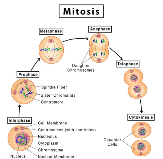
MITOSIS- its Occurrence, Stages and Significance.
Unlocking knowledge one post at a time! Check out our latest notes on www.microscopiaiwm.com – a treasure trove of insights, ideas, and inspiration. Dive in, explore, and let the journey of discovery begin! 📚
INTRODUCTION: Mitosis is a type of cell division that takes place in living organisms and it is commonly defined as the process of duplication of chromosomes in eukaryotic cells and distributed during cell division. The process where a single cell divides resulting in two identical cells, each resulted cell contains the same number of chromosomes and…
View On WordPress
#anaphase#asexual reproduction#cell cycle#cell cycle stages#cell division#cell plate#chromosome#cleavage furrow#contractile ring#cytokinesis#daughter cell#daughter cells#division of cytoplasm#division of nucleus#dna#DNA double#duplication of chromosome#fragmoplast#genetics#germ cells#homologous chromosome#homologous recombination#homologous recombination repair#karyokinesis#kinetochore#kinetoplast#meiosis#metaphase#MicroScopia IWM#mitos
5 notes
·
View notes
Text

The Science Research Manuscripts of S. Sunkavally. Page 116.
#electrophoresis#DNA#negative charge#electric current#zygote#division#immunoglobulins#charge#size-exclusion chromatography#glycocalyx#macromolecule#viscosity#oxygen diffusion#march hemoglobinuria#caffeine#brain#cytoplasm#fragile X chromosome#cursive#handwriting#notebooks#manuscripts
1 note
·
View note
Text
Miss Ortega | j.o

part 7
—The cell is made up of the nucleus and the cytoplasm and is enclosed by the cell membrane, which regulates passage in and out. The nucleus contains chromosomes, the cell's genetic material, and the nucleolus, which produces ribosomes. My eyes shift to Olivia, who was jotting down my words in her notebook.
In the late afternoon, I was at Olivia's house to help her study science, primarily about what a cell is and its functions. Olivia nods, giving me a nervous smile.
—One last question... what's cell division?— She puts the tip of her pen between her teeth, thoughtful.
—Cell division is the process by which a cell multiplies, splitting into two. In prokaryotes, it happens through binary fission (DNA filament duplication and subsequent division into two identical new individuals). In eukaryotes, it occurs through mitosis and, in reproductive cells, meiosis.— I say, shrugging casually.
Olivia writes it all down and then closes her notebook with a soft thud, sighing with satisfaction and tiredness.
—We're done,— she murmurs weakly, looking at me with a smile on her lips.
—We're done,— I repeat, and she stretches, slightly tense from maintaining an uncomfortable position for a long time.
—I'm not surprised you never get a failing grade, you're a book,— she says as she gets up from her desk, flopping onto her bed's mattress.
—Don't exaggerate...— I chuckle and give her a playful look. —Now... will you let me hear something you've written?— I nervously bite my lip, accepting the invitation to sit beside her on the bed.
Olivia sighs and reluctantly agrees to my request, blushing as she looks at me. —Wait,— she murmurs softly, leaning towards the edge of the bed, picking up a guitar case from the floor. Olivia glances at me sideways, holding the guitar in her hands.
—I'll sing you a little snippet of the song, okay? Also... I haven't finished it yet,— she says, toying with the guitar strings, likely tuning it.
I gaze in awe at her profile. Olivia had her head tilted down, holding the guitar in her lap. Her eyes briefly meet mine for a split second before she looks away with flushed cheeks.
Taking a breath, she closes her eyes, focusing.
—And I won't fight for love if you won't meet me halfway...— she begins to sing. And I say that I'm through but this song's still for you–
Her voice sounds angelic, surprising me with her talent. Olivia glances at me briefly, giving me a small smile.
—All I want is love that lasts— her eyes glisten, still looking at me.
—Is all I want too much to ask?— her fingers pause, interrupting the sweet melody. Olivia sets the guitar aside and looks at me with embarrassment, accepting my applause.
—Oh my god... you have an amazing voice,— I admit, and she tucks a strand of hair behind her ear, staring at a fixed point on her lap. —Thank you,— she offers a shy smile, and I reciprocate.
A knock on the door draws our attention to the entrance of her room. Olivia's mother, Emma, is standing there with a smile on her lips.
—T/N, dear, why don't you stay for dinner with us?— Mrs. Rodrigo suggests.
With a smile, I look at Olivia's reaction. She's looking at me with bright eyes and a smile, nodding enthusiastically.
—That would be fantastic,— I reply, and immediately, two arms wrap around my neck, hugging me. The force makes me lie back on the bed, and amid laughter, I return the hug, smiling shyly at Emma, who watches us with tenderness as I hold Olivia in my arms.
(...) —So... how's it going with the girl you like?— Enid asks, hugging a pillow in her arms.
After helping Olivia study, I received an invitation from Enid to have a pajama party at her house, inviting Olivia as well since she was with me. The blonde only knew that I liked someone, but she didn't know who, and for obvious reasons, she was really mad at me. I know she's my best friend, but I still couldn't tell her that I was in love with Professor Ortega.
—Actually, it's all going wrong... she said it's better if I forget what happened,— I lower my head towards my lap, sadly biting my lower lip. —Well, what a jerk...— Enid makes a face. —If only I knew who she was, I would have given her a piece of my mind,— she says absentmindedly, pulling at the corners of the pillow in her hands.
—You tried your best,— I smile sideways, and Enid throws the pillow at my face, messing up my hair. I chuckle slightly and wink at her.
—What do you think about Olivia, though?— she suddenly asks, lying down on the bed. I turn toward the door, relieved when I see that the subject of conversation is still downstairs preparing popcorn for the movie.
—Are you crazy? She's here...— I whisper, and she rolls her eyes at my comment.
—I don't see her,— Enid turns toward me, focusing her attention on me.
I sigh and shake my head. —She's nice...— I shrug indifferently, smiling at the blonde. Enid raises an eyebrow and gives me a smile, silently asking me to tell her more.
—She's beautiful... there's no doubt... but you know I'm in love with someone else,— I play with my fingers, embarrassed by the situation.
—She'd be perfect for you, you know? Plus... she really likes you,— Enid confesses. She adjusts her pajamas and gets under the covers, getting ready to watch the movie on her room's TV.
—I know... but for now... I only see her as a friend,— I tuck a strand of hair behind my ear and look confused at Enid's reaction, who is looking with panic over my shoulder.
I turn to her line of sight and pale when I see Olivia near the door. The brunette awkwardly leaves the popcorn bowl on the shelf and, with tears in her eyes, looks at me, shaking her head with regret. I stand up and bite my lips, mentally scolding myself for being so stupid.
I close the door behind me.
—Liv, wait,— I quickly descend the stairs, trying to catch up with Olivia. The brunette ignores me and walks toward the couches, searching for her jacket. I quicken my pace and grab her wrist. Olivia turns around and looks at me with tear-streaked cheeks, making me feel guilty.
—What do you want? You've said enough,— she says with venom, clenching her jaw.
—Liv...— I whisper, and her eyes glisten. Her shoulders relax, and she tentatively shuffles in place, wanting to hear what I have to say.
—Tell me...— her voice tone is clearly broken, showing that my confession has hurt her. I step closer, placing my hands around her face, wiping away some tears. Her eyes look at me sweetly despite the pain she's feeling. She places a hand against mine, giving me a comforting squeeze.
—Right now... I'm in love with someone else,— she nods, with bitterness in her mouth. —But it doesn't mean that in the future, I can't be with you... if you heard the whole conversation... and I'm pretty sure you did... I said that for now, I see you as a friend,— I smile sidelong, stroking her cheek. Olivia tilts her chin up and licks her lips, looking at me seriously. Suddenly, we're at the same height level since she's on tiptoes. My breath catches in my throat, and I timidly observe what the brunette wants to do.
—Kiss me...— she whispers, closing her eyes and clenching her jaw. —I just want to kiss you... at least once,— she confesses, making my chest tighten. I remove one hand from her face and trail it down her back, stopping at her waist, pulling Olivia closer to me.
—This...— I swallow, nervous due to the proximity. —This I can do— I lean toward her face and close the minimal distance between our lips. The kiss is sweet and at the same time salty from her tears. Olivia wraps her arms around my neck, sighing against my lips, receiving the long-desired kiss. The rhythm of the kiss is slow; we're simply enjoying the contact between our mouths. Olivia taps her tongue against my lower lip, asking for permission to enter. I part my lips, and our tongues meet, tentatively exploring each other's mouths.
I press my forehead against hers after ending the kiss. The brunette has a smile on her lips, looking at me with shining eyes of happiness. She leans in and briefly connects our lips for a split second before pulling away.
—That was... wow,— I admit, and she nods, completely agreeing.
I have to admit that the kiss was beautiful, I really enjoyed it. Her lips were sweet, inviting in a different way from Jenna's. Just mentioning the brunette makes me grimace, and I try to erase the image of her eyes from my mind so as not to ruin the moment. Olivia looks at me smiling, happy about what just happened.
—So... shall we go upstairs to watch the movie?— I suggest, and she nods slowly, starting to climb the stairs, our hands still intertwined.
—So... can you wait? I know it sounds horrible to ask, but I want to know, I want to find out if it's truly over with... the other person. I swear, if she's convinced that our... relationship? I don't know what to call it... is completely over... I'll give myself a chance to be with you,— I timidly ask, nervous about making this proposition. Olivia sighs and nods her head with both sadness and excitement at having a chance with me.
—Yes... you're... you're right, you know? I understand... it's not easy to choose between two girls you like... I'll wait... and if you choose me... I promise I'll never leave, T/N,— she admits, making me shiver slightly at the intensity of her gaze. I blush.
—Alright... because I was already getting ready to chase after you to talk,— I joke, and she chuckles softly, tilting her head back.
Her fingers tighten around my hand, stroking the back of my hand with her thumb.
—I wouldn't have gone anywhere... not in pajamas, obviously,— she raises her head with pride, and I burst out laughing at the expression on her face.
—Well... now let's go watch the movie? Enid's waiting for us,— I suggest, and she nods, starting to climb the stairs while still holding my hand, our fingers entwined.
It was late, but I was still awake, studying for the English literature exam I had the next day. The words on the pages were blurry, and I was unsure if I'd remember half of what I was reading due to how tired I was. But I had to keep going to be able to say that I had at least tried.
The vibration of the phone on the desk pulled my attention away from the book. With a sigh of relief, I picked up the device, thanking my lucky stars for the break. I looked at the screen, puzzled, when I saw that both Olivia and Jenna had messaged me.
I decided to read Olivia's message first.
Liv: heyyy (1:13 AM)
Damn, was it already one in the morning?
Yo: Hey Liv!
Liv: Are you done studying?
I furrowed my brows and nervously bit my lower lip.
Yo: Not really.
Yo: But if you need help, I'm here.
Liv: Great! Actually, you'd do me a huge favor if you could open the window.
I closed the chat and walked over to a corner of my room, spotting Olivia in front of my house, holding her phone. I opened the window and leaned out, smiling at the girl standing on the street.
—What are you doing here?— I whispered, not wanting to wake up the rest of my family.
Olivia looked up from her phone and smiled at me.
As a response, she moved closer to stand right beneath my window, gazing up at the tree near my house. With a swift but careful movement, she started climbing its branches, eventually reaching out to touch the edge of my window with her fingertips.
—Are you crazy or something?— I looked at my friend with concern.
—If you help me, you'd be doing me a favor,— she panted, not being able to hold on much longer.
I extended my hand and grabbed hers, helping her into my room. With a little jump, she made it all the way in, looking at me with a nervous smile.
—So, spill it,— I absentmindedly stared at the lamp light that was focused on the book on my desk. I sighed in frustration.
—In a few days, there's the end-of-semester dance... you know, the start of the Christmas break...— she put her hands in her pockets, blushing as she looked at me.
Oh... I knew where this was going.
—T/N... would you like to come to the dance with me?— she asked, sounding hopeful.
I opened my mouth in surprise and remained silent for a few seconds, wanting to think about her proposal. In reality... I wasn't even sure if I wanted to go, as I didn't want to be a third wheel between Enid and Ajax... but if I had to choose someone to go with... besides Jenna, of course... it would definitely be Olivia Rodrigo.
—Yes...— I whispered, and she leaned slightly forward, not having heard my response. I widened my eyes when I saw the living room light shining through my slightly open door. Quickly, I grabbed Olivia by the shoulders and motioned for her to move towards the window, needing to get out of here immediately. Olivia placed a foot on a tree branch before turning back in my direction.
—So? — my eyes darted towards the door as I used my hands to urge Olivia. I looked at her with wide eyes before nodding repeatedly. —Yes?— she asked, with a smile on her lips.
—Yes! Now go before you get caught— I muttered under my breath, looking at Olivia. She nodded and leaned towards my face, briefly connecting our lips for a split second. I looked at her in surprise but didn't say anything, watching closely as she jumped down from the tree, landing on her feet.
—Goodnight— she smiled at me, waving her hand, and ran down the sidewalk towards her house on the other side of the neighborhood.
With a yawn, I returned to my desk, picked up my phone, turned off the lamp, and collapsed onto my bed. A sigh of relief escaped my mouth as I heard the sound of the toilet flushing.
Well, it was just a bathroom break.
I turned on my phone and went on WhatsApp, reading Jenna's message. I couldn't deny that I was quite nervous; I didn't expect her to message me after days... maybe a week or two without hearing from her.
Ortega: Are you awake?
Yo: Yes.
Jenna's smile appeared on my screen, and I responded to her call with confusion.
—Hello?— I asked, hearing a breath on the other end. Jenna remained silent for a few seconds before speaking.
—Is it true?— she asked, leaving me completely stunned. I got under the covers, trying to figure out what to say.
—What?— I inquired, not exactly sure what she was talking about. She sighed in frustration before gritting her teeth.
—There are rumors at school that you and Rodrigo are together... is it true?— she muttered, sounding both annoyed and curious.
—Excuse me?— I was rather incredulous, not being able to believe what I was hearing. Jenna Ortega had called me in the middle of the night to ask me something like this.
—Is it true or not? ANSWER— she raised her voice, noticeably angry. I could hear her heavy breathing, making me feel uncomfortable and slightly afraid.
—No... We're not together... we're just getting to know each other... that's it,— I confessed, nervously biting my lower lip. —But anyway, isn't it none of your business who I'm dating? After all, you were the one who wanted distance a few days ago,— I retorted, annoyed by her attitude.
Jenna sighed loudly and ended the call, leaving me feeling both triumphant and confused. Whatever had gotten into her, I didn't know, but in any case, she had no right to treat me like this, especially after she wanted to pull away.
I placed the phone on the bedside shelf and closed my eyes, trying to fall asleep. The ghost of Olivia's kiss lingered on my lips, while Jenna's voice echoed in my head.
To say that I'm confused is an understatement.
#wednesday addams x reader#jenna ortega x reader#jenna ortega#jenna ortega x you#miércoles addams#jenna ortega x y/n#wednesday x you#jenna ortega x fem!reader#wednesday addams x you#professor#olivia rodrigo#kisses
155 notes
·
View notes
Text
This post is about:
The evolution of plants!
Plant evolution is a bit complex but beautiful. This post serves to educate you without losing your interest! [And it serves as a method of studying for me too :)]
Let's start simple!
The plant kingdom:
Plants are multicellular (are made of multiple cells) organisms (living beings) that contain chlorophyll. This is the green pigment that absorbs sunlight, allowing the plant to make their own food by photosynthesis [and it gives the plant its signature green colour!].
All plant cells have a cell wall found outside of their cell membrane. It is non-living and made of cellulose. It allows liquids and dissolved substances to pass through it. Thus, it is permeable. The cell membrane only allows certain substances through as it is partially permeable.
Most mature plant cells have a large permanent vacuole filled with the fluid sap. This is an aqueous solution of sugars and salts. The vacuole also produced turgor pressure on the cytoplasm and cell wall, making the cell firm/turgid.
Advantages of plant life in water:
Water provided:
Support, allowing for more exposure to sunlight.
Constant supply of water.
Readily available minerals.
A medium for spore/gamete transfer [necessary for reproduction].
Advantages of plant life on land:
Land provides:
Abundant sunlight.
Higher concentration of carbon dioxide.
Space availability as land had not yet been colonised by animals.
But land also provided many challenged for plants like:
Exposure to UV radiation which could cause mutations.
Exposure to air increases the chances of dessication (drying).
Structures needed support to replace the buoyancy previously offered by water.
Male gametes [sex cells] required a watery medium to reach female gametes.
Thus, plants had to develop structure that could:
Provide ways of capturing and filtering light.
Avoid dessication.
Offer structural support.
Facilitate transfer of gametes.
Now on to a bit more complicated stuff...
Plant evolution is grouped into five main divisions:
Bryophyta, pteridophyta, gymnosperms, monocotyledonous angiosperms and dicotyledonous angiosperms.
[Gymnosperms and angiosperms and collectively known as Spermatophytes]
Bryophyta:
This includes liverworts like Pellia and mosses like Funaria.
They have a very simple structure known as the thallus, with no proper roots, leaves, and stems.
They have hair-like structures called rhizoids on the lower surface to absorb moisture.
Their size is limited due to the absence of vascular tissue (xylem - structures that transport water and minerals, and phloem - structures that transport food).
Their spreading is also limited as they are heavily dependent on water.
Pteridophyta:
Ferns are pteridophyta.
They have roots, stems and leaves.
They possess vascular tissues (xylem + phloem), and thus, they can reach larger sizes.
Their leaves are called fronds, and these are made of smaller leaves called pinnae. These have a waxy layer that prevents water loss, allowing them to colonise drier areas. [Reproduction still requires a damp environment].
They have sporangia (capsules that produce spores) on the lower side of leaves, typically grouped in compact groups called sori.
Gymnosperms (conifers):
These are able to conserve water, allowing them to survive in both cold and dry climates. They have small leaves adapted to help them withstand drought on poor soils or in freezing/hot conditions.
Many of these plants are evergreen (keep their leaves all year round).
They reproduce via seeds formed in cones. [Gymnosperms - naked seeds. The seeds are not enclosed in an ovary.].
They are woody plants with true roots, stems and leaves.
Many conifers possess both male and female cones on the same tree, with the female cone being larger than the male and different in appearance.
Spores no longer require water and are dispersed by wind. Gametes do not require water for fertilisation anymore and instead occurs via a pollen tube. [This shows how evolution was in favour of life on land without the need for water for fertilisation].
An example of a gymnosperm is the Aleppo pine.
Angiosperms:
These are plants with seed bearing flowers. The seeds are formed in the ovaries of flowers.
Seeds may be protected by being in a fruit.
These range from small herbs to large trees.
This division is split into two groups:
Monocotyledons:
These tend to have a fibrous root system and long parallel-veined leaves.
Their floral parts are in multiples of three [3..6..9].
They have vascular bundles throughout the stem.
The seed has one cotyledon [food store].
Ex: Daffodils.
Dicotyledons:
These tend to have a tap root system and broad net-veined leaves.
Their floral parts are in multiples of fours or fives. [4..8..12] [5..10..15].
They have vascular bundles at the periphery (edges) of the stem.
The seed has two cotyledons (food stores).
Ex: wallflower, daisy.
Plant colonisation of land provided food, prompting aquatic organisms to move to land, too.
That's the end of my first post! I hope it was interesting and relatively understandable. If you have any comments you'd like to leave to add onto this information, any questions, or any suggestion, please go right ahead!
Thank you for reading, and have a good day or night!

8 notes
·
View notes
Text
Propaganda!


A megakaryocyte is a large bone marrow cell with a lobated nucleus that produces blood platelets (thrombocytes), which are necessary for normal clotting. Once the cell has completed differentiation and become a mature megakaryocyte, it begins the process of producing platelets. The maturation process occurs via endomitotic synchronous replication whereby the cytoplasmic volume enlarges as the number of chromosomes multiplies without cellular division. The cell ceases its growth at 4N, 8N or 16N, becomes granular, and begins to produce platelets.
Cytokines are a broad and loose category of small proteins (~5–25 kDa) important in cell signaling. Due to their size, cytokines cannot cross the lipid bilayer of cells to enter the cytoplasm and therefore typically exert their functions by interacting with specific cytokine receptors on the target cell surface. Cytokines have been shown to be involved in autocrine, paracrine and endocrine signaling as immunomodulating agents. They act through cell surface receptors and are especially important in the immune system; cytokines modulate the balance between humoral and cell-based immune responses, and they regulate the maturation, growth, and responsiveness of particular cell populations. Cytokines are important in health and disease, specifically in host immune responses to infection, inflammation, trauma, sepsis, cancer, and reproduction.
#Megakaryocytes#Cytokines#poll#polls#tumblr poll#tumblr polls#tournament poll#wikipedia#cells of the human body#science tournament#biochemistry
17 notes
·
View notes
Text
Organelles
Nucleus -- located near the center of the cell -- contains the genetic material of the cell (DNA) and nucleoli -- site of ribosome and messenger RNA synthesis
Nucleolus -- located in the nucleus -- site of ribosomal subunit assembly
Ribosomes -- located in the cytoplasm -- site of protein synthesis
Rough Endoplasmic Reticulum -- located in the cytoplasm -- has many ribosomes attached -- site of protein synthesis
Smooth Endoplasmic Reticulum -- located in the cytoplasm -- site of lipid synthesis -- participates in detoxification
Golgi Apparatus -- located in the cytoplasm -- modifies protein structure -- packages proteins in secretory vesicles
Secretory Vesicle -- located in the cytoplasm -- contains materials produced in the cell -- formed by the Golgi apparatus -- secreted by exocytosis
Lysosome -- located in the cytoplasm -- contains enzymes that digest material taken into the cell
Mitochondria -- located in the cytoplasm -- site of aerobic respiration -- major site of ATP synthesis
Microtubule -- located in the cytoplasm -- supports cytoplasm -- assists in cell division -- forms components of cilia and flagella
Centrioles -- located in the cytoplasm -- facilitate the movement of chromosomes during cell division
Cilia -- located on the cell surface with many on each cell -- move substances over the surface of certain cells
Flagella -- located on sperm cells -- one per cell -- propels sperm cell
#medblr#studyblr#notes#my notes#anatomy and physiology#anatomy#physiology#biology#bio#bio notes#biology notes#anatomy notes#physiology notes#cells#cells notes#microbiology#microbiology notes#studyblr notes
64 notes
·
View notes
Text
I wanted to talk a bit about how Gexgean land plants work on a structural level (something I’ve mentioned in passing before).
Cellulose isn’t a thing on Gexge. The cells of its plants owe their stiffness to a different compound (unnamed). Their cell walls take time to fully develop. When the plant forms a new structure, whether it’s a stem from a seed or a new leaf, it is initially as soft as the flesh of an animal and hardens in layers from the inside out.
This is what an un-walled Gexgean plant cell looks like under a microscope:

[Image description: A diagram of a round cell. It has a nucleus, large squarish mitochondria, “roseoplasts” instead of chloroplasts, a large organelle called a “cleaner subcell”, an oval organelle called a “reticustructum”, and several lysosomes floating around. End ID.]
Nucleus, mitochondria, lysosomes: You know these ones… more or less.
Roseoplasts: You know the equivalents of these ones.
Cleaner subcell: The equivalent of a Golgi body, if it also budded off lysosomes (and later consumed and repurposed them for protein-packaging material). The lysosome-production function was observed first, hence the cell being named for that.
Reticustructum: A unique organelle that produces the cellulose equivalent for the cell wall.

[Image description: A three-part diagram of the same cell. In the first image, the reticustructum is starting to produce a dark purple cell wall that builds up around the edge of the cell. In the second image, the cell wall encircles the entire cell, thicker near the reticustructum and thinner opposite it; the organelle is smaller, and is being eaten by lysosomes. In the third image, the reticustructum is gone and the cell wall has formed a square, with the cytoplasm having expanded to fill the new shape. End ID.]
As the plant’s new structure begins to come into its shape, the process of cell wall construction begins. The reticustructum secretes two substances: one that becomes the cell wall and another that dissolves the old cell membrane. As the process continues, lysosomes are attracted to it and begin to eat it as a way to ensure the cell wall doesn’t get too thick.
There are two methods of cell division. If a branch is making more branch cells, for example, a new reticustructum will form, enabling the cell to split in the regular plant manner. If a branch is growing a leaf, the lysosomes eat the cell wall as the nucleus starts to split and a cell membrane forms around it. The new cell kind of oozes out.
#Gexge#Celestial Community#speculative biology#xenobiology#speculative botany#this is probably a mess but I’d already decided that Gexgean plant tissues are soft for a while after forming#and came up with this when trying to reason how it might work
3 notes
·
View notes
Text
STRATEGIES NEED TO FOLLOW FOR HESI
Some effective strategies for practicing and succeeding on the HESI:
The HESI A2 typically includes:
Math (basic algebra, conversions, dosage calculations)
Reading Comprehension
Vocabulary & Grammar
Anatomy & Physiology
Biology, Chemistry, Physics (depending on school requirements)
Critical Thinking and Personality/Behavioral assessments
Feel free to join us at our private study group: HESI and TEAS Study Group 👈.

Here are some important objective-type questions related to Anatomy and Physiology that are commonly encountered in the HESI A2 exam.
GET FULL SERIES OF VIDEO TUTORIAL 👈.
Focused on Cell Biology, covering cell structure, organelles, functions, and basic processes — ideal for HESI A2 or general biology
What is found only in plant cells and allows photosynthesis? A) Mitochondria B) Ribosomes C) Chloroplasts D) Golgi bodies Answer: C) Chloroplasts
What is mitosis? A) Division of cytoplasm B) Formation of sex cells C) Division of the nucleus into two identical nuclei D) Breakdown of glucose Answer: C) Division of the nucleus into two identical nuclei
In which phase of mitosis do chromosomes line up at the cell's equator? A) Prophase B) Anaphase C) Telophase D) Metaphase Answer: D) Metaphase
GET COMPLETE VIDEO LESSON FOR HESI TEST PREP 👈.
#digital learning#education#higher education#learning#study inspiration#study motivation#study tips#career#math class#math test prep#nursing home care#nursing student#nursing#healthcare#treatment#hesi a2 practice test#hesi practice test#hesi a2 practice#hesi test prep#hesi online classes#hesi a2 test#nclex 2025#nclex practice test#nclex test prep#nclex study tips#nclexrn
0 notes
Text
Cellbiology

I. Introduction to Cell Biology
Cell biology is the study of the structure, function, and behavior of cells, which are the basic units of life. Cells are the building blocks of all living organisms, and they are responsible for carrying out the functions necessary for life.
II. Cell Structure
A cell consists of several organelles, each with its own unique structure and function. The main components of a cell include:
1. Plasma Membrane: The plasma membrane is the outermost layer of the cell that separates the cell from its environment. It is semi-permeable, allowing certain substances to pass through while keeping others out.
2. Cytoplasm: The cytoplasm is the jelly-like substance inside the cell membrane where many of the cell's metabolic reactions take place.
3. Nucleus: The nucleus is the control center of the cell where DNA is stored.
4. Mitochondria: The mitochondria are the powerhouses of the cell where energy is produced.
5. Endoplasmic Reticulum: The endoplasmic reticulum is a network of membranous tubules and cisternae that is responsible for protein synthesis and transport.
6. Ribosomes: The ribosomes are small organelles found throughout the cytoplasm where protein synthesis takes place.
7. Lysosomes: The lysosomes are membrane-bound organelles that contain digestive enzymes and are responsible for cellular digestion and recycling.
8. Golgi Apparatus: The Golgi apparatus is a complex organelle that is responsible for protein modification and transport.
III. Cell Function
Cells perform a wide range of functions necessary for life, including:
1. Metabolism: Cells carry out metabolic reactions to produce energy and synthesize the molecules needed for life.
2. Protein Synthesis: Cells synthesize proteins using the genetic information stored in DNA.
3. Cell Division: Cells divide to produce new cells, which is necessary for growth, repair, and reproduction.
4. Cell Signaling: Cells communicate with each other through cell signaling pathways, which allow them to coordinate their behavior and respond to their environment.
5. Cell Movement: Cells move using various mechanisms, such as muscle contraction and cytoskeletal rearrangement.
IV. Cell Cycle
The cell cycle is the series of events that a cell goes through from one cell division to the next. The cell cycle consists of three main stages:
1. Interphase: During interphase, the cell grows, replicates its DNA, and prepares for cell division.
2. Mitosis: During mitosis, the replicated DNA is divided equally between two daughter cells.
3. Cytokinesis: During cytokinesis, the cytoplasm divides and the cell splits into two daughter cells.
V. Cell Signaling
Cell signaling is the process by which cells communicate with each other and respond to their environment. Cell signaling pathways involve a series of molecular interactions that allow cells to transmit and receive signals.
A. Types of Cell Signaling
There are several types of cell signaling, including:
1. Endocrine Signaling: Endocrine signaling involves the release of hormones into the bloodstream, which then bind to receptors on target cells.
2. Paracrine Signaling: Paracrine signaling involves the release of signaling molecules that diffuse through the extracellular space and bind to receptors on nearby cells.
3. Autocrine Signaling: Autocrine signaling involves the release of signaling molecules that bind to receptors on the same cell.
4. Juxtacrine Signaling: Juxtacrine signaling involves the release of signaling molecules that bind to receptors on adjacent cells.
VI. Cell Movement
Cell movement is the ability of cells to change their position in response to various stimuli. There are several types of cell movement, including:
1. Muscle Contraction: Muscle contraction involves the sliding of actin and myosin filaments past each other to produce force.
2. Cytoskeletal Rearrangement: Cytoskeletal rearrangement involves the reorganization of the cytoskeleton to produce force and movement.
3. Cell Migration: Cell migration involves the movement of cells from one location to another in response to various stimuli.
VII. Cell Death
Cell death is the process by which cells die and are removed from the body. There are several types of cell death, including:
1. Apoptosis: Apoptosis is a form of programmed cell death that involves the activation of a series of molecular pathways that lead to the death of the cell.
2. Necrosis: Necrosis is a form of cell death that involves the uncontrolled death of cells, often as a result of injury or infection.
VIII. Conclusion
In conclusion, cell biology is the study of the structure, function, and behavior of cells.
1 note
·
View note
Text
Understanding Cancer Cytopathology: A Beginner’s Guide
Introduction
Cancer cytopathology is a critical field in medical science that focuses on diagnosing cancer at the cellular level. This branch of pathology helps detect malignancies early, guiding treatment decisions and improving patient outcomes. For beginners, understanding cancer cytopathology involves learning about its techniques, significance, and role in modern medicine.
What is Cancer Cytopathology?
Cytopathology is the study of cells extracted from tissues or bodily fluids to identify abnormalities. When applied to cancer detection, cytopathology examines the microscopic structure of cells to determine whether they are benign, precancerous, or malignant. This method is widely used in screenings and diagnostic procedures.
Importance of Cancer Cytopathology
Early cancer detection significantly increases the chances of successful treatment. Cancer cytopathology offers a minimally invasive way to assess cellular changes, allowing doctors to diagnose cancer before it spreads. This field plays a vital role in preventive medicine and personalized treatment plans.
Common Techniques in Cancer Cytopathology
Several techniques are used to collect and analyze cellular samples:
Fine Needle Aspiration Cytology (FNAC) – A thin needle extracts cells from lumps or masses for microscopic examination.
Exfoliative Cytology – Cells naturally shed from body surfaces (e.g., Pap smear for cervical cancer screening) are analyzed.
Liquid-Based Cytology (LBC) – A refined method that preserves and processes cells in a liquid medium for more accurate results.
How Cytopathologists Identify Cancerous Cells
Cytopathologists look for specific cellular changes that indicate malignancy, including:
Irregular cell shapes and sizes
Increased nuclear-to-cytoplasm ratio
Abnormal chromatin patterns
High mitotic activity (rapid cell division)
Advantages of Cytopathology in Cancer Diagnosis
Minimally Invasive: Requires only small tissue or fluid samples.
Quick Results: Faster diagnosis compared to traditional biopsies.
Cost-Effective: Less expensive than surgical procedures.
Early Detection: Helps in identifying cancers before symptoms appear.
Challenges in Cancer Cytopathology
While highly effective, cytopathology has some limitations:
False Positives or Negatives: Errors may occur due to sample quality or interpretation.
Limited Tissue Information: Unlike biopsies, cytology provides fewer details about tissue structure.
Need for Skilled Experts: Proper analysis requires experienced cytopathologists.
Future of Cancer Cytopathology
With advancements in artificial intelligence (AI), digital pathology, and biomarker discovery, cancer cytopathology is becoming more precise and efficient. AI-powered image analysis can enhance accuracy, reducing human error and improving early detection rates.
Conclusion
Cancer cytopathology is a powerful tool in diagnosing and managing cancer. By understanding its methods, benefits, and challenges, we can appreciate its role in saving lives through early detection and effective treatment planning. As technology advances, cytopathology will continue to evolve, offering even greater accuracy and hope for cancer patients worldwide.
0 notes
Text
Cell division is a fundamental process in both plants andanimals’ growth and development as well as recreation. This paper investigates the mitosis process in both plants and animals’ cells to identify distinct stages of mitosis and the structure of cells at each stage. The experiment identifies similarity in mitotic stages for cells in both plants and animals. The mitotic process leads to division of one parent cell to form two identical daughter cells that can further undergo subsequent mitotic divisions. Introduction Cells are the fundamental elements of living things, both plants and animals. Consequently, realized mechanisms originate from cells and manifest in organs and the entire organism. Mitosis and meiosis are some of the fundamental processes that take place at the cell level. They refer to cell divisions that lead to generation of new cells to replace dead or worn out cells, generation of cells for development of organs, and cell division towards growth. Mitosis leads to generation of identical daughter cells for growth or replacement of cells in organs. It takes place in a number of stages, interphase, prophase, metaphase, anaphase, telophase, and cytokinesis (Goldberg and Goldberg, p. 77- 80). The interphase is a preliminary stage in cell division that precedes the mitotic stages. It is fundamental as it forms the largest percentage of a cell’s life. The major activity at this stage is replication of cell proteins within cells and developments to visibility of cell nucleoli. Mitosis however has four stages that result into cell division. The first stage is the prophase, followed by metaphase, anaphase, and telophase respectively (Goldberg and Goldberg, p. 79). At the prophase, “strands of chromosomes begin to condense” and can be seen with the aid of a powered microscope. Visibility of the nucleoli however disappears while fibers develop in the cell’s cytoplasm. The developed fibers emanates from a pair of centrosomes that stretch to opposite poles of the cytoplasm. Disintegration of the “nuclear membrane begins” at this stage and marks the end of the prophase and the process moves to the metaphase (Goldberg and Goldberg, p. 79). At the metaphase stage, the chromosomes are arranged along a plate that is perpendicular to the centrioles’ plane and the “spindle fibers” interlink the centrosomes and the chromosomes (Goldberg and Goldberg, p. 79). A new phase, the anaphase is then marked by disintegration of chromosomes into centromeres that are then attracted to the centrosomes along the fibers (Goldberg and Goldberg, p. 79). The cell then enters a new phase, the telophase, where the pulled chromosomes converge at the opposite sides of the cell, along the fibers and the nuclear membrane begins to reappear. Each set of chromosomes assumes the normal thread like structure and the nuclear membrane develop around each group of chromosomes to form two nucleuses within the cytoplasm (Toole and Toole, p. 139). The cytokinesis process, where the cytoplasm divides to form two different cells then follows this (Goldberg and Goldberg, p. 79- 80). The structure of the cells in plants and in animals may however be different. This is because of a number of factors such as the lack of significance of the spindle fibers in the plant cells. The nucleus, rather than spindle fibers in plant cells, moves to define the new position of chromosomes in the mitosis process (Cassimeris, Plopper and Lingappa, p. 952). T Read the full article
0 notes
Text
Zoology Practical Sem 4
Q1. Root Tip Slide Preparation
Aim: To study the various stages of mitotic division using onion root tip.
Principle:
Mitosis is an equational cell division occurring in vegetative cells of plants and animals where in a parent cell divides into two daughter cells of same genetic makeup as that of the parent cells.
Actively growing somatic regions of living organisms undergo mitosis at a good rate, leading to the growth and development of the body.
Onion root tips are a good source to study the various stages of mitosis under a microscope.
Acetocarmine is routinely used to stain root tips after properly squashing them. They are then placed on the slide. Acetocarmine can specifically stain the chromosomes.
A typical eukaryotic cell mainly divides by mitosis, which comprises the following stages - Interphase - Prophase - Metaphase - Anaphase - Telophase and Cytokinesis.
Procedure:
Take a small onion root tip on a clean slide and add a drop of acetocarmine stain. Warm gently by passing the slide over a flame a few times.
Tease the stained material with a dissection needle. Place a coverslip and keep a tissue paper over it. Tap or press the material from above so that the stained cells spread out.
Locate a suitable area using a magnification of 10X and 40x. Observe the slide and identify the stage of mitosis based on their features described.
Prophase:
The first stage of mitosis is prophase. The important events during this phase are:
A) Nuclear Changes - Chromatin material of nucleus gradually condenses into distinct chromatin threads by losing water. - The chromatin threads coil like cylindrical springs, and in doing so they gradually becomes shorter and thicker, forming chromosomes. - The proteinaceous matrix gets deposited around the chromosomes, so that this gradually becomes shorter and thicker to form chromosomes. - Each chromosome is already doubled due to the doubling of DNA in interphase. - By the end of prophase, two chromatids of each chromosome become more distinct and appears to be split up length wise. - The nucleus and the nuclear membrane disappear by the end of prophase.
B) Cytoplasm Events - The centriole divides into two and then one of the daughter centrioles moves towards the opposite pole. - Astral rays radiate out from each daughter centriole.
--------------------------------------------------------
Spotters
Lampbrush Chromosome
The largest known chromosomes, can be seen with naked eye and are characterized by their lateral loops.
Found in the yolk rich oocyte nuclei of certain vertebrates like fish, amphibians, reptiles, and birds during first prophase of meiosis.
Loops give it a brush like appearance
Discovered by Elemming in 1882.
Can reach up to 5900μ in length
Consists of a longitudinal axis formed by a single DNA molecule along with several hundred bead like chromosomes distributed in a linear fashion
From each chromosome, there emerge 2 symmetrical lateral loops which can expand or contract in response to external stimuli
5-10% of DNA is in the lateral loops
Functions: RNA syntheses and formation of yolk material
---
Polytene Chromosome
Giant banded chromosomes, smaller than lampbrush chromosomes.
Found in the larvae of certain dipterans.
First discovered by E.G. Balbiani in Chironimus in 1881.
Can occur in the larval salivary glands, midgut epithelium, rectum, and malpighian tubules of various genera like drosophila.
The salivary cells are so large they can be easily seen with a dissecting microscope.
Nuclei very large, around 25μ in diameter, chromosomes 50-200 times larger than typical chromosomes.
Tightly coiled chromatin: dark bands, loosely coiled: light bands.
Puffs: uncoiled regions of active RNA synthesis.
Balbiani rings: regions with larger puffs than others.
Primary function is to carry genes which ultimately control physiology of an organism.
---
T.S. of Testis
Round or oval in shape, externally surrounded by visceral peritoneum, a layer of fibrous connective tissue, and the tunica albuginea.
Each testis has a mass of coiled microscopic seminiferous tubules held together by connective tissue.
The connective tissue has small patches of interstitial cells which secretes the hormone testosterone responsible for male secondary sexual characters.
Stratified germinal epithelium lines the seminiferous tubules.
Germinal cells give rise to sperm.
Sertoli cells between germinal cells, nourish the developing sperm.
---
T.S. of Ovary
Ovary is solid and oval in shape.
It consists of the stroma that is differentiated into peripheral cortex and central medulla.
Lying in the cortex are ovarian follicles, corpora lutea, corpora albicans and interstitial cells. Medulla only contains blood vessels
Primary follicles arise from germinal epithelium into the cortex and each contains a central oocyte surrounded by a single layer of follicle cells.
Sec.follicles contain zona pellucida, several layers of follicle cells and thin connective tissue layer called theca having capillaries.
Mature/Graffian follicle contains large central follicular cavity, antrum externally lined by membrana granulosa.
Corpus luteum - ruptured follicle has solid mass of cells - secretes progesterone.
---
Cleavage of Zygote of Frog - 2 celled stage
The first cleavage plane is meridional.
The pattern of cleavage is holoblastic and unequal type.
The first cleaving furrow appears near the animal pole and extends towards the vegetal pole.
The cleaving furrow cuts the egg through its medium animal vegetal polar axis vertically into two equally sized blastomeres.
The mode of division of the zygote is mitotic.
---
Cleavage of Zygote of Frog - 4 celled stage
The second set of cleavage is also meridional which starts before the completion of the first.
The second cleavage is at right angle to the first, resulting in 4 blastomeres.
All the 4 blastomeres are equal-sized.
The grey crescent, animal pole, and vegetal pole show no change in their position and size.
---
Cleavage of Zygote of Frog - 8 celled stage
The third cleavage results into 8-celled stage.
The third cleavage is holoblastic but unequal.
The third cleavage is horizontal but well above the equator towards the animal pole.
It results into 8-blastomeres of two distinct types.
The upper 4 smaller pigmented blastomeres at the animal pole are called micromeres.
The lower 4 larger yolk-ladder cells at vegetal pole are called macromeres.
---
Cleavage of Zygote of Frog - 16 celled stage
The fourth cleavage plane is oriented from animal to vegetal pole.
The fourth set of cleavage is also vertical, therefore, produces a 16-celled stage.
The micromeres divide more rapidly.
The yolk-ladder macromeres divide slowly.
The yolk shows its influence on cleavage rate.
---
Cleavage of Zygote of Frog - Morula
The fifth cleavage is irregular but it is usually double oriented parallel to third cleavage plane resulting into 32-celled stage.
The fifth cleavage separates the micromeres and macromeres in complete two sets of 16 each.
The blastomeres assume spherical shape.
The surfaces of blastomeres in contact to each other are flattened due to their mutual pressure.
The whole embryo acquires a characteristic appearance reminiscent of a mulberry, hence the name morula.
---
Blastula of Frog
It is a hollow ball of different sized cells representing blastula stage in the development of frog.
The cavity is known as blastocoel.
Blastocoel is eccentric and placed towards the animal pole
Blastocoel is surrounded by micromeres and macromeres.
Micromeres are smaller, found in the anterior animal pole, and have dark pigments.
Macromeres are larger, found in the posterior vegetative pile, and are larger in the yolk.
---
Gastrula
Embryo developed from blastula
Oval in shape, contains three germ layers: ectoderm, endoderm, and mesoderm.
Internally the gastrula has a cavity called the archenteron.
It opens to the exterior through blastopore.
A mass of endoderm cells protrudes through the blastopore, these cells constitute the yolk plug.
The floor and the lateral sides of the archenteron are formed from the endoderm.
The roof of the archenteron is formed from the chordomesoderm.
Externally the gastrula is covered by ectoderm.
The ectoderm that lies on the mid-dorsal line develops into the nervous system, hence it is called neurectoderm.
The remainder of the ectoderm is the epidermal ectoderm.
---
Chick Embryo - 18 hours of incubation
After 18 hours of incubation the notochord has become markedly elongated, forming a prominent structure.
Notochord extends towards cephalic region in the mid-line from Hensen's node.
18 hours incubated embryo is spoken of as being in the head process stage.
As the tip of the notochord neural plate develops, caudal and cephalic ends are seen.
In front of notochord and neural plate there is a space called as pro-amnion.
Embryonic area, anterior border of mesoderm area pellucida and area opaca become more prominent.
Primitive streak gradually decreases in size.
---
Chick Embryo - 24 hours of incubation
The folding of the neural plate is clearly marked. In stained and transparent preparation of entire embryo neural folds appear as a pair of dark bands formed of neural groove.
Neural folds at cephalic are more prominent than at caudal end.
Foregut is established, the gut caudal to foregut called as midgut and the anterior intertinal portal appear.
In the middle, four pairs of somites are seen.
Hensen's nod is pushed caudally and primitive streak is further reduced.
---
Chick Embryo - 33 hours of incubation
Shows some of the fundamental steps in the formation of central nervous system and circulatory system.
Various neuromeric enlargements from brain regions.
Brain differentiated into prosencephalon, mesencephalon, and rhombencephalon.
Optic vesicles established.
Infundibulum formed.
12 pairs of somites formed.
Mid-region of heart is considerably dilated and bent to the sight.
Primitive streak becomes shorter due to lengthening of neural tube.
---
Chick Embryo - 48 hours
Embryo shows cranial flexure and twisting of head over the right side.
Fore, mid, and hind brain distinct, fore brain takes the anterior most places.
Heart differentiated into ventricular arterial, and sinus region.
Second aortic arched develop.
Vitelline arteries and veins are distinct.
Somites are 26 pairs.
---
Chick Embryo - 96 hours
The entire body bends 90°, the embryo lies with it's left side on the yolk.
At the end of the 96 hours, the body folds have undercut the embryo so it only remains attached to the yolk by a slender stalk.
The yolk stalk becomes elongated, allowing the embryo to become straight first in the mid-dorsal region and then convex dorsally.
Optic cub shows more developed lens.
Endolymphatic duct arises from the auditory vesicle.
Visceral arches have become very much thickened.
Appendage bugs increase rapidly in size, become elongated.
No of somites increases to 41 pairs.
---
Mitosis: Prophase
The first stage of mitosis.
Chromosomes appear as long coiled threads called chromatids.
Chromatin becomes shorter, thicker and more visible due to the condensation of DNA to form chromosomes.
Stability of nucleus increases.
Each chromosome starts to splits longitudinally into two sister chromatids. These sister chromatids are attached with each other at centromere.
Nuclear membrane and nucleolus disappear by the end of prophase.
---
Metaphase
The 2nd phase of mitosis
Appearance of spindle fibres
Spindle fibres attached to the centromere of chromosome.
The chromosomes are arranged on the equatorial plane.
The process of gathering of chromosomes in equator is called congression and plate formed is called metaphasic plate.
---
Anaphase
The third and shortest phase of mitosis
The centromere of each chromosome splits into two sister chromatids and forms two daughter chromosomes.
The daughter chromosomes are pulled towards the poles due to the contraction of spindle fibers.
During polar movement, the chromosomes shows different shapes like J,U,V,L or I shaped in appearance.
At the end of anaphase, each pole will get one set of daughter chromosomes.
---
Telophase
The fourth and final stage of mitosis.
The daughter chromosomes reach respective poles, they uncoil and become thin, long and visible.
The spindle fibers start to and finally disappear.
The nuclear membrane and the nucleolus reappear.
Two nuclei are formed at the end of telophase. Both the nuclei have the same number of chromosome as parent cell.
---
Meiosis: Prophase-I
The longest stage of meiosis-I
Divided into 5 sub-stages:
Leptotene: size of cell and nucleus increases, fine thread like chromomeres present
Zygotene: Synapsis occurs, the pairing of homologous chromosomes.
Pachytene: Tetrads form from bivalents, crossing over starts.
Diplotene: Crossing over occurs, homologous chromosomes begin to sperate but are held together by chiasmata.
Diakinesis: Chiasmata move towards the end of chromosomes, nuclear membrane and nucleolus disappear
---
Metaphase-I
2nd phase of meiosis
The spindle fibers organize between two poles and get attached to the centromere of chromosomes.
Chromosome moves to equator
The bivalent chromosomes are arranged in the equatorial plate in such a way that 2 metaphasic plates are formed
---
Anaphase-I
3rd phase of meiosis
Spindle fibers contract, pulls chromosomes to the polar region.
The separated chromosome is known as dyads
No splitting of chromosomes occurs so the centromere of each homologous chromosome does not divide. Thus, the chromosome number of the daughter nuclei is reduced to half.
Now the separated chromosome moves toward opposite poles
---
Telophase-I
Two groups of chromosome formed at each pole and organized into nuclei.
The nuclear membrane and nucleolus reappears.
The chromosomes get uncoiled into chromatin thread.
The spindle fibers disappear.
---
Prophase-II
First stage of meiosis-II
The dyads chromosome becomes thicker and shorter
Nuclear membrane and nucleolus disappear
Spindle fibre starts to form
---
Metaphase-II
The dyads chromosomes comes to equatorial plane
Spindle fibres organize between poles and attaches to centromere of chromosome.
---
Anaphase-II:
Centromere of each chromosome divides and sister chromatids separates to form two daughter chromosome
Spindle fibre contracts and pull the daughter chromosome apart towards opposite pole.
---
Telophase-II:
Chromosome become organize at respective pole into nuclei
Chromosome elongates to form thin networks of chromatin
Nuclear membrane and nucleolus reappears
0 notes
Text
As a community, aquarists tend to become paranoid about 'hitch hikers', such as aiptasia (glass or triffid) anemones, and the entire clade of vermetid snails. As an online subculture, the community talks of these creatures as indestructible and existential threats, something more like fictional, movie xenomorphs, than what they are, real animals. Usually there exists a kernel of truth behind such fears, although aquarists folklore ignores the basic knowledge, that is relevant to their management - something more realist than a 'cure'. It seems strange today, but in the 1990s, any non-sedentaey polychaete could cause the majority of aquarists to panic. Of course the majority of them are harmless, and even helpful to the aquarist, because the common species are detrivores.
If the world of macroscopic plants has a counterpart in the 'reefer' imagination, it is surely that curious seaweed called bubble algae, or in other sorts of literature, sailor's eyeballs. We often call them all Valonia, and - with the caveat that I am not a botanist - I see no basis to doubt this. However the junior synonym Ventricaria is sometimes in use. These peculiar seaweeds - for they are unusual, green macroalgae - are all quite similar to one another, although some invest more energy in a larger 'bubble' - a spheroid to avoid body (or thallus), that is attached firmly to an underlying substrate - whilst others form a more creeping mat of smaller 'bubbles', because they may generate a stolon. It is the latter growth morphs that becomes invasive in our aquariums.
An curious and only half true bit of folklore about Valonia sp. is that they are the world's largest single celled organisms. Wether or not they are single celled is a matter of opinion, since they are miltinucleate, as are other green seaweeds of 'siphonous' type. Siphonous seaweeds are known to exist only among the green algae, with no counterparts among the brown or red algae, and to complicate things more, true multicellular algae have also evolved from non-siphonous, green algal ancestors. The 'giant cells' of siphonous algae contain numerous nuclei, each with its own 'cytoplasmic domain', whereas typical cells - of classically unicellular or multicellular organisms - possess but one cell nucleus. No cell wall separates the nuclei in the siphonous species, which is a basic difference from the truly multicellular plants.
Siphonous macroalgae thus exist outside of the familiar division of organisms into single or multiple cells, as we tend to take it for granted. For over one hundred years it has been pointed out, anyway, that the idea of a 'unicellular' organisms is flawed, on both semantic and empirical grounds. To begin with an organisms without cell walls is undivided, and therefore it cannot contain cells; also the 'cells' of what we call 'unicellular life' do not function entirely like the cells which act as the mere compartments of larger, living entities.
It is a frustrating description, but it is one that has stuck, and siphonous algae - and similarly multinucleate organisms, such as the famous slime molds -are clumsily expressed as 'giant cells'. If we view them as unicellular, then they surely are the biggest 'single celled organisms' in the world, but it is not the bubble algae would hold that title. Much larger siphonous macroalgae exist - such as Caulerpa sp and Halimeda sp. - that would wear that particular crown, sharing as they do the same siphonous nature as Valonia, and yet growing much, much larger.
Some people think that crabs which eat Valonia actively spread or, by releasing it's spores which are stored inside the globular thallus. This is nonsense because no spores are inside the plant, due to its structure making this impossible. This is not how the bubble algae multiply, although any intact 'cytoplasmic domain' can regenerate to form a complete Valonia organism. This is true of other siphonous algae such as Caulerpa, and neither makes bubble algae unique nor invincible. Without interference the bubbles multiply through division, which is also normal for such 'giant cells'. They also release new individuals via pores on their cell wall, and Valonia actually resembles, in its organization, the reproductive organs of some other siphonous algae
Incidentally the Mithrax group of crabs, including the emerald crab, M. sculptus, effectively consume Valonia; the problem is, their diets are broader than algae alone, and they are opportunistic foragers of sessile animals such as corals, and even slow, motile prey, such as snails. The popular focus on the emerald crab as a control agent of Valonia, is because few other herbivores find them palatable. In the wild they seem to be held in check by other, competing algae, and Valonia is commonest where these competitors are relatively absent. Some of the saccoglossan snails may be found on bubble algae, but most saccoglossan species will not consume it.
This is not to say that no other animals eat Valonia, but their extracts are known to be unappetising to herbivorous fishes. The fact they seem to profit where sea urchins graze on other algae, also points to their general unpalatability. It's curious that Siganus argenteus, a strictly vegetarian siganid, shows an actual preference for Valonia as food, whilst to another siganid, S. spinosus, the extracts are distasteful. Two species of the acanthurid genus Naso - N. lituratus and N. unicornis - are also verified to consume these macroalgae, whereas Acanthurus nigrofuscus and Zebrasoma flavescens do not It seems that the will to eat Valonia has evolved in fishes inhabiting those habitats, where they are relatively common as a potential food source.
Valonia is an adaptable enough genus, and can be found in harsh situations such as the intertidal zone, where it might be exposed to tropical air for hours of the day. Unsurprisingly, hardy algae of this kind easily survive transport on damp but emersed 'live rock' whilst it is transported by aeroplane - more fragile organisms are much less likely to survive the long process of shipment and 'curing'. They can also be found as deep as 80 meters, showing a preference for relatively shaded and flow deficient microhabitats, for example between stones. They are common on solid substrates for this reason, and are found growing in the wild, in association with certain species of Caulerpa. Bubble algae may grow, in the wild, as epiphytes on larger marine plants, including an association with the mangroves.
Valonia sp. ought not to be conflated with the 'grape'-forming morphs within the genus Caulerpa, nor the red grape or bubble algae of the genus Botryocladia, which belongs to the classically multicellular red seaweeds. Newcomers to the hobby are confused by the grape-like bubbles of these plants. Both these genera have a very different growth habit and appearance, when they are compared to the Valonia - although the latter, too, are marine plants, despite their noteworthy growth form. And when they are viewed as what they are, siphonous algae, the genus Valonia are not so unusual or perplexing as they might at first seem.
#bubble algae#valoniaceae#green algae#seaweeds#macroalgae#valonia#genus valonia#siphonous algae#sea grape#sailors eyeballs#misunderstood fish#qaautomation#oddballs#coldwater tropicals
1 note
·
View note
Text

On Mitosis
I don't know a lot about mitosis aside from the fact that it occurs after the Interphase and once DNA has been replicated, and that it is a continuous process but is divided into four stages: prophase (from the greek pro- before), metaphase, anaphase and telophase.
During prophase, chromatin is in its condensed form: heterochromatin. During metaphase, chromosomes are pushed to the center of the nucleus. Following metaphase, there is migration during the anaphase, when microtubules that make up the mitotic spindle separate chromatides and pull them to either sides of the nucleus. In the final phase, the telophase, microtubules progressively disappear, sister chromatides are decondensed and the contraction of myosine and actine filaments splits cytoplasm for cell division.
0 notes
Text
Propaganda!


Granulocytes are cells in the innate immune system characterized by the presence of specific granules in their cytoplasm. There are four types of granulocytes (full name polymorphonuclear granulocytes): Basophils, eosinophils, neutrophils, and mast cells. Except for the mast cells, their names are derived from their staining characteristics; for example, the most abundant granulocyte is the neutrophil granulocyte, which has neutrally staining cytoplasmic granules.
In cell biology, the spindle apparatus is the cytoskeletal structure of eukaryotic cells that forms during cell division to separate sister chromatids between daughter cells. It is referred to as the mitotic spindle during mitosis, a process that produces genetically identical daughter cells, or the meiotic spindle during meiosis, a process that produces gametes with half the number of chromosomes of the parent cell.
#Granulocytes#Spindle fibers#tournament poll#polls#wikipedia#cells of the human body#science tournament#biochemistry#poll#tumblr poll#tumblr polls
10 notes
·
View notes
Text
Eukaryote cell have specialized organelle in cytoplasm such as plant, animal, fungi, protozoa and algae and define as a membrane bound nucleus enclosing genetic materials into chromosomes, so it cell divisions occurs by mitosis and meiosis divisions #geneticteacher
0 notes