#eosinophilic gastrointestinal disease
Text
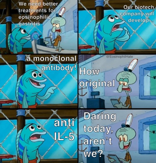
#eosinophilic gastritis#chronic illness#eosinophilic disorders#chronic illness meme#rare disease#EoG#eosinophilic disease#eosinophilic gastrointestinal disorders#eosinophilic gastrointestinal disease#monoclonal antibody#immune system disorder#immune system#pharmacology#biotech#biotechindustry#medication#medicine#rare disorders#il-5#fasenra#interleukin-5#benralizumab
7 notes
·
View notes
Text
Eosinophilic Gastrointestinal Disease
According to the localization of the gastrointestinal tract, primary eosinophilic gastrointestinal diseases include five variants such as esophagitis, gastritis, gastroenteritis, enteritis, and colitis.
0 notes
Text
Complementary therapies for autism/Autism Spectrum Disorder (ASD) - Overview and recommendations for biological therapies

Autism spectrum disorder (ASD) is a genetically based neurodevelopmental disorder characterized by persistent impairments in social interaction and restricted, repetitive patterns of behavior, interests, and activities.
它是一种因大脑区域功能障碍而引起的精神障碍。
这也是一种精神表现异常的综合症。
It is a neurodevelopmental disorder of neural connections.
Excluding brain trauma, infection, tumors, etc., most causes of ASD are caused by inherited metabolic disorders (IEMs).
1. Signs and symptoms of ASD
Listed below are some common types of behaviors seen in people with autism/ASD . Not all people with autism will exhibit all of the behaviors, but most will exhibit some of the following.
1. Social communication/interaction behaviors may include:
Little or infrequent eye contact
Does not seem to be looking at or listening to others
Rarely shares interest, affection, or enjoyment of objects or activities (including rarely pointing to or showing things to others)
No response or slow response to others' name or other verbal requests for attention
Difficulty in back-and-forth conversation
Frequently talks at length about favorite topics without noticing that others are not interested or without giving others a chance to respond
Displaying facial expressions, movements, and gestures that are inconsistent with what is being said
Unusual intonation, which may sound sing-song or flat and robotic
Difficulty understanding other people’s perspectives, or being unable to predict or understand other people’s behavior
Difficulty adjusting behavior to social situations
Difficulty engaging in imaginative play or making friends
2. Restrictive/repetitive behaviors may include:
Repeating certain behaviors or having unusual behaviors, such as repeating words or phrases (a behavior called echolalia)
A persistent interest in a particular subject, such as numbers, details, or facts
Shows an exaggerated interest, such as in moving objects or parts of objects
Feeling uneasy about minor changes in daily life and having difficulty adjusting to transitions
Being more or less sensitive than other people to sensory input (such as light, sound, clothing, or temperature)
3. Complications
ASD is often associated with multiple physical and psychiatric disorders:
Feeding problems: eating disorders, anorexia nervosa, aversion to certain types of food, only eating foods with a certain taste/texture, which may cause abnormal weight, gastrointestinal symptoms such as diarrhea and constipation, or lack of nutrition due to unreasonable eating
Sleep disorders: sleep anxiety, tossing and turning, difficulty falling asleep, inability to lie flat to sleep, insomnia, repeated awakenings at night, sleepwalking and other abnormal sleep structures
Gastrointestinal (GI) problems: Gastroesophageal reflux, eosinophilic esophagitis, nausea and vomiting, constipation, diarrhea, flatulence, colitis, food intolerances
Allergic diseases: asthma, nasal allergies, allergic diseases (IgE-mediated), food allergies and intolerances
Toileting problems: difficulty learning to use the toilet during the day and night, not knowing when to use the toilet, communicating the need to use the toilet, being able to use the toilet independently or promptly, learning to use different and unfamiliar toilets, self-wiping, sensory differences (dislike of noises made by the toilet, the sensation of urine/faeces, cold toilet seats or excessive concern about water in the toilet), fecal smearing, a range of specific continence difficulties including bowel or bladder control difficulties
Abnormal responses to sensory stimuli: Over-, under-, or abnormal responses to environmental stimuli such as noise, touch, smells, tastes, or visual stimuli. Examples include looking out of the corner of the eye, excessive focus on edges, spinning objects, shiny surfaces, lights, or smells, refusing or only eating foods with certain tastes/textures, obsessive sniffing or licking of non-food objects, tactile defensiveness or resistance to being touched, or hypersensitivity to certain types of touch; light touch may be painful, while deep pressure may be soothing. There may be resistance to wearing certain textures or colors of clothing against the skin, apparent indifference to pain, a strong preference and/or persistence for certain textures and a strong aversion to other textures, and hypersensitivity to certain frequencies or types of sounds (e.g., a fire truck in the distance) but no response to nearby sounds or sounds that would scare other children.
For more information about this type of auditory and visual abnormalities, please refer to previous articles:
Symptoms of autism and CVI cranial neurological visual impairment
Mental health problems: epilepsy, intellectual disability, learning disabilities, attention deficit hyperactivity disorder (ADHD), tics, anxiety and depression, obsessive-compulsive disorder (OCD), personality and mood changes, visual and auditory hallucinations, phobias, schizophrenia and bipolar disorder.
4. Other clinical features:
Motor deficits: Children with ASD may have motor deficits, including abnormal gait, clumsiness, walking on tiptoes, or other signs of abnormal movement, such as low muscle tone. Motor deficits are common but are not a key feature of ASD.
Head deformity : About 15% of children with ASD have microcephaly, which is common in people with related diseases (such as Angelman syndrome and Smith-Lemli-Opitz syndrome). Among children with ASD, about 1/4 have head circumference greater than the 97th percentile. Up to 70% of children with ASD have accelerated head growth in the first year after birth, but these children do not necessarily become macrocephalic. People with ASD and macrocephaly may have PTEN gene mutations and are therefore at risk of developing hamartoma syndrome.
Special skills : Some people with ASD have special skills (i.e., "scholar" skills) in areas such as memory, mathematics, music, art, or puzzles, even though they have significant deficits in other areas. Other special skills include calendar calculation (calculating the day of the week on a given date) and precocious reading (spontaneous and premature mastery of single-word reading). This reading is usually formal, with little comprehension of meaning or understanding of the purpose of reading.
2. Causes of ASD: Impaired brain function
Excluding brain damage caused by brain trauma, tumors, and infectious diseases, most brain functional abnormalities in ASD patients are caused by IEM, an inherited metabolic disorder.
The introduction to Brodmann's brain partitioning shows the functions of various brain areas. When a patient has a problem in a certain area, the function of that area will be abnormal. Some areas are connected throughout the functional areas. Damage to these areas will cause the communication and expression of information in several areas.
The functional areas of the brain include the intelligence area, mental thinking and emotional area, muscle control, limb movement area, etc. These functional areas are interconnected with the eyes, ears, and body perception, and transmit the information seen by the eyes, heard by the ears, and felt by the body (cool, painful, hot and other limb sensory signals) to the various functional areas of the brain for analysis and response, and then respond to commands. This is accomplished by a whole set of brain functions. The neurons in some areas are interconnected and none of them can be missing.
This type of disease affects different areas of the brain in some patients, so the symptoms shown by each patient are different:
Some patients have normal or superior intelligence and logical thinking, but are not good at handling interpersonal relationships, that is, they lack emotional intelligence.
Some patients have normal intelligence but impaired logical thinking, and will show excellent stereotyped memory, but poor logical ability such as mathematical thinking.
Some patients have normal brain functions elsewhere, only poor sense of direction and prosopagnosia, which do not affect their daily life and work. However, they may have consumed large amounts of food and alcohol that their bodies cannot metabolize during adolescence or an infection, which triggered a metabolic crisis and caused brain damage. Therefore, if a metabolic disease exists, even if it is only a slight influence, it is still necessary to diagnose the disease and conduct disease management.
Articles that affect brain function:
Brodmann area
Brodmann area
Visual information travels through two communication streams: the dorsal stream and the ventral stream
3. Causes of ASD: Inherited Metabolic Disorders (IEM)
1. Common types of inherited metabolic disorders (IEM) :
Small molecule diseases: (1) Protein (amino acid) metabolism disorders and organic acidemias ; (2) Carbohydrate metabolism disorders ; (3) Fatty acid metabolism disorders ; (4) Porphyrin and heme metabolism disorders ; (5) Mineral absorption and transport disorders .
Organelle diseases : (1) Lysosome and lysosome-related organelle diseases; (2) Peroxisome diseases ; (3) Mitochondrial encephalomyopathy .
Others: (1) Purine, pyrimidine and neurotransmitter metabolism disorders ; (2) Vitamin and non-protein cofactor metabolism and transport disorders ; (3) Energy metabolism disorders ; (4) Cholesterol synthesis and metabolism disorders ; (5) Creatine metabolism disorders
For the types and diagnosis of IEM, please refer to previous articles:
Types and diagnostic methods of inherited metabolic disorders (IEMs)
Pathogenesis and clinical features of inherited metabolic disorders (IEMs)
Metabolic emergencies in SWAN syndrome (suspected inherited metabolic disorder): Presentation, evaluation, and treatment
Blood ammonia, blood sugar, blood lipids, lactic acid, blood gas analysis, cerebrospinal fluid analysis
2. Biochemical signs and symptoms of IEM
The signs and symptoms are grouped into organ systems and conditions (autonomic, cardiovascular, dental, dermatologic, digestive, malformation, ear, endocrine, eye, genitourinary, hair, blood, immune, metabolic, muscular, neurological, psychiatric, renal, respiratory , skeletal, neoplastic, etc.)
2.1 Eating disorders nervosa (anorexia and dietary avoidance):
Stereotyped and monotonous eating habits ;
Avoidant/Restrictive Food Intake Disorder (ARFID) ;
Mitochondrial neurogastrointestinal encephalomyopathy ( MNGE)
Migraine Complications: Irritable Bowel Syndrome (IBS)
2.2 Epileptic disorders: Epileptic disorders caused by IEM
2.3 Phenotypes of cerebral palsy: spastic type (including diplegia, hemiplegia and quadriplegia), involuntary movement type (including dystonia and choreoathetosis), ataxia type
2.4 Mental disorders: depression, autism, cognitive impairment, Alzheimer's disease
2.5 Movement disorders: dystonia, myoclonus, chorea, tremor, tics, Parkinsonism or ataxia, eye movement disorders, progressive myoclonic epilepsy, stroke and stroke-like seizures
2.6 Metabolic myopathy: muscle weakness, hypotonia, exercise intolerance, myoglobinuria (soy sauce urine), rhabdomyolysis
2.7 Metabolic kidney diseases: renal tubules, glomeruli, renal cysts, kidney stones, renal malformations
2.8 Metabolic liver disease MLD: Hepatomegaly; hepatocellular disease with elevated transaminases or frank acute liver failure; cholestasis; steatosis; fibrosis or cirrhosis; and liver tumors
2.9 Gastrointestinal disorders: loss of appetite, gastroesophageal sphincter dysfunction, constipation, dysphagia, vomiting, gastroparesis, intestinal pseudo-obstruction, diarrhea, rare manifestations of gastrointestinal cavities, pancreatitis, liver disease
2.10 Respiratory system diseases: interstitial lung disease, lower respiratory tract infection, chronic airway aspiration, pulmonary hypertension, alveolar hypoventilation, upper airway obstruction
2.11 Immunodeficiency: Innate and Adaptive Immunity
2.12 Metabolic ear disease: sensorineural hearing loss, mixed hearing loss, congenital external ear anomaly, congenital external ear anomaly, inner ear or retrocochlear involvement
2.13 Metabolic eye diseases: retinitis pigmentosa (RP), choroideremia, Stargardt's disease, cone-rod dystrophy (CRD), Leber congenital amaurosis (LCA)
2.14 CVI cortical visual impairment (cortical blindness): agnosia, visual field loss, visual stimulation
2.15 Tumors: Accumulation of toxic metabolites, tumor metabolites, mitochondrial dysfunction, metabolic reprogramming
2.16 Metabolic skin diseases and hair abnormalities: papules, skin nodules, xanthomas, melanin pigmentation, photosensitive dermatitis, pellagra, acrodermatitis enteropathica, angiokeratoma, short, sparse and/or brittle hair, alopecia nodularis
2.17 Metabolic cardiovascular diseases: cardiomyopathy, arrhythmia, vascular disease, cardiometabolic
2.18 Metabolic hypertension: H-type hypertension , endothelial-dependent hypertension
The following collection summarizes this type of genetic metabolic disease series represented by clinical symptoms such as ophthalmology, myopathy, and mental disorders in this collection and continues to update it, hoping to provide some clinical tips for undiagnosed patients. It is hoped that the patients' families will summarize these clues and submit them to professional metabolic doctors for analysis, so as to strive for early diagnosis and standardized treatment, thereby avoiding secondary organ damage caused by the disease, as well as preventing family-related diseases and early warning of reproduction.
Collection:
Inherited metabolic disorders affecting the eyes, liver, kidneys, heart, and muscles
Genetic metabolic diagnostic tests: blood and urine tandem mass spectrometry, blood gas, lactate, blood ammonia, liver and kidney function, etc.
IV. Introduction and evaluation of complementary and alternative therapies (CAM) for ASD
Complementary and alternative medicine (CAM) is defined as: "a diverse group of medical and health care systems, practices, and products that are not generally considered part of conventional medicine"
Traditional treatments for ASD include behavioral therapies, such as intensive behavioral therapies such as applied behavior analysis. Complementary and alternative therapies (biological therapies) are an integrative approach.
Some parents choose CAM (hereafter referred to as biological therapy) because they believe that traditional treatments are ineffective for ASD and they hope to find a cure, while others use CAM as a supplement to traditional treatments to help their children as much as possible. Parents' reasons for choosing CAM may include: treating a variety of symptoms, including the core symptoms of ASD (such as deficits in social communication and social interaction, as well as restricted, repetitive behaviors, interests and activity patterns), inattention, gastrointestinal symptoms and sleep disorders; promoting a healthy lifestyle; and concerns about the safety or side effects of traditional allopathic treatments (such as prescription drugs). Some treatments are used to address the biological conditions that parents believe contribute to ASD, regardless of whether there is scientific evidence to support the hypothesis. Parents who use such treatments have their own circle, which is commonly .
Biological therapies are classified according to their benefits and risks as follows:
1. No benefit
Secretin: Secretin is not recommended for children with ASD. Secretin does not improve the core features of ASD.
Secretin is a gastrointestinal hormone that inhibits intestinal motility and gastric acid release and stimulates the secretion of pancreatic juice and bicarbonate. Secretin may be used to treat children with ASD based on the hypothesis that autism is associated with abnormalities of the gastrointestinal system. However, there is little evidence to support this hypothesis. A 2012 systematic review of 16 randomized controlled trials involving more than 900 children failed to confirm that secretin improves the core features of autism. No serious side effects have been reported.
Facilitated communication: Facilitated communication is not recommended for communicating with children with ASD.
In assistive communication technology, a helper physically guides a nonverbal child to communicate using an output device (such as a keyboard or mouse) with their hands. An analysis of published research on assistive communication found no evidence to support its use for people with communication disabilities. Controlled studies show that it is the helper, not the child, who provides the information.
Augmentative communication must be distinguished from augmentative communication, which is an established nonverbal method of communication that uses gestures, picture exchanges, transducers, or sound output devices to communicate without the help of a facilitator. Augmentative communication is appropriate for some children with ASD.
2. Potentially beneficial but with potential risks
Gluten-free and casein-free diet: A gluten-free and casein-free (GFCF) diet is not recommended for children with ASD unless celiac disease or gluten sensitivity is confirmed. Evidence for the efficacy of a GFCF diet is weak and limited; strict adherence to a GFCF diet is difficult and may result in nutritional deficiencies (eg, calcium, vitamin D, amino acid deficiencies) unless monitored by a registered dietitian.
Despite the lack of evidence, many caregivers put children with ASD on a GFCF diet. Children with ASD need to get enough vitamin D, calcium, and protein. Rice milk, almond milk, and potato milk do not provide adequate protein. (See: Nutrient Protein )
The rationale for the use of a GFCF diet in children with ASD is that increased intestinal permeability allows gluten and casein peptides to leak from the intestine, resulting in excessive opioid activity and thus contributing to the behaviors seen in ASD. However, most children with ASD do not have an increased incidence of celiac disease or excess opioid compounds in their urine.
Multiple systematic reviews evaluating GFCF diets in children with ASD using randomized trials have found limited and weak evidence supporting the benefits of GFCF diets. Limitations include lack of blinding, small sample sizes, and differences in patient populations, interventions, and outcome measures.
A meta-analysis of six randomized trials with a total of 143 participants found that children following a GFCF diet had similar physician-reported core ASD symptoms, caregiver-reported levels of functioning, and behavioral disturbances compared with children following a conventional diet. The quality of this evidence was limited due to inconsistency, imprecision, and risk of bias. A subsequent randomized trial of 66 children aged 36 to 69 months found no differences in autism symptoms, maladaptive behaviors, and intelligence after following a study diet for 6 weeks.
Therefore, the GFCF diet may be effective for ASD patients with celiac disease and gluten allergy. Therefore, it is recommended that such patients can use a gluten-free diet. Of course, before using a gluten-free diet, it is necessary to test whether there is celiac disease or gluten allergy.
Symptoms of Celiac Disease Diagnosis and Gluten-Free Diet Introduction:
Diagnosis of Celiac Disease
Gluten-free diet
3. Unclear benefits and possible risks
The treatments described in this section have little evidence of effectiveness and may be harmful. Using these treatments may divert time, energy, and money from proven treatments. Given their potential harm, their use in treating ASD is discouraged.
Intravenous immune globulin: Intravenous immune globulin (IVIG) is not recommended for the treatment of ASD unless there is another indication for IVIG, such as a proven immunodeficiency or if the patient has immunodeficiency caused by an inherited metabolic disorder (IEM).
Immune deficiency caused by inherited metabolic diseases: innate immunity and adaptive immunity
The use of IVIG and other immunotherapies in children with ASD is based on the hypothesis that ASD is associated with a dysregulated immune system. Data to support this hypothesis are limited. Although small, open-label trials have shown that IVIG improves some areas in children with ASD, the available evidence is mixed, and adverse effects are common. The only randomized, placebo-controlled, blinded trial of 12 males with ASD found similar physician-rated outcomes in the IVIG and placebo groups, but greater improvements from baseline in scores on the Aberrant Behavior Checklist (ABC) or some subscales of the symptom inventory were observed in the IVIG group; the researchers noted that IVIG may only be beneficial in certain patients and cautioned against its misuse.
IVIG is expensive. Potential adverse effects of IVIG and other immunotherapies include transmission of blood-borne pathogens.
Chelation therapy: Chelation therapy is not recommended for ASD patients without a confirmed metal metabolism disorder. Chelation therapy has no evidence of efficacy and may cause serious harm.
Chelation therapy is used in metal metabolism disorders. Patients with such disorders will show abnormal manifestations of autism. Therefore, chelation therapy has flowed into the biosphere. Some parents do not diagnose ASD children with metal metabolism disorders and use chelation therapy, which is highly unrecommended. Moreover, the diagnosis of metal metabolism disorders is rigorous. It is necessary to test the excretion and metabolism of metal trace elements in patients (hair, blood, urine) in regular and specialized hospitals, and also to conduct genetic diagnosis, rather than diagnosing by measuring trace elements in fingertip blood in some institutions.
Metal metabolism disorders: Copper metabolism disorders: Wilson's disease, Menkes' disease, Iron metabolism disorders: familial hyperferremia, neurodegeneration with brain iron deposition, Magnesium metabolism disorders: magnesium-dependent epilepsy, magnesium transporter mutation disease, magnesium malabsorption syndrome, Manganese metabolism disorders: hypermanganeseemia.
Introduction to the diagnosis of metal metabolism disorders: Metal metabolism disorders and chelation therapy
Chelation is the process of removing heavy metals from the body by administering a substance such as ethylenediaminetetraacetic acid (EDTA), 2,3-dimercaptosuccinic acid (DMSA), or 2,3-dimercapto-1-propanesulfonate (DMPS).
The rationale for using chelation to treat ASD is based on the hypothesis that the behaviors of children with ASD are secondary to mercury or other heavy metal poisoning that these children are unable to effectively excrete. However, there is little evidence to support this hypothesis. The clinical manifestations of mercury poisoning do not resemble those of ASD, and there is no evidence to support a causal relationship between thimerosal and autism.
The safety and efficacy of chelation therapy (including over-the-counter oral or rectal products) for ASD have not been adequately studied in controlled trials. The only randomized trials have methodological limitations. Chelating agents bind ions nonspecifically and, in addition to reducing toxic heavy metals, can also reduce calcium, iron, and magnesium levels. Close monitoring is required when chelating agents are used to treat proven heavy metal poisoning. Fatal hypocalcemia has been reported with chelation therapy using sodium EDTA (but not calcium EDTA).
Hyperbaric oxygen: Hyperbaric oxygen therapy (HBOT) is not recommended for the treatment of ASD. HBOT is expensive and the evidence of its effectiveness is not sufficient.
The rationale for using HBOT to treat autism is that increasing atmospheric pressure increases the amount of oxygen delivered to the brain, thereby reducing swelling and promoting brain recovery.
A 2016 systematic review included the only randomized trial with 60 children and found that HBOT did not improve social interaction, behavioral problems, communication, and cognitive function in children with ASD. A subsequent review included literature from 2015-2021 and concluded that the effect of HBOT on children with ASD has not been proven. Adverse effects of HBOT include: barotrauma of the middle ear, sinuses, or lungs, reversible myopia, pulmonary oxygen toxicity, and seizures.
Antimicrobial agents: We do not recommend the use of antimicrobial agents, including antifungals (eg, nystatin, fluconazole), antibiotics, or antivirals, for the treatment of ASD. These agents have unproven efficacy and may cause adverse effects.
The use of antimicrobial agents is based on the hypothesis that individuals with ASD have an imbalance in the gut microbiota, such as overgrowth of yeast or bacteria.
Few controlled trials have evaluated antimicrobial therapy in children with ASD. Adverse effects include hypersensitivity reactions, hepatotoxicity, anemia, diarrhea, exfoliative dermatitis, and promotion of antimicrobial resistance.
Vitamin B6 and Magnesium: Vitamin B6 (pyridoxine) and magnesium supplements are not recommended for the treatment of ASD.
Although vitamin B6 and magnesium (magnesium is added to reduce the side effects of vitamin B6) have long been used to treat psychiatric disorders, few high-quality studies have specifically evaluated vitamin B6-magnesium supplementation for ASD. A 2010 systematic review included three small randomized trials (n = 33) with methodological flaws and found that the current evidence was inconclusive.
High doses of vitamin B6 (>100 mg/d) may cause neuropathy.
Vitamin B6 and magnesium are clinically used to treat some inherited metabolic disorders IEM. For example, magnesium is used to treat magnesium-dependent epilepsy and migraine, and vitamin B6 is used to treat vitamin B6-dependent epilepsy and hydroxykynureninuria, among which patients with hydroxykynureninuria have autistic symptoms.
Familial hypomagnesemic epilepsy is a hereditary disease of epilepsy and hypomagnesemia caused by mutations in magnesium transporters. The cause is due to mutations in magnesium transporters (MgT1) or magnesium transporters 2 (MgT2), which are responsible for the absorption and excretion of magnesium in the intestines and kidneys. Clinical manifestations include epilepsy, hypomagnesemia, hypocalcemia, and increased .
Neurotrophic drugs: mouse nerve growth factor, brain protein hydrolysate injection, etc. are promoted as neuroprotectants for treating brain nerves, but they cannot improve the symptoms of ASD because these neuroprotectants cannot penetrate the blood-brain barrier, and most ASDs are caused by genetic metabolic disorders. Repairing brain nerves directly without diagnosing the specific cause and resolving these metabolic disorders is like the broken barrel effect of patching while leaking.
Fecal microbiota transplantation (FMT): There is a hypothesis that the gut-brain axis causes autistic symptoms. Such diseases can be treated by improving the intestinal flora of autistic patients. However, some autistic patients do not see improvement in their symptoms after such treatment. There is currently insufficient evidence to prove this hypothesis. For example, some studies have shown that intestinal flora disorders are caused by the stereotyped diet of ASD patients. Some IEM diseases can cause the production of intestinal ammonia and propionic acid, which in turn cause a series of gastrointestinal symptoms such as abdominal pain, vomiting and refusal to eat. Therefore, some ASD patients have such intestinal flora imbalance and eating disorders caused by IEM diseases. Therefore, the disease should be diagnosed and treated instead of treating such abnormal gastrointestinal symptoms. A radical treatment method is needed.
4. Other interventions
Other interventions that have not been shown to be beneficial for ASD and their potential adverse effects include:
Vitamin A: Taking large amounts of vitamin A can cause toxicity
Vitamin D: A small randomized trial showed that vitamin D supplementation did not improve the core symptoms of ASD. Excessive use of vitamin D can also have adverse effects
Homeopathy: May contain undeclared toxic ingredients
Vagus nerve stimulation: It is a recognized intervention for epilepsy, but its effectiveness in ASD has not been proven.
Stem cell transplantation is currently being studied to treat certain central nervous system diseases that require nerve regeneration, such as spinal cord injury. However, since most ASDs are caused by genetic metabolic disorders, it is physiologically meaningless to use stem cells directly for regenerative treatment without confirming the specific cause. In addition, stem cell transplantation to repair nerve damage is still in the research stage and has not yet entered clinical treatment.
picture
Given the lack of demonstrated benefit and potential for harm, these therapies are not recommended for individuals with ASD.
5. Possibly beneficial and low risk
With the exception of melatonin for sleep disturbances, these therapies are not strongly encouraged for ASD given the lack of clear benefit. If used, these therapies should be monitored for side effects, interactions, and impact on the prescribed/recommended treatment.
Music therapy: Music therapy may be beneficial for children with ASD and is unlikely to be harmful. Until further research is done, we do not strongly encourage the use of music therapy to treat core features of ASD unless it is included in a broader comprehensive behavioral treatment program.
The use of music therapy for children with ASD is based on the hypothesis that engaging in musical interaction (sometimes interpreted as a form of nonverbal or preverbal language) can help individuals with autism develop communication skills and social interaction abilities.
A systematic review of 26 clinical trials with 1165 participants comparing music therapy with standard care or 'placebo' treatment (similar treatment that does not include music) found that music therapy was likely to improve participants' overall condition soon after the end of the intervention, with improvements in overall autism severity and quality of life, and did not appear to increase adverse events, with moderate confidence. It is uncertain whether music therapy improves social interaction, non-verbal communication, and verbal communication. Although no differences were found between the groups, the quality of the evidence was low due to risk of bias and lack of precision.
Melatonin: Open observational studies and randomized placebo-controlled trials have found that melatonin is beneficial for falling asleep and maintaining sleep in children with ASD. A meta-analysis included 5 randomized crossover trials with a total of 57 children with ASD. Compared with placebo, melatonin prolonged sleep time by 73 minutes and shortened sleep latency by 66 minutes, but did not affect nighttime awakenings. It has very few side effects. In a single study, the melatonin dose was 0.75-10 mg/d, and the duration of treatment was 14 days to 6 months.
These results suggest that melatonin 1-10 mg given 30 minutes before bedtime to children with ASD may help with sleep onset and sleep duration in the short term (i.e., up to 6 months). However, there are no guidelines for its use, nor is there information on long-term use and side effects. Side effects of melatonin may include difficulty awakening, daytime sleepiness, and enuresis.
If children with ASD have difficulty falling asleep and staying asleep despite appropriate sleep hygiene and behavioral or environmental interventions, melatonin is recommended. The starting dose of melatonin is usually 0.5 to 1 mg (depending on age), which can be increased by 1 mg each time as needed, to a maximum dose of 10 mg (although higher doses may be used clinically).
Melatonin is an over-the-counter drug that is not regulated by the FDA. When parents or caregivers purchase melatonin, they should choose preparations that contain melatonin as the only active ingredient.
Oxytocin: Small or open studies have shown that oxytocin may improve social interaction or function in children with ASD, but a blinded randomized trial did not find any benefit. Further randomized trials are needed to determine whether oxytocin can be recommended for improving social interaction in children with ASD.
Therapeutic Horseback Riding: Therapeutic horseback riding (hippotherapy) is not actively encouraged as a core treatment for children with ASD, but may be useful if used within a broader comprehensive behavioral/educational treatment program and monitored appropriately. Although some evidence suggests that hippotherapy is beneficial, further research is needed before this therapy can be actively recommended.
It has been hypothesized that therapeutic horseback riding may stimulate multiple domains of functioning in children with ASD, such as cognition, socialization, and gross motor skills. A randomized trial of 116 children and adolescents with ASD compared 10 weeks of therapeutic horseback riding with 10 weeks of barn-based activities without horseback riding and showed that therapeutic horseback riding improved measures of hyperactivity and irritability after approximately 5 weeks. Parent-report assessments showed that therapeutic horseback riding also improved social cognition and social communication. In a smaller nonrandomized study, 19 children with autism who participated in 12 weeks of therapeutic horseback riding had improvements in attention, distractibility, and social motivation compared with 15 wait-list controls.
The risk of injury from therapeutic horseback riding is similar to that of other "limited contact" recreational activities (eg, baseball, ice skating). If caregivers choose this type of therapy, they must emphasize the use of helmets and appropriate supervision.
Other types of animal/pet therapy: Observational studies suggest that the presence of animals can reduce social stress and increase social behavior in individuals with ASD, but further research is needed before animal therapy can be recommended.
A preliminary randomized trial comparing canine-assisted occupational therapy with standard occupational therapy (wait-list control) in 22 children with ASD found a trend toward improved on-task behavior and goal achievement in the canine-assisted occupational therapy group, but more research is needed.
Sulforaphane: Sulforaphane supplementation is not recommended for children with ASD. Further research is needed to confirm the benefits of sulforaphane.
Sulforaphane is an antioxidant derived from broccoli sprout extract that increases the activity of specific genes to protect aerobic cells from oxidative stress, inflammation, and DNA damage. It has been hypothesized that sulforaphane may reverse abnormalities associated with ASD, including neuroinflammation, oxidative stress, and decreased glutathione synthesis, mitochondrial function, and oxidative phosphorylation.
One trial evaluated the effect of sulforaphane in ASD, in which young men aged 13 to 27 years were randomized to receive daily sulforaphane (n=29) or placebo (n=15) for 18 weeks. During the treatment period, the ABC, Social Responsiveness Scale, and Clinical Effectiveness Summary-Improvement Scale rated by parents and doctors showed that the behavior of the sulforaphane group improved compared with baseline. After discontinuation of sulforaphane, the subject's behavior returned to baseline. A subsequent placebo-controlled randomized trial included 57 children with ASD aged 3 to 12 years, with 45 followed after 15 weeks (22 in the sulforaphane group and 23 in the placebo group). Clinical effects varied by outcome measure (no benefit on the Ohio Autism Clinical Global Rating Scale and Social Responsiveness Scale 2nd Edition, improvement on the ABC Scale) and were less significant than in the trial in young men. Sulforaphane was well tolerated in both trials, but the sample sizes were too small to adequately assess adverse effects. While these results are encouraging, more research is needed.
Until further research is done, broccoli sprouts or other foods rich in sulforaphane (such as Brussels sprouts and cauliflower) would not be specifically recommended for children with ASD, but given their other health benefits, there is no objection to their consumption. The subjects consumed 50-150 μmol of sulforaphane, which is much higher than the amount that can be obtained from food.
Transcranial magnetic stimulation: Transcranial magnetic stimulation (TMS) is not recommended for children with ASD unless used as a clinical trial protocol. Although TMS appears to be safe in clinical trials, further research is needed before TMS can be recommended for the treatment of children with ASD.
TMS is an energy-based therapy that may work through electromagnetic induction, which can alter neural excitability. Controlled and uncontrolled studies have shown that TMS may improve depression, bipolar disorder, schizophrenia, epilepsy, and Tourette syndrome.
The use of TMS in children with ASD is based on the hypothesis that autism is associated with disturbances in cortical modularity. A 2016 literature review found some evidence that TMS may reduce symptoms of ASD (core and associated symptoms), but more rigorous research is needed. TMS is generally considered safe for use in children and adults. Seizures are the most serious adverse event of TMS, but they are very rare.
Other interventions:
Other interventions that lack clear scientific evidence to support benefits for children with ASD but are unlikely to be harmful include:
●Yoga.
●Body manipulation and energy therapies such as Chi massage, therapeutic touch, healing touch, Reiki.
●Biofeedback/neurofeedback – In biofeedback/neurofeedback, behavioral therapy focuses on achieving self-regulation of cortical electrical activity measured by electroencephalography (EEG), often with the use of visual feedback. Reported side effects are minimal but may include headaches. Although safe, neurofeedback can be expensive and has not been proven to improve core symptoms of ASD.
●Hypnosis therapy.
Vitamin C (up to 2 g/day).
●Vitamin B12.
●Folinic acid and folic acid.
Given the lack of clear evidence of benefit, we discourage the use of these interventions in individuals with ASD unless the individual has a documented inherited metabolic disorder.
6. Benefits are unclear but risks are low
Given the unclear benefits of these therapies, their use in the treatment of ASD is discouraged. If caregivers insist on using these therapies, it is recommended that they monitor for side effects, interactions, and effects on the prescribed/recommended treatment.
Auditory Integration Training: Auditory Integration Training (AIT) is discouraged for children with ASD. Although AIT has a low risk of adverse effects, its efficacy is unproven and it is expensive.
The use of AIT in children with ASD is based on the hypothesis that repeated exposure to changing sounds through headphones can functionally alter central auditory processing, affecting language and behavior.
A systematic review of randomized trials showed that AIT did not improve language outcomes in children with ASD. The evidence is limited by inconsistent results and small sample sizes.
Omega-3 fatty acids: Although they may have some benefits, omega-3 fatty acids are not recommended for treating the core symptoms of ASD. Omega-3 fatty acids are essential fatty acids that may have cardiovascular benefits and include eicosapentaenoic acid (EPA) and docosahexaenoic acid (DHA). Some studies have shown reduced plasma omega-3 fatty acid concentrations in children with ASD, but no clear clinical association has been found.
Introduction to fats: Energy-supplying nutrients for the human body: fatty acids and MCT oil
Several systematic reviews have evaluated the use of omega-3 fatty acids for ASD through randomized trials, with inconsistent results. Most trials found that omega-3 fatty acid supplementation did not improve core symptoms or associated symptoms of ASD, but was unlikely to be harmful. A meta-analysis showed that omega-3 fatty acid supplementation could slightly improve language and social deficits and related symptoms (such as inattention, irritability, behavioral disorders, and cognitive impairment), but these results were limited by the low quality of the included studies.
Common side effects of omega-3 fatty acid supplementation include gastrointestinal upset, such as nausea and diarrhea. Studies evaluating omega-3 fatty acid supplementation in individuals with ASD have not reported major side effects. Omega-3 fatty acid supplementation should be used with caution in patients with bleeding disorders or those with fish allergies.
There are no specific dosing guidelines for omega-3 fatty acids. Studies in children with ASD have used doses of 1.3 g/d and 1.5 g/d.
Probiotics: It is hypothesized that probiotics can correct the imbalance of intestinal flora in individuals with ASD. FDA regulation of probiotics depends on the type of product, such as biologics, drugs, dietary supplements, medical foods, and food ingredients. Probiotic products that are not strictly regulated may contain undeclared ingredients or strains.
Although the relationship between intestinal flora and ASD remains a hot topic and related research is underway, there is insufficient evidence to determine whether there is a causal relationship between the two and which is the cause and which is the effect. For example, studies have suggested that restricted diet and eating disorders in ASD patients affect the intestinal flora of many ASD people, rather than the intestinal flora causing ASD symptoms.
Eating disorders nervosa (anorexia and food avoidance):
Stereotyped and monotonous eating habits ;
Avoidant/Restrictive Food Intake Disorder (ARFID) ;
Mitochondrial neurogastrointestinal encephalomyopathy ( MNGE)
Migraine Complications: Irritable Bowel Syndrome (IBS)
The use of probiotics for ASD is discouraged. Although probiotics are unlikely to be harmful, their use in children with ASD has not been rigorously studied. A systematic review of six clinical trials concluded that probiotics have limited efficacy for gastrointestinal or behavioral symptoms in children with ASD. The trials used different strains, concentrations, and durations of treatment. Some evidence suggests that probiotics may be beneficial for other conditions, such as acute gastroenteritis and functional abdominal pain.
Cannabinoids: The use of medical marijuana or cannabinoids for the treatment of ASD or related symptoms is discouraged. Cannabinoids include cannabidiol (CBD; the non-psychoactive component of marijuana), tetrahydrocannabinol (THC; the psychoactive component of marijuana), dronabinol (synthetic THC), etc. Although CBD can be used to treat certain types of childhood epilepsy and appears to be safe, its benefits for children with ASD are not clear.
Although animal models suggest that endocannabinoid signaling may influence genetic disorders associated with ASD (e.g., Fragile X syndrome), few studies have evaluated the direct effects of medical marijuana in individuals with ASD. A randomized trial enrolled 150 individuals aged 5–21 years with ASD and found inconsistent efficacy. The study was limited by the lack of pharmacokinetic data and the wide range of age and functional levels of the participants. Other randomized and open-label trials are currently underway to evaluate the effects of cannabinoids on behavior in children with ASD. Observational studies have found that cannabinoids can lead to subjective improvements in behavioral problems (self-injury, hyperactivity), anxiety, and sleep, and reduce the need for other psychoactive medications. Limitations of these studies include the lack of objective assessment tools, poor follow-up, participant attrition, and inconsistent cannabinoid dosing regimens.
In randomized trials and observational studies of cannabinoid use in individuals with ASD, adverse effects have included somnolence, decreased appetite, irritability, and restlessness. Randomized trials of CBD in children with Lennox-Gastaut syndrome and Dravet syndrome have shown adverse effects including somnolence, fever, decreased appetite, diarrhea, and vomiting. However, some children discontinued CBD because of elevated liver transaminase concentrations.
Other interventions: Other interventions with unclear benefits but unlikely to cause harm include:
●Methylcobalamin and N-acetylcysteine.
●Zinc.
●Herbal products.
●Amino acids, including dimethylglycine (DMG; a derivative of glycine), and other amino acids such as taurine, lysine, and gamma-aminobutyric acid (GABA).
●Digestive enzymes.
Mindfulness-based approaches have been studied for children with ASD and their parents, but the quality of the evidence is low due to methodological limitations (eg, small sample size, use of self-reported outcome measures); mindfulness-based approaches are unlikely to be harmful.
●Acupuncture: Acupuncture is generally safe, but it can cause injury in uncooperative patients, and acupuncture of the brain carries the risk of inducing epilepsy.
●Craniosacral therapy: Although systematic reviews have shown that the risks of craniosacral therapy are low, its adverse effects may be underreported.
●Chiropractic (usually safe for children without spinal abnormalities).
7. Other risks of biological therapies for ASD
Biological therapy is not recommended without analyzing the patient's metabolic disorder range and without the guidance of a doctor and nutritionist, because sometimes the operation is reversed, which can lead to many risks:
Dietary risks: If an autistic patient has ASLD argininosuccinic aciduria (a type of urea cycle disorder), some patients with this disease will show mild intellectual disability and mental abnormalities. Undiagnosed patients will be clinically diagnosed with autism. If this patient follows a gluten-free diet without being diagnosed, the diet will be high in protein. ASLD itself is a protein metabolism problem. This kind of diet will aggravate the patient's metabolic disorder, thereby inducing a metabolic crisis and causing a series of disease risk problems.
Supplement risks: For example, some ASD patients will supplement arginine (an amino acid), but if the patient has hyperargininemia, supplementing arginine is the opposite operation. Some supplement therapies for inherited metabolic disorders (IEM) are used in excess of the instructions. Without a confirmed diagnosis of a specific metabolic disorder, large doses of supplements beyond the instructions, without a doctor's monitoring and management, can cause many health risks. For example, some parents take ASD patients to informal private institutions to test for methyl metabolism disorders, and take large doses of folic acid on the advice of these private institutions. These are not recommended behaviors because the diagnosis of the disease is rigorous. Even if a patient has a certain type of gene mutation during genetic testing, a doctor is needed to check the clinical symptoms with metabolite or enzyme results for diagnosis, and post-diagnosis drug treatment management also requires a doctor's guidance and monitoring.
5. Treatment of IEM diseases and biological treatment of ASD
Most of the alternative/complementary therapies (biological therapies) for ASD are based on IEM therapies. Many of these complementary therapies are not managed by a professional medical system and are basically a kind of therapy that parents in the biological therapy circle communicate privately. Therefore, the treatment is very similar to a hodgepodge therapy. Therefore, it is not recommended that parents carry out this kind of complementary therapy without the guidance of a professional nutritionist or doctor. ASD patients should not try to explore this kind of complementary therapies like Shennong tasting a hundred herbs, because without the supervision of a doctor, many therapies may bring risks. It is recommended that parents use this kind of therapy and carry out corresponding treatment after the disease is confirmed. For undiagnosed patients, the scope of metabolic disorders of ASD patients can be analyzed under the guidance of professional doctors, as well as corresponding nutritional management.
1. Current treatments for inherited metabolic disorders (IEMs)
1.1 Diet therapy
Low-protein, high-carb diet: For example, maple syrup urine disease restricts leucine, isoleucine, and valine + low-protein, high-carb diet, because maple syrup urine disease is a metabolic disorder of the three amino acids: leucine, isoleucine, and valine. Therefore, by restricting these three amino acids and a low-protein diet, early treatment of patients can achieve normal growth and development.
Dietary management of genetic metabolic disorders: low protein + high carbohydrate + moderate high fat
Ketogenic diet: A diet for GLUT-1 deficiency glucose metabolism disorder and partial mitochondrial metabolism disorder ( PDH ). GLUT-1 deficiency syndrome is a genetic disease (caused by SLC2A1 mutations) characterized by impaired glucose transport across the blood-brain barrier, leading to generalized epilepsy, developmental delay, and related movement disorders. It may also manifest as early-onset absence epilepsy. The ketogenic diet is an effective first-line treatment for this disease, providing ketones to the brain as an alternative source of energy. The ketogenic diet can also provide an alternative energy source for the brain in patients with pyruvate dehydrogenase (PDH) deficiency; PDH deficiency is a mitochondrial disease characterized by lactic acidosis, severe neurological impairment, and occasionally intractable epilepsy. In non-controlled reports, patients who received a ketogenic diet seemed to have improved neurological outcomes.
Dietary therapy for drug-resistant epilepsy and mitochondrial and sugar metabolism disorders: ketogenic diet therapy
Gluten-Free Diet: A gluten-free diet is currently the only way to treat people with celiac disease. Since there are no available medications or therapies, the only way to manage celiac disease is with a strict 100% gluten-free diet. People with non-celiac gluten allergies can also benefit from a gluten-free diet. Celiac disease is an autoimmune disease that is triggered by consuming gluten and causes damage to the small intestine. When someone with celiac disease eats gluten, the immune system sees gluten as a threat and attacks. However, it ends up damaging the villi in the intestines that help digest food. The damaged villi make it nearly impossible for the body to absorb nutrients, leading to malnutrition and many other problems. Mental problems caused by celiac disease: Schizophrenia, autism, brain fog, headaches.
Gluten-free diet
Celiac disease (non-tropical sprue and gluten-sensitive enteropathy)
1.2 Coenzyme factor supplementation
Vitamin B12: Methylcobalamin (a form of B12) is a cofactor for methionine synthase, an essential enzyme that forms methionine from homocysteine. B12 is used to treat methylmalonic acidemia and combined methylmalonic acidemia and homocystinemia.
Coenzymes, enzyme cofactors, and antioxidants that activate metabolic pathways or provide energy for the treatment of inherited metabolic diseases
Mitochondrial cocktail therapy: The term “mitochondrial cocktail” refers to a large number of vitamins, minerals, and other nutrients (L-carnitine) that play an important role in energy metabolism. This type of pharmaceutical supplement is designed to promote key enzymatic reactions, reduce the putative sequelae of excess free radicals, and clear out the toxic acyl-coenzyme A (acyl-CoA) molecules that accumulate in mitochondrial diseases .
Primary mitochondrial disease (PMD) Cocktail therapy of coenzymes, enzyme cofactors, and antioxidants used to activate metabolic pathways or supply energy
Creatine: Creatine is a nitrogenous organic acid that is mainly produced in the kidneys and liver and stored in high-energy demand tissues such as skeletal muscle and brain tissue. Creatine supplementation is often used to treat: creatine deficiency syndrome CDS and some mitochondrial metabolic disorders. CDS is a group of inborn errors of creatine metabolism and transport that cause intellectual disability, behavioral problems, autism, speech delay, epilepsy and movement disorders .
Creatine deficiency syndrome (CDS) causes autism, intellectual disability, intractable epilepsy, and myopathy and movement disorders
Folic acid (vitamin B9): It is necessary for one-carbon transfer reactions; the active form folinic acid (5-formyltetrahydrofolate) can cross the blood-brain barrier and is often used to treat some mitochondrial diseases and cerebral folate deficiency CFD and methylmalonic acid combined with homocystinemia. Folic acid is especially important for mitochondrial diseases associated with cerebral folate deficiency (such as polymerase gamma (POLG) deficiency, mtDNA) deletion, Kearns Sayre syndrome), which may cause white matter changes. This change may be reversible after supplementation with folinic acid (folinic acid), which is suitable for mitochondrial DNA depletion (including POLG) and Kearns Sayre syndrome.
Cerebral folate deficiency (CFD) causes autism, epilepsy, schizophrenia, and depression
1.3 Substrate supplementation and nitrogen removal agents
This type of therapy is common in the treatment of urea cycle disorders (UCD), which is a group of metabolic disorders that cause hyperammonemia. If patients are not treated in time, they will suffer from brain nerve damage such as intellectual and mental abnormalities.
Urea cycle substrates: arginine, citrulline
Nitrogen-removing agents: sodium benzoate, sodium phenylbutyrate, glyceryl phenylbutyrate oral solution
Guidelines for the diagnosis and management of urea cycle disorders and hyperammonemia
1.4 Liver transplantation
Inherited metabolic disorders (IEMs) are a significant cause of morbidity and mortality in children. Their clinical presentation is diverse and can include end-stage liver disease, hepatocellular carcinoma, renal tubular acidosis, epilepsy, encephalopathy, myopathy, and others. Liver transplantation (LT) is the treatment of choice for many metabolic diseases. LT is contraindicated for mitochondrial disease with significant extrahepatic involvement. Combined liver-kidney transplantation is indicated for disease in which the underlying defect severely impairs both organs. LT has excellent results in metabolic diseases. Adjuvant partial orthotopic LT is an attractive option as it delivers the defective enzyme, leaves the native liver intact, and has the option to remove immunosuppression in case gene therapy can be offered in the future.
Liver diseases caused by inherited metabolic disorders: the evolution of liver transplantation and the future prospects of hepatocyte transplantation
1.5 Future treatments for inherited metabolic disorders (IEMs)
The above therapies are the current treatments for IEM, none of which can cure this type of disease. In addition, some patients suffer from sequelae of brain damage due to failure to receive timely treatment. Because brain damage is irreversible, current therapies cannot cure brain damage. Future therapies will mainly focus on curative therapies: gene editing, enzyme replacement therapy, stem cell therapy, etc. Among them, gene editing and enzyme replacement therapy can cure this type of metabolic disorder in patients, and stem cell therapy can repair human nerves (brain nerves and muscle nerves, etc.). The combined treatment of these future therapies can change the current medical predicament of patients. Most of these therapies are currently in the research and development stage, and it is believed that they will be clinically used for patient treatment in the near future. At this stage, patients need to manage their diseases and nutrition and wait for the day of medical progress.
Future curative therapies for inherited metabolic disorders
2. Treatment of IEM disease and biological treatment of ASD
Combining the above introduction, we can find that there are many similarities between biological therapy for ASD and therapy for IEM diseases, because most genetic metabolic disorders will have manifestations of autism. After some autistic patients are diagnosed with IEM diseases in clinic, these patients can get timely treatment, and some patients' autism symptoms can be alleviated. Therefore, there are some hypotheses that IEM therapy can treat autism. However, there are thousands of IEMs, and there are many kinds of treatment methods involved. Some treatments are used in large doses beyond the instructions. Therefore, it is not recommended for ASD patients to try Shennong's tasting of herbs without being diagnosed with IEM disease.
Some ASD parents report that their children have made a lot of progress after receiving a certain type of biological therapy. In such cases, it is necessary to analyze whether the child is in a progressive stage of development. If the child does indeed benefit from progress and emotional stability through the use of a certain biological therapy, then it is necessary to analyze whether the benefits of these biological therapies mean that the patient has a metabolic disorder. For example, if the patient benefits from a low-protein diet, it is necessary to analyze whether the patient has amino acid or urea cycle disorders.
A patient had methylmalonic acid combined with homocysteine (an inherited metabolic disorder IEM) and showed symptoms of intellectual regression, autism, and mental disorders. He was treated with B12 injections and his abnormal neurological symptoms were alleviated. This does not mean that B12 can treat autism, but because this patient had methylmalonic acid combined with homocysteine and showed symptoms of autism, but B12 can treat methylmalonic acid combined with homocysteine, and the patient's clinical mental abnormalities were alleviated through correct treatment.
end
The treatment of IEM, an inherited metabolic disorder, has many similarities with the biological treatment of ASD. The treatment of IEM is to carry out treatment and monitoring under the guidance of a doctor after the disease is diagnosed. Through correct treatment, the disease of IEM patients is controlled and some abnormal mental symptoms are alleviated. ASD biological treatment is an experimental treatment for patients with autism/autism spectrum disorder ASD without a confirmed diagnosis. Because there is no monitoring by the medical management system, most treatments are private actions of ASD parents. This article systematically introduces the risks and benefits of biological treatments for ASD. For biological treatments that may be beneficial and have lower risks, it is recommended to be carried out under the guidance of doctors and nutritionists.
The current recommendations for people with autism/autism spectrum disorder (ASD) are:
The cause of ASD symptoms should be investigated based on the family situation, because at this stage a small number of ASD patients have been diagnosed with IEM genetic metabolic disorders. Through diagnosis and management of the disease, the abnormal manifestations of autism in these patients are alleviated, especially the core symptoms. Of course, the prognosis is related to whether the disease is treated early and in time. The earlier the diagnosis and treatment, the lighter the neurological sequelae, or even no neurological sequelae.
If the cause of ASD patients cannot be found, we can wait until IEM metabolomics research is more advanced in the future to find out the cause. For ASD patients who have undergone genetic testing, the genetic data can be saved and provided to other doctors for re-analysis in the future, or genetic companies can be asked to re-analyze them regularly.
At this stage, it is necessary to manage the nutritional management of ASD patients, as well as appropriate individual intervention training and suitable outdoor sports activities.
Nutritional management of inherited metabolic disorders
The important role of genetic testing in disease diagnosis, family inheritance pattern analysis and reproductive planning
Genetic testing and sequencing: whole genome (WGS), whole exome (WES), next-generation sequencing (NGS), mRNA sequencing (RNA-Seq)
Reference for this article: UPTODATE
0 notes
Text
Churg strauss syndrome

Churg Strauss Syndrome, also known as eosinophilic granulomatosis with polyangiitis (EGPA), is a rare autoimmune disease that affects multiple organs, particularly the lungs, skin, and blood vessels. In this blog post, we will delve into the various aspects of Churg Strauss Syndrome, including its causes, symptoms, diagnosis, treatments, and potential complications. We will also explore the lifestyle changes and self-care measures that can help patients manage their condition. Furthermore, we will discuss the support groups and resources available for individuals with Churg Strauss Syndrome, as well as the latest research and advancements in understanding this syndrome. Finally, we will compare it with other similar autoimmune diseases, discuss the prognosis and long-term outlook for patients, and examine prevention and risk reduction strategies for Churg Strauss Syndrome. Stay tuned for a comprehensive overview of this complex syndrome and how it affects those diagnosed with it.
What Is Churg Strauss Syndrome?
Churg Strauss Syndrome, also known as eosinophilic granulomatosis with polyangiitis (EGPA), is a rare autoimmune disorder that causes inflammation of the blood vessels. It primarily affects the small and medium-sized blood vessels, especially those in the lungs, skin, nerves, and gastrointestinal tract. The syndrome is characterized by the accumulation of eosinophils, a type of white blood cell, in various organs and tissues. These eosinophils release harmful substances that cause damage to the blood vessels, leading to a wide range of symptoms and complications.
One of the key features of Churg Strauss Syndrome is the presence of asthma and allergic rhinitis in most patients. Many individuals with the syndrome have a long history of asthma before the onset of other symptoms. Asthma symptoms can worsen along with the progression of the syndrome. In some cases, previously well-controlled asthma may become more difficult to manage. Churg Strauss Syndrome can also affect other organs, such as the heart, kidneys, and nervous system. This can result in symptoms such as chest pain, heart palpitations, kidney problems, and nerve damage.
In terms of its cause, the exact triggers for Churg Strauss Syndrome remain unclear. However, research suggests that a combination of genetic and environmental factors may play a role in its development. Some studies have identified specific genetic mutations that are more common in individuals with the syndrome. Environmental factors, such as exposure to certain chemicals or allergens, may trigger an abnormal immune response in susceptible individuals. Further research is needed to fully understand the underlying mechanisms.
Eosinophilic granulomatosis with polyangiitis (EGPA) is a rare autoimmune disorder
The syndrome primarily affects the small and medium-sized blood vessels
Churg Strauss Syndrome is characterized by the accumulation of eosinophils
Common Symptoms:
• Asthma
• Allergic rhinitis
• Skin rashes
• Nerve damage
• Chest pain
Causes Of Churg Strauss Syndrome
The exact cause of Churg Strauss Syndrome is still unknown. However, researchers believe that it is an autoimmune disorder, meaning that the body's immune system mistakenly attacks healthy tissues and organs. It is believed that certain genetic factors play a role in the development of the syndrome, as it tends to run in families. Environmental factors, such as exposure to allergens or certain medications, may also contribute to the development of the condition.
Additionally, it has been observed that individuals with certain conditions, such as asthma or allergies, have a higher risk of developing Churg Strauss Syndrome. This suggests that there may be a link between these conditions and the development of the syndrome. However, more research is needed to fully understand the relationship between these factors and the development of Churg Strauss Syndrome.
Furthermore, it is important to note that Churg Strauss Syndrome is a rare condition. It is estimated to affect only a small number of individuals worldwide. While the exact causes are still unclear, researchers are continuously studying the syndrome in order to gain a better understanding of its underlying mechanisms and factors that contribute to its development.
Symptoms And Manifestations Of The Syndrome
Churg Strauss Syndrome, also known as eosinophilic granulomatosis with polyangiitis (EGPA), is a rare autoimmune disease that causes inflammation in blood vessels. This condition primarily affects small and medium-sized arteries, leading to a range of symptoms and manifestations.
One of the hallmark symptoms of Churg Strauss Syndrome is asthma. Many patients with this syndrome have a history of asthma or allergic rhinitis. The asthma may become more severe and difficult to manage as the syndrome progresses. Patients may also experience recurrent sinusitis, which can cause facial pain and congestion.
Another common manifestation of Churg Strauss Syndrome is eosinophilia. Eosinophils are a type of white blood cell that play a role in allergic reactions. In EGPA, the body produces an excess of eosinophils, leading to high levels of these cells in the blood. Eosinophilia can cause damage to organs and tissues throughout the body.
In addition to asthma and eosinophilia, patients with Churg Strauss Syndrome may develop vasculitis. Vasculitis is inflammation of blood vessels, which can restrict blood flow and damage organs. Symptoms of vasculitis can vary depending on the organs affected, but may include skin rashes, nerve damage, gastrointestinal problems, and kidney dysfunction.
Other manifestations of Churg Strauss Syndrome can include cardiac involvement, such as myocarditis or heart rhythm abnormalities, and pulmonary complications, such as pulmonary infiltrates and nodules. Patients may also experience musculoskeletal symptoms like muscle pain and joint inflammation.
It is important to note that the symptoms and manifestations of Churg Strauss Syndrome can vary greatly from person to person. Some individuals may have mild symptoms and a relatively benign disease course, while others may experience more severe symptoms and complications. Early recognition and diagnosis of the syndrome is crucial to initiate appropriate treatment and management options.
Diagnosis And Medical Tests For Churg Strauss Syndrome
When it comes to diagnosing Churg Strauss Syndrome, a thorough medical evaluation is essential. As an autoimmune disease that affects the blood vessels, accurate diagnosis is crucial for determining the appropriate treatment plan. Doctors often rely on a combination of medical tests and clinical symptoms to diagnose this syndrome.
One of the key tests used for the diagnosis of Churg Strauss Syndrome is a blood test. This test helps in assessing the levels of eosinophils, which are a type of white blood cells. Elevated levels of eosinophils can indicate the presence of inflammation in the body, which is a characteristic feature of this syndrome. Additionally, the blood test also helps in measuring other markers of inflammation and evaluating the overall health of the patient.
In addition to the blood test, a biopsy may also be performed to confirm the diagnosis. A small sample of the affected tissue, such as a sample from the lungs or skin, is taken and examined under a microscope. This can help identify the presence of eosinophils and confirm the diagnosis of Churg Strauss Syndrome.
- Furthermore, imaging tests such as X-rays, CT scans, and MRIs may be used to visualize the affected organs and detect any abnormalities. These tests are particularly useful in assessing the extent of organ damage or the presence of complications associated with Churg Strauss Syndrome.
Medical Tests for Diagnosis of Churg Strauss Syndrome
Purpose
Blood test
To assess eosinophil levels and measure markers of inflammation
Biopsy
To confirm the diagnosis by examining tissue samples under a microscope
Imaging tests
To visualize affected organs and detect abnormalities
Overall, the diagnosis of Churg Strauss Syndrome requires a comprehensive approach involving various medical tests and clinical evaluations. Prompt and accurate diagnosis is crucial for starting appropriate treatment and managing the condition effectively.
Treatments And Management Options
Treatments And Management Options for Churg Strauss Syndrome
Churg Strauss Syndrome, also known as eosinophilic granulomatosis with polyangiitis (EGPA), is a rare autoimmune disease that affects multiple organs. It is characterized by the inflammation of blood vessels, causing damage to various tissues in the body. While there is no cure for Churg Strauss Syndrome, there are several treatment options available to manage the symptoms and improve the quality of life for patients.
Medication:
One of the primary goals in the treatment of Churg Strauss Syndrome is to suppress the immune system and reduce inflammation. This is typically achieved through the use of medications such as corticosteroids and immunosuppressants. Corticosteroids, such as prednisone, help to reduce inflammation and control symptoms. However, long-term use of corticosteroids can lead to side effects, so it is important to find the lowest effective dose. Immunosuppressants, such as azathioprine or methotrexate, may be prescribed in combination with corticosteroids to further suppress the immune response.
Biologic Therapy:
In some cases, biologic therapies may be recommended for individuals with Churg Strauss Syndrome. Biologic drugs, such as mepolizumab and rituximab, target specific molecules or cells involved in the immune response. Mepolizumab, an anti-interleukin-5 antibody, has shown effectiveness in reducing the frequency of asthma exacerbations and improving lung function in patients with EGPA. Rituximab, a monoclonal antibody, can selectively deplete B cells, which play a role in the immune response. These biologic therapies are usually used when other treatments have not provided sufficient relief.
Monitoring and Supportive Care:
Regular monitoring of Churg Strauss Syndrome is crucial to assess the response to treatment and detect any potential complications. This may involve blood tests, imaging studies, and lung function tests. Supportive care measures, such as maintaining a healthy lifestyle, managing symptoms, and preventing infections, are also essential for the overall management of the disease. It is important for individuals with Churg Strauss Syndrome to work closely with a healthcare team, including rheumatologists and pulmonologists, to ensure optimal management of the condition.
Conclusion:
While Churg Strauss Syndrome can be a challenging and complex disease to manage, there are treatment options available to help control symptoms and improve the quality of life for patients. Medication, including corticosteroids and immunosuppressants, is commonly used to reduce inflammation and suppress the immune response. Biologic therapies, such as mepolizumab and rituximab, may be considered when other treatments are ineffective. Regular monitoring and supportive care are essential components of managing this rare autoimmune disease.
Potential Complications And Risks
Churg Strauss syndrome, also known as eosinophilic granulomatosis with polyangiitis (EGPA), is a rare autoimmune disease that causes inflammation of blood vessels. While the exact cause of this syndrome is yet to be determined, it is believed to be a combination of genetic and environmental factors. It primarily affects the small and medium-sized blood vessels, leading to a wide range of symptoms and manifestations.
Churg Strauss syndrome can potentially lead to various complications and pose certain risks to individuals diagnosed with the condition. One of the most significant complications is the potential damage to vital organs as a result of vasculitis. The inflammation of blood vessels can restrict blood flow, leading to organ dysfunction and even organ failure. The organs most commonly affected include the lungs, heart, gastrointestinal tract, skin, and nervous system. It is crucial for individuals with Churg Strauss syndrome to be aware of these potential complications and seek immediate medical attention if they experience any symptoms related to organ dysfunction.
In addition to organ involvement, individuals with Churg Strauss syndrome may also experience respiratory complications. Asthma, which is often present in patients with this syndrome, can worsen and become difficult to manage. Severe asthma attacks may require intensive medical intervention and can be life-threatening if not promptly treated. Furthermore, the inflammation in the respiratory system can lead to other complications such as chronic sinusitis, recurrent bronchitis, and lung scarring, which can impair lung function over time.
- Table: Potential Complications and Risks of Churg Strauss Syndrome
Complications
Risks
Organ dysfunction/failure
Restricted blood flow due to vasculitis
Respiratory complications
Worsening asthma, chronic sinusitis, lung scarring
Cardiovascular complications
Inflammation of blood vessels in the heart, heart attack
Gastrointestinal complications
Inflammation of blood vessels in the gastrointestinal tract, digestive issues, bowel perforation
Neurological complications
Inflammation of blood vessels in the nervous system, nerve damage, stroke
Another potential complication associated with Churg Strauss syndrome involves the cardiovascular system. Inflammation of blood vessels in the heart can lead to the development of coronary artery disease and increase the risk of heart attack. It is important for individuals with this syndrome to manage their cardiovascular health through regular check-ups and appropriate interventions, such as medication and lifestyle modifications.
Gastrointestinal complications may also arise in people with Churg Strauss syndrome. Inflammation of blood vessels in the gastrointestinal tract can cause abdominal pain, digestive issues, and in severe cases, bowel perforation. Regular monitoring of gastrointestinal symptoms, along with appropriate medical management, is crucial in preventing and managing these complications.
Additionally, Churg Strauss syndrome can affect the nervous system, leading to neurological complications. Inflammation of blood vessels in the nervous system can result in nerve damage and increase the risk of stroke. Neurological symptoms may include numbness, tingling, weakness, difficulty in coordination, and changes in vision. Close monitoring and early intervention are essential to prevent further deterioration and minimize the impact on the individual's quality of life.
In conclusion, Churg Strauss syndrome carries potential complications and risks that individuals should be aware of. Organ dysfunction, respiratory complications, cardiovascular issues, gastrointestinal problems, and neurological manifestations are among the possible consequences of this rare autoimmune disease. By understanding these potential complications and seeking appropriate medical care, individuals with Churg Strauss syndrome can better manage their condition and mitigate the risks associated with it.
Lifestyle Changes And Self-Care For Patients
Living with Churg Strauss Syndrome can be challenging, but making certain lifestyle changes and taking self-care measures can greatly improve the quality of life for patients. These changes and practices can help patients manage symptoms, prevent flare-ups, and reduce the overall impact of the syndrome on their daily lives.
1. Follow a Healthy Diet:
Adopting a healthy and balanced diet is crucial for patients with Churg Strauss Syndrome. Consuming a variety of fruits, vegetables, whole grains, lean proteins, and healthy fats can support the immune system and help reduce inflammation. It is important to limit processed foods, sugary snacks, and foods high in saturated fats, as they can exacerbate symptoms and contribute to overall health issues.
2. Regular Exercise:
Engaging in regular physical activity is highly beneficial for patients with Churg Strauss Syndrome. Exercise can help improve cardiovascular health, strengthen muscles, and enhance overall well-being. However, it is important to consult with a healthcare professional before starting any exercise regimen to ensure it is suitable for individual circumstances.
3. Stress Management:
Managing stress is crucial for patients with Churg Strauss Syndrome, as stress can trigger flare-ups and worsen symptoms. Engaging in stress-reducing activities such as meditation, deep breathing exercises, yoga, or spending time in nature can help alleviate stress and promote relaxation. It is also beneficial to establish a support network of family, friends, or support groups to share experiences and receive emotional support.
4. Adequate Rest and Sleep:
Getting enough rest and quality sleep is essential for patients with Churg Strauss Syndrome. Fatigue is a common symptom of the syndrome, and ensuring adequate rest can help manage its impact. Establishing a regular sleep schedule, creating a comfortable sleep environment, and practicing good sleep hygiene can contribute to better sleep quality.
5. Medication Management:
Strictly adhering to prescribed medications and treatment plans is crucial for managing Churg Strauss Syndrome. Patients should follow the recommended dosage and schedule for medications, and communicate any concerns or side effects to their healthcare provider. It is important not to alter or stop medication without medical advice.
6. Avoidance of Triggers:
Identifying and avoiding triggers that can worsen symptoms or trigger flare-ups is important for patients with Churg Strauss Syndrome. Common triggers include allergens, environmental pollutants, and certain medications. By recognizing and avoiding these triggers, patients can reduce the frequency and intensity of symptoms.
By implementing these lifestyle changes and practicing self-care, patients with Churg Strauss Syndrome can take an active role in managing their condition and improving their overall well-being. It is important to work closely with healthcare professionals to develop personalized approaches and strategies that meet individual needs and circumstances.
Support Groups And Resources For Individuals With Churg Strauss Syndrome
Support groups and resources play a crucial role in providing emotional support, information, and resources to individuals diagnosed with Churg Strauss Syndrome. Being diagnosed with such a rare and complex autoimmune disease can be overwhelming and isolating.
Read the full article
1 note
·
View note
Text
How to Tell If you’re Baby Has a NonIgE Allergy
If your baby's eczema clears after you remove a food from their diet and reappear when the food is reintroduced, they might have non-IgE mediated allergy. However, the symptoms are very similar to those of a food intolerance.
Allergy symptoms usually occur within minutes to hours after a food is eaten, but the symptoms of these syndromes appear several hours later. This delayed presentation makes these disorders more difficult to diagnose.
Symptoms

In non ige mediated allergy symptoms are delayed and often not present at all upon ingesting the offending food. These reactions include FPIES (Food Protein Induced Enteropathy in Infants), FPIAP, and Eosinophilic Gastrointestinal Disorders (EGIDs) such as eosinophilic oesophagitis (EoE). These diseases are poorly understood and it is unclear what causes the immunological reaction that results in these disorders.
During an allergic reaction, mast cells (immune system cells) identify the allergen as something that is dangerous and release histamine and other substances to protect us from it. Histamine dilates blood vessels, increases the permeability of them and stimulates nerves. This leads to a variety of allergy symptoms including runny nose, itchy eyes, itching of the skin and hives.
Symptoms of non IgE mediated food allergies are similar to those of foods that cause intolerance, so it can be hard to diagnose them. It is important to remove the offending food from your baby’s diet and observe if their symptoms improve. If they reappear, the doctor will test for a food allergy using a double blind placebo controlled food challenge.
Diagnosis
If your baby has an IgE mediated food allergy they will most likely have a skin reaction (urticaria or angioedema) and respiratory symptoms such as asthma, rhinitis or conjunctivitis. They will also have gastrointestinal symptoms such as abdominal pain, vomiting or diarrhea.
Non-IgE mediated food allergies may be harder to diagnose. This is because the symptoms do not appear as quickly after eating the food. They may take hours or even days to develop. Blood or skin prick tests do not detect these reactions so it is important to have your baby eat the food they are allergic to and then see how they react.
Some examples of non-IgE mediated food allergies are FPIES, FPIAP and EoE. The pathophysiology of these diseases is not fully understood but they all involve food sensitization, barrier dysfunction and immune dysregulation.
Treatment
The symptoms of non-IgE mediated food allergies are usually much more mild than the IgE mediated food allergies. Reactions from this type of food allergy are also less likely to be life-threatening and hardly ever require an injection of epinephrine (Epipen).
Skin prick tests or blood tests cannot diagnose a non-IgE mediated food allergy because these types of tests are designed to look for IgE antibodies in the body. Symptoms of a non-IgE mediated allergy often take hours to days to appear after a person eats the food that they are allergic to, which makes it harder to diagnose.
Most people with a non-IgE mediated foods allergy outgrow it by adulthood. For those who do not, avoiding the offending food can help ease symptoms. The symptoms of this type of food allergy can be similar to those of a food intolerance, so it is important to consult with a doctor before completely removing any food from your baby’s diet.
Prevention
Non ige mediated food allergy reactions are rare and mainly affect the gastrointestinal tract. These diseases include eosinophilic esophagitis (EoE), food protein-induced enterocolitis syndrome (FPIES), and food protein-induced allergic proctocolitis (FPIAP). Little is known about the immunology of these diseases. It is likely that innate immunity drives these reactions.
Unlike IgE-mediated food allergies, non-IgE triggered reactions usually occur within several hours of eating the offending food. This is because of the faster onset of symptoms caused by mast cells, which are immune system cells that are found in the skin, nose, eyes, mouth and gut. When exposed to an allergen, these cells identify it as an enemy and release histamine and other chemicals that induce allergy symptoms. When babies suffer from FPIES, EoE or FPIAP they must remove the offending food from their diet for a few days. Then slowly add the food back into their diet and monitor for symptoms.
0 notes
Text
EvoEndo® Announces Ambulatory Payment Classification (APC) Reassignments for Flexible Transnasal Esophagogastroduodenoscopy (EGD)
DENVER, CO, EvoEndo®, Inc. (“EvoEndo”), a medical device company developing systems for Unsedated Transnasal Endoscopy (TNE), is pleased to share recent decisions announced by the Centers for Medicare and Medicaid Services (CMS) in its CY 2023 Hospital Outpatient Prospective Payment System (OPPS) final rule. Based on information submitted to CMS including the cost of the EvoEndo® Model LE Single-Use Gastroscope, and its recent 510(k) FDA clearance, CMS decided to modify the APC assignments for CPT codes 0652T, 0653T and 0654T. Effective July 1, 2023, the APC assignment for CPT 0652T (transnasal diagnostic EGD) and 0653T (transnasal EGD with biopsy) will change from APC 5301 (Level 1 Upper GI Procedures) to APC 5302 (Level 2 Upper GI Procedures). Additionally, CPT 0654T (transnasal EGD with insertion of intraluminal tube or catheter) will be move from APC 5302 (Level 2 Upper GI Procedures) to APC 5303 (Level 3 Upper GI Procedures).1
“We thank CMS for its consideration. We look forward to sharing this new payment alignment with healthcare providers as they consider the clinical and financial value of unsedated transnasal endoscopy,” said Heather Underwood, Chief Executive Officer at EvoEndo®.
“Unsedated transnasal endoscopy with the EvoEndo® Single-Use Endoscopy System is designed to reduce preparation and recovery time for upper endoscopy procedures and to eliminate potential risks associated with sedation. This is especially important for our pediatric patients needing diagnostic endoscopy with Eosinophilic Gastrointestinal Disorders,” remarks Dr. Ali Mencin, Chief of Pediatric Gastroenterology, Hepatology, and Nutrition at Columbia University Medical Center.
The EvoEndo® Single-Use Endoscopy System received FDA 510(k) clearance in February 2022. The EvoEndo® System includes a sterile, single-use, flexible gastroscope designed for unsedated transnasal upper endoscopy, and a small portable video controller. The EvoEndo® Comfort Kit includes virtual reality (VR) goggles for patient distraction during the unsedated transnasal endoscopy procedure. Unsedated TNE can be used to evaluate and diagnose a wide range of upper GI conditions that may require frequent monitoring including eosinophilic esophagitis (EoE), dysphagia, celiac disease, gastroesophageal reflux disease, Barrett’s esophagus, malabsorption, and abdominal pain. The EvoEndo® System is only intended for use by medical professionals. Physicians and other medical providers interested in learning more about EvoEndo’s TNE system or scheduling demonstrations and training can contact the company here.
1As is always the case, providers should independently verify procedure and product codes.
About EvoEndo®
EvoEndo®, Inc. is a medical device company developing sterile single-use, flexible endoscopes that enable unsedated endoscopic procedures. EvoEndo’s technology allows pediatric patients and adults alike to consider an unsedated option for routine endoscopies in a clinic setting without the use of general anesthesia or sedation. To learn more, please visit: https://evoendo.com
News Source: https://www.evoendo.com/news/evoendo-announces-ambulatory-payment-classification-apc-reassignments-for-flexible-transnasal-esophagogastroduodenoscopy-egd
#Single Use Gastroscope#Flexible Transnasal Esophagogastroduodenoscopy (EGD)#Single Use Endoscopy#Single-use Gastroenterology Endoscopes#FDA Cleared Endoscopy
0 notes
Photo

Immunological Gastrointestinal Tract Diseases
The CME/CPD accredited 13th World Gastroenterology, IBD & Hepatology Conference, which will take place on December 15-17, 2023 in Dubai, UAE. This is the best ways to network with fellow professionals, earn educational credits.
https://gastroenterology.universeconferences.com/submit-abstract/
Email: [email protected]
WhatsApp No: +442033222718
Contact No- +1 2073070027
This chapter discusses some of the most common immunological GI diseases seen in clinic, such as autoimmune gastritis, chronic Helicobacter pylori gastritis, celiac disease, inflammatory bowel diseases (IBDs; Crohn disease, ulcerative colitis [UC], microscopic colitis).
The gastrointestinal (GI) tract has the greatest concentration of immune cells of any organ in the body. The gut employs a number of strategies to promote mucosal structural integrity and maintain homeostatic immune regulation in response to continuous exposure to environmental and dietary antigens, as well as commensal microbes, pathogens, and their metabolic products. Despite these defences, the gastrointestinal tract is vulnerable to chronic immune-mediated inflammation from pathogenic organisms, as well as autoimmunity and immunodeficiency.
This chapter discusses some of the most common immunological diseases of the GI tract that are likely to be encountered in the clinic, such as autoimmune gastritis, chronic Helicobacter pylori gastritis, celiac disease, Crohn disease, ulcerative colitis, microscopic colitis, and eosinophilic esophagitis, and GI complications of primary immunodeficiency. The coverage focuses on clinical manifestations, disease mechanisms, and approaches to diagnosis and treatment for these conditions.
More than 80 different autoimmune diseases affect nearly 4% of the world's population, the most common of which are type 1 diabetes, multiple sclerosis, rheumatoid arthritis, lupus, Crohn's disease, psoriasis, and scleroderma.
There are two types of immunity: active and passive:
When our own immune system is in charge of protecting us from a pathogen, this is referred to as active immunity.
Passive immunity occurs when we are protected from a pathogen by the immunity of another person.
Immunity is classified into two types: innate and adaptive.
The innate immune system: rapid and all-around effectiveness. The skin and mucous membranes provide protection. Immune system cells (defence cells) and proteins provide protection. The adaptive immune system: Directly combating germs. T lymphocytes are immune cells.
Immunologic tests use an antigen to detect the presence of antibodies to a pathogen in the specimens, or an antibody to detect the presence of an antigen in the specimens. Laboratory immunological tests are created by creating artificial antibodies that exactly "match" the pathogen in question.
Primary: These are genetic disorders that are usually hereditary and are usually present at birth.
Secondary: These disorders usually appear later in life and are often caused by the use of certain medications or by another disorder, such as diabetes.
0 notes
Text
Mpv normal range

Mpv normal range skin#
Mpv normal range plus#
The main symptoms are asthma, cough, rhinitis and/or sinusitis, pulmonary infiltrates (opacities), pleural effusion… Digestive issuesĪbdominal pain, vomiting or diarrhea may occur when the digestive system is affected. When the lungs are affected, the patient wheezes and suffers from shortness of breath.
Mpv normal range skin#
When the skin is affected, pruritus (itching related to skin conditions), skin rashes, subcutaneous edema, urticaria, mucosal erosions may occur.
Neoplasia (2-7% of cases): lympho or myeloproliferative diseases (abnormal production of certain cells in the bone marrow), Hodgkin’s disease (form of cancer of the lymphatic system).
Certain systemic inflammatory diseases: Churg-Strauss syndrome (characterized by inflammatory destruction of blood vessels), sarcoidosis, rheumatoid arthritis.
Idiopathic hypereosinophilic syndromes (severe eosinophilia).
Infestations and infections: parasitosis and helminths (worms).
Respiratory, food allergies and dermatitis account for up to 80% of eosinophilia cases in industrialized countries: allergic rhinitis (mild eosinophilia), asthma, eczema.
The causes of secondary eosinophilia are varied:
Mpv normal range plus#
Idiopathic hypereosinophilia: with no identifiable cause, is defined as a level of eosinophils greater than 1,500 per mm3 of blood for at least six months, plus organ involvement.
Secondary or reactive: in more than 95% of cases, it is secondary to an acquired pathology.
Primary: in less than 5% of cases, it is genetic, linked to a defect on chromosome 5 and transmitted in an autosomal dominant mode.
Three types of eosinophilia can be distinguished: They play an important role in the immune response -especially parasitic- and in hypersensitivity reactions. The majority of eosinophils are found in tissues at mucosal interfaces with the environment, particularly in the lungs and gastrointestinal tract.
Severe: the level of eosinophils is greater than 5,000 per mm3 of blood.
Moderate: the eosinophil count is between 1,500 and 5,000 per mm3 of blood.
Mild: eosinophil count is between 600 and 1,500 per mm3 of blood.
0 notes
Photo

Kirigami-Inspired, Snakeskin-Like Stent With Pop-Out Needles Offers New Drug Delivery Method
Stretchable, snakeskin-like design features pop-out needles to allow circumferential and longitudinal delivery of therapeutics in challenging situations.
Diseases that affect tubular structures in the body, such as the gastrointestinal (GI) system, vasculature and airway, present a unique challenge for delivering local treatments. Vertically oriented organs, such as the esophagus, and labyrinthine structures, such as the intestine, are difficult to coat with therapeutics, and in many cases, patients are instead prescribed systemic drugs that can have immunosuppressive effects.
To improve drug delivery for diseases that affect tubular organs, like eosinophilic esophagitis and inflammatory bowel disease, a multidisciplinary team from Brigham and Women’s Hospital, Massachusetts General Hospital and Massachusetts Institute of Technology (MIT) designed a stretchable stent based on the principles of kirigami that is capable of supporting rapid deposition of drug depots. The research is described in Nature Materials.
Read more.
47 notes
·
View notes
Text

#eosinophilic gastritis#eosinophilic disorders#rare disease#eosinophilic disease#eosinophilic gastrointestinal disorders#eosinophilic gastrointestinal disease#chronic illness#chronic illness meme#rare disorders#EoG#food#food intolerances#food allergies#elimination diet
6 notes
·
View notes
Link
2020 Yale-G’s Monthly Clinical Updates According to www.uptodate.com
(As of 2020-11-12, updated in Yale-G’s 6th-Ed Kindle Version; will be emailed to buyers of Ed6 paper books)
Chapter 1: Infectious Diseases
Special Viruses: Coronaviruses
Coronaviruses are important human and animal pathogens, accounting for 5-10% community-acquired URIs in adults and probably also playing a role in severe LRIs, particularly in immunocompromised patients and primarily in the winter. Virology: Medium-sized enveloped positive-stranded RNA viruses as a family within the Nidovirales order, further classified into four genera (alpha, beta, gamma, delta), encoding 4-5 structural proteins, S, M, N, HE, and E; severe types: severe acute respiratory syndrome coronavirus (SARS-CoV), Middle East respiratory syndrome coronavirus (MERS-CoV), and novel coronavirus (2019-nCoV, which causes COVID-19). Routes of transmission: Similar to that of rhinoviruses, via direct contact with infected secretions or large aerosol droplets. Immunity develops soon after infection but wanes gradually over time. Reinfection is common. Clinical manifestations: 1. Coronaviruses mostly cause respiratory symptoms (nasal congestion, rhinorrhea, and cough) and influenza-like symptoms (fever, headache). 2. Severe types (2019-nCoV, MERS-CoV, and SARS-CoV): Typically with pneumonia–fever, cough, dyspnea, and bilateral infiltrates on chest imaging, and sometimes enterocolitis (diarrhea), particularly in immunocompromised hosts (HIV+, elders, children). 3. Most community-acquired coronavirus infections are diagnosed clinically, although RT-PCR applied to respiratory secretions is the diagnostic test of choice.
Treatment: 1. Mainly consists of ensuring appropriate infection control and supportive care for sepsis and acute respiratory distress syndrome. 2. In study: Chloroquine showed activity against the SARS-CoV, HCoV-229E, and HCoV-OC43 and remdesivir against 2019-nCoV. Dexamethasone may have clinical benefit.
Prevention: 1. For most coronaviruses: The same as for rhinovirus infections, which consist of handwashing and the careful disposal of materials infected with nasal sec retions. 2. For novel coronavirus (2019-nCoV), MERS-CoV, and SARS-CoV: (1) Preventing exposure by diligent hand washing, respiratory hygiene, and avoiding close contact with live or dead animals and ill individuals. (2) Infection control for suspected or confirmed cases: Wear a medical mask to contain their respiratory secretions and seek medical attention; standard contact and airborne precautions, with eye protection.
Hepatitis A: HAV vaccine is newly recommended to adults at increased risk for HAV infection (substance use treatment centers, group homes, and day care facilities for disabled persons), and to all children and adolescents aged 2 to 18 years who have not previously received HAV vaccine.
Hepatitis C: 8-week glecaprevir-pibrentasvir is recommended for chronic HCV infection in treatment-naive patients. In addition to the new broad one-time HCV screening (17-79 y/a), a repeated screening in individuals with ongoing risk factors is suggested.
New: Lefamulin is active against many common community-acquired pneumonia pathogens, including S. pneumoniae, Hib, M. catarrhalis, S. aureus, and atypical pathogens.
New: Cefiderocol is a novel parenteral cephalosporin that has activity against multidrug-resistant gram-negative bacteria, including carbapenemase-producing organisms and Pseudomonas aeruginosa resistant to other beta-lactams. It’s reserved for infections for which there are no alternative options.
New: Novel macrolide fidaxomicin is reserved for treating the second or greater recurrence of C. difficile infection in children.
Vitamin C is not beneficial in adults with sepsis and ARDS.
Chapter 2: CVD
AF: Catheter ablation is recommended to some drug-refractory, paroxysmal AF to decrease symptom burden. In study: Renal nerve denervation has been proposed as an adjunctive therapy to catheter ablation in hypertensive patients with AF. Alcohol abstinence lowers the risk of recurrent atrial fibrillation among regular drinkers.
VF: For nonshockable rhythms, epinephrine is given as soon as feasible during CPR, while for shockable rhythms epinephrine is given after initial defibrillation attempts are unsuccessful. Avoid vasopressin use.
All patients with an acute coronary syndrome (ACS) should receive a P2Y12 inhibitor. For patients undergoing an invasive approach, either prasugrel or ticagrelor
has been preferred to clopidogrel. Long-term antithrombotic therapy in patients with stable CAD and AF has newly been modified as either anticoagulant (AC)
monotherapy or AC plus a single antiplatelet agent.
Long-term antithrombotic therapy (rivaroxaban +/- aspirin) is recommended for patients with AF and stable CAD. Ticagrelor plus aspirin is recommended for some patients with CAD and diabetes.
VTE (venous thromboembolism): LMW heparin or oral anticoagulant edoxaban is the first-line anticoagulants in patients with cancer-associated VTE.
Dosing of warfarin for VTE prophylaxis in patients undergoing total hip or total knee arthroplasty should continue to target an INR of 2.5.
Chapter 3: Resp. Disorders
Asthma: Benralizumab is an IL-5 receptor antibody that is used as add-on therapy for patients with severe asthma and high blood eosinophil counts.
Recombinant GM-CSF is still reserved for patients who cannot undergo, or who have failed, whole lung lavage.
Pulmonary embolism (PE): PE response teams (PERT, with specialists from vascular surgery, critical care, interventional radiology, emergency medicine, cardiac surgery, and cardiology) are being increasingly used in management of patients with intermediate and high-risk PE.
Although high-sensitivity D-dimer testing is preferred, protocols that use D-dimer levels adjusted for pretest probability may be an alternative to unadjusted D-dimer in patients with a low pretest probability for PE.
Non-small cell lung cancer (NSCLC): Newly approved capmatinib is for advanced NSCLC associated with a MET mutation, and selpercatinib for those with advanced RET fusion-positive. Atezolizumab was newly approved for PD-L1 high NSCLC.
Circulating tumor DNA tests for cancers such as NSCLC are increasingly used as “liquid biopsy”. Due to its limited sensitivity, NSCLC patients who test (-) for the biomarkers should undergo tissue biopsy.
Cystic Fibrosis (CF): Tx: CFTR modulator therapy (elexacaftor-tezacaftor-ivacaftor) is recommended for patients ≥12 years with the F508del variant.
Vitamin E acetate has been implicated in the development of electronic-cigarette, or vaping, product use associated lung injury.
Chapter 4: Digestive and Nutritional Disorders
Comparison of Primary Biliary Cholangitis (PBC) and Primary Sclerosing Cholangitis (PSC):
Common: They are two major types of chronic cholestatic liver disease, with fatigue, pruritus, obstructive jaundice, similar biochemical tests of copper metabolism, overlapped histology (which is not diagnostic), destructive cholangitis, and both ultimately result in cirrhosis and hepatic failure. (1) PBC: Mainly in middle-aged women, with keratoconjunctivitis sicca, hyperpigmentation, and high titer of antimitochondrial Ab (which is negative for PSC). (2) PSC: Primarily in middle-aged men, with chronic ulcerative colitis (80%), irregular intra- and extra-hepatic bile ducts, and anti-centromere Ab (+).
CRC: Patients with colorectal adenomas at high risk for subsequent colorectal cancer (CRC) (≥3 adenomas, villous type with high-grade dysplasia, or ≥10 mm in diameter) are advised short follow-up intervals for CRC surveillance. Pembrolizumab was approved for the first-line treatment of patients with unresectable or metastatic DNA mismatch repair (dMMR) CRC.
UC and CRC: Patients with extensive colitis (not proctitis or left-sided colitis) have increased CRC risk.
Eradication of H. pylori: adding bismuth to clarithromycin-based triple therapy for patients with risk factors for macrolide resistance.
Thromboelastography and rotational thromboelastometry are bedside tests recommended for patients with cirrhosis and bleeding.
Pancreatic cancer: Screening for patients at risk for hereditary pancreatic cancer (PC): Individuals with mutations in the ataxia-telangiectasia mutated gene and one first-degree relative with PC can be screened with endoscopic ultrasound and/or MRI/magnetic retrograde cholangiopancreatography.
Olaparib is recommended for BRCA-mutated advanced pancreatic cancer after 16 weeks of initial platinum-containing therapy.
HCC (unresectable): New first-line therapy is a TKI (sorafenib or sunitinib) or immune checkpoint inhibitor atezolizumab plus bevacizumab, +/- doxorubicin. Monitor kidney toxicity for these drugs.
UC: Ustekinumab (-umab) anti-interleukin 12/23 antibody, is newly approved for the treatment of UC.
Crohn disease: The combination of partial enteral nutrition with the specific Crohn disease exclusion diet is a valuable alternative to exclusive enteral nutrition for induction of remission.
Obesity: Lorcaserin, a 5HT2C agonist that can reduce food intake, has been discontinued in the treatment of obesity due to increased malignancies (including colorectal, pancreatic, and lung cancers).
Diet and cancer deaths: A low-fat diet rich in vegetables, fruits, and grains experienced fewer deaths resulted from many types of cancer.
Note that H2-blockers (-tidines) are no longer recommended due to the associated carcinogenic N-nitrosodimethylamine.
Gastrointestinal Stromal Tumors (GIST):
GIST is a rare type of tumor that occurs in the GI tract, mostly in the stomach (50%) or small intestine. As a sarcoma, it’s the #1 common in the GI tract. It is considered to grow from specialized cells in the GI tract called interstitial cells of Cajal, associated with high rates of malignant transformation.
Clinical features and diagnosis: Most GISTs are asymptomatic. Nausea, early satiety, bloating, weight loss, and signs of anemia may develop, depending on the location, size, and pattern of growth of the tumor. They are best diagnosed by CT scan and mostly positive staining for CD117 (C-Kit), CD34, and/or DOG-1.
Treatment: Approaches include resection of primary low-risk tumors, resection of high-risk primary or metastatic tumors with a tyrosine kinase inhibitor (TKI) imatinib for 12 months, or if the tumor is unresectable, neoadjuvant imatinib followed by resection. Radiofrequency ablation has shown to be effective when surgery is not suitable. Newer therapies of ipilimumab, nivolumab, and endoscopic ultrasound alcohol ablation have shown promising results. Avapritinib or ripretinib (new TKI) is recommended for advanced unresectable or metastatic GIST with PDGFRA mutations.
Anal Cancer:
Anal cancer is uncommon and more similar to a genital cancer than it is to a GI malignancy by etiology. By histology, it is divided into SCC (#1 common) and adenocarcinoma. Anal cancer (particularly SCC among women) has increased fast over the last 30 years and may surpass cervical cancer to become the leading HPV-linked cancer in older women. A higher incidence has been associated with HPV/HIV infection, multiple sexual partners, genital warts, receptive anal intercourse, and cigarette smoking. SCCs that arise in the rectum are treated as anal canal SCCs.
Clinical features and diagnosis: 1. Bleeding (#1) and itching (often mistaken as hemorrhoids). Later on, patients may develop focal pain or pressure, unusual discharges, and lump near the anus, and changes in bowel habits. 2. Diagnosis is made by a routine digital rectal exam, anoscopy/proctoscopy plus biopsy, +/- endorectal ultrasound.
Treatment: Anal cancer is primarily treated with a combination of radiation, chemotherapy, and surgery—especially for patients failing the above therapy or for true perianal skin cancers.
Chapter 5: Endocrinology
Diabetes (DM):
Liraglutide can be added as a second agent for type-2 DM patients who fail monotherapy with metformin or as a third agent for those who fail combination therapy with metformin and insulin.
Metformin is suggested to prevent type 2 DM in high-risk patients in whom lifestyle interventions fail to improve glycemic indices.
Metabolic (bariatric) surgery improves glucose control in obese patients with type 2 DM and also reduce diabetes-related complications, such as CVD.
Teprotumumab, an insulin-like growth factor 1 receptor inhibitor, can be used for Graves’ orbitopathy if corticosteroids are not effective. Subclinical hypothyroidism should not be routinely treated (with T4) in older adults with TSH <10 mU/L.
Chapter 6: Hematology & Immunology
Anticoagulants: Apixaban is preferred to warfarin for atrial fibrillation with osteoporosis because it lowers the risk of fracture. Rivaroxaban is inferior to warfarin for antiphospholipid syndrome.
Cancer-associated VTE: LMW heparin or oral edoxaban is the first-line anticoagulant prophylaxis.
NH-Lymphoma Tx: New suggestion is four cycles of R(rituximab)-CHOP for limited stage (stage I or II) diffuse large B cell non-Hodgkin lymphoma (DLBCL) without adverse features. New suggestions: selinexor is for patients with ≥2 relapses of DLBCL, and tafasitamab plus lenalidomide is for patients with r/r DLBCL who are not eligible for autologous HCT.
Chimeric antigen receptor (CAR)-T (NK) immunotherapy is newly suggested for refractory lymphoid malignancies, with less toxicity than CAR-T therapy. Polatuzumab + bendamustine + rituximab (PBR) is an alternative to CAR-T, allogeneic HCT, etc. for multiply relapsed diffuse large B-C NHL.
Refractory classic Hodgkin lymphoma (r/r cHL) is responsive to immune checkpoint inhibition with pembrolizumab or nivolumab, including those previously treated with brentuximab vedotin or autologous transplantation.
Mantle cell lymphoma: Induction therapy is bendamustine + rituximab or other conventional chemoimmunotherapy rather than more intensive approaches. CAR-T cell therapy is for refractory mantle cell lymphoma.
AML: Gilteritinib is a new alternative to intensive chemotherapy for patients with FLT3-mutated r/r AML.
Oral decitabine plus cedazuridine is suggested for MDS and chronic myelomonocytic leukemia.
Multiple myeloma (MM): Levofloxacin prophylaxis is suggested for patients with newly diagnosed MM during the first three months of treatment. For relapsed MM: Three-drug regimens (daratumumab, carfilzomib, and dexamethasone) are newly recommended.
Transplantation: As the transplant waitlist continues to grow, there may be an increasing need of HIV-positive to HIV-positive transplants.
Porphyria:
Porphyria is a group of disorders (mostly inherited) caused by an overaccumulation of porphyrin, which results in hemoglobin and neurovisceral dysfunctions, and skin lesions.
Clinical types, features, and diagnosis: I. Acute porphyrias: 1. Acute intermittent porphyria: Increased porphobilinogen (PBG) causes attacks of abdominal pain (90%), neurologic dysfunction (tetraparesis, limb pain and weakness), psychosis, and constipation, but no rash. Discolored urine is common. 2. ALA (aminolevulinic acid) dehydratase deficiency porphyria (Doss porphyria): Sensorimotor neuropathy and cutaneous photosensitivity. 3. Hereditary coproporphyria: Abdominal pain, constipation, neuropathies, and skin rash. 4. Variegate porphyria: Cutaneous photosensitivity and neuropathies. II. Chronic porphyrias: 1. Erythropoietic porphyria: Deficient uroporphyrinogen III synthase leads to cutaneous photosensitivity characterized by blisters, erosions, and scarring of light-exposed skin. Hemolytic anemia, splenomegaly, and osseous fragility may occur. 2. Cutaneous porphyrias–porphyria cutanea tarda: Skin fragility, photosensitivity, and blistering; the liver and nervous system may or may not be involved. III. Lab diagnosis: Significantly increased ALA and PBG levels in urine have 100% specificity for most acute porphyrias. Normal PBG levels in urine can exclude acute porphyria.
Treatment: 1. Acute episodes: Parenteral narcotics are indicated for pain relief. Hemin (plasma-derived intravenous heme) is the definitive treatment and mainstay of management. 2. Avoidance of sunlight is the key in treating cutaneous porphyrias. Afamelanotide may permit increased duration of sun exposure in patients with erythropoietic protoporphyria.
Chapter 7: Renal & UG
Membranous nephropathy (MN): Rituximab is a first-line therapy in patients with high or moderate risk of progressive disease and requiring immunosuppressive therapy.
Diabetes Insipidus (DI): Arginine-stimulated plasma copeptin assays are newly used to diagnose central DI and primary polydipsia, often alleviating the need for water restriction, hypertonic saline, and exogenous desmopressin.
Prostate cancer: Enzalutamide (new androgen blocker) is available for metastatic castration-sensitive prostate cancer. Cabazitaxel, despite its great toxicity, is suggested as third-line agent for metastatic prostate cancer. Either early salvage RT or adjuvant RT is acceptable after radical prostatectomy for high-risk disease.
UG cancers: Nivolumab plus ipilimumab is suggested in metastatic renal cell carcinoma for long-term survival.
Enfortumab vedotin is suggested in locally advanced or metastatic urothelial carcinoma. Maintenance avelumab is recommended with other chemotherapy in advanced urothelial bladder cancer. Pyelocalyceal mitomycin is suggested for low-grade upper tract urothelial carcinomas.
Chapter 8: Rheumatology
Janus kinase (JAK) inhibitors (upadacitinib, filgotinib) are new options for active, resistant RA and ankylosing spondylitis.
Graves’ orbitopathy: new therapy–teprotumumab, an insulin-like growth factor 1 receptor inhibitor.
Chapter 9: Neurology & Special Senses
Epilepsy: Cenobamate, a novel tetrazole alkyl carbamate derivative that inhibits Na-channels, provides a new treatment option for patients with drug-resistant focal epilepsy. A benzodiazepine plus either fosphenytoin, valproate, or levetiracetam is recommended as the initial treatment of generalized convulsive status epilepticus.
Migraine: Lasmiditan is a selective 5H1F receptor agonist that lacks vasoconstrictor activity, new therapy for patients with relative contraindications to triptans due to cardiovascular risk factors.
Stroke: New recommendation for cerebellar hemorrhages >3 cm in diameter is surgical evacuation.
TBI: Antifibrolytic agent tranexamic acid is newly recommended for moderate and severe acute traumatic brain injury (TBI).
Ofatumumab is a new agent that may delay progression of MS.
Chapter 10: Dermatology
Minocycline foam is a new topical drug option for moderate to severe acne vulgaris.
Melanloma: Nivolumab plus ipilimumab in metastatic melanoma has confirmed long-term survival. With sun-protective behavior, melanoma incidence is decreasing.
New: Tazemetostat is suggested in patients with locally advanced or metastatic epithelioid sarcoma (rare and aggressive) ineligible for complete surgical resection.
Psoriasis: New therapies for severe psoriasis and psoriatic arthritis: a TNF-alpha inhibitor (infliximab or adalimumab, golimumab) or IL-inhibitor (etanercept or ustekinumab) is effective. Ixekizumab is a newly approved monoclonal antibody against IL-17A. Clinical data support vigilance for signs of symptoms of malignancy in patients with psoriasis.
Chapter 11: GYH
Breast cancer:
Although combined CDK 4/6 and aromatase inhibition is an effective strategy in older adults with advanced receptor-positive, HER2-negative breast cancer, toxicities (myelosuppression, diarrhea, and increased creatinine) should be carefully monitored. SC trastuzumab and pertuzumab is newly recommended for HER2-positive breast cancer.
Whole breast irradiation is suggested for most early-stage breast cancers treated with lumpectomy. Accelerated partial breast irradiation can be an alternative for women ≥50 years old with small (≤2 cm), hormone receptor-positive, node-negative tumors.
Endocrine therapy is recommended for breast cancer prevention in high-risk postmenopausal women.
Uterine fibroids: Elagolix (oral gonadotropin-releasing hormone antagonist) in combination with estradiol and norethindrone is for treatment of heavy menstrual bleeding (HMB) due to uterine fibroids.
Chapter 12: OB
Table 12-6: Active labor can start after OS > 4cm, and 6cm is relatively more acceptable but not a strict number.
Table 12-7: Preeclampsia is a multisystem progressive disorder characterized by the new onset of hypertension and proteinuria, or of hypertension and significant end-organ dysfunction with or without proteinuria, in the last half of pregnancy or postpartum. Once a diagnosis of preeclampsia is established, testing for proteinuria is no longerdiagnostic or prognostic. “proteinuria>5g/24hours” may only indicate the severity.
Mole: For partial moles, obtain a confirmatory hCG level one month after normalization; for complete moles, reduce monitoring from 6 to 3 months post-normalization.
Chapter 14: EM
SHOCK RESUSCITATION
Emergency treatment—critical care!
“A-B-C”: Breathing: …In mechanically ventilated adults with critical illness in ICU, intermittent sedative-analgesic medications (morphine, propofol, midazolam) are
recommended.
Chapter 15: Surgery
Surgery and Geriatrics: Hemiarthroplasty is a suitable option for patients who sustain a displaced femoral neck fracture.
Chapter 16: Psychiatry
Depression: Both short-term and maintenance therapies with esketamine are beneficial for treatment-resistant depression.
Schizophrenia: Long-term antipsychotics may decrease long-term suicide mortality.
Narcolepsy: Pitolisant is a novel oral histamine H3 receptor inverse agonist used in narcolepsy patients with poor response or tolerate to other medications. Oxybate salts, a lower sodium mixed-salt formulation of gamma hydroxybutyrate is for treatment of narcolepsy with cataplexy.
Chapter 17: Last Chapter
PEARLS—Table 17-9: Important Immunization Schedules for All (2020, USA)
Vaccine Birth 2M 4M 6M 12-15M 2Y 4-6Y 11-12Y Sum
HAV 1st 2nd (2-18Y) 2 doses
HBV 1st 2nd 3rd (6-12M) 3 doses
DTaP 1st 2nd 3rd 4th (15-18M) 5th + Td per 10Y
IPV 1st 2nd 3rd (6-18M) 4th 4 doses
Rotavirus 1st 2nd 2 doses
Hib 1st 2nd (3rd) (3-4th) 3-4 doses
MMR 1st 2nd 2 doses
Varicella 1st 2nd + Shingles at 60Y
Influenza 1st (IIV: 6-12Y; LAIV: >2Y (2nd dose) 1-2 doses annually
PCV 1st 2nd 3rd 4th PCV13+PPSV at 65Y
MCV (Men A, B) 1st Booster at 16Y
HPV 9-12Y starting: <15Y: 2 doses (0, 6-12M); >15Y or immunosuppression: 3 doses (0, 2, 6M).
Chapter 17 HYQ answer 22: No routine prostate cancer screening (including PSA) is recommended and answer “G” is still correct–PSA
screening among healthy men is not routinely done but should be indicated in a patient with two risk factors.
9 notes
·
View notes
Text
Children Ages 0-18 With Gastrointestinal Problems
A belly ache or rumbling tummy is quite common in children and teenagers of all ages. But what happens when the aches and pains occur on a regular basis? If your child requires paediatric digestive care in Aurangabad,it is critical to contact a gastroenterology provider to find relief. Adult Gastroenterology Associates can guide you in the right direction. Continue reading to learn more about common childhood digestive conditions, as well as some helpful hints.
How Frequently Are Kids Affected by GI Disorders?
According to the International Foundation for Gastrointestinal Disorders, functional gastrointestinal disorders account for 40 to 50% of paediatric gastroenterology physician visits. Encopresis or constipation occurs in approximately 10 to 25% of children referred to a pediatric GI specialist, and approximately 17% of high school students report symptoms consistent with IBS (irritable bowel syndrome). Our gastroenterology providers in Aurangabad find treatment options for pediatric GI conditions.
GI Conditions that Affect Teens, Children and Infants
A number of the pediatric gastrointestinal conditions that are commonly identified in infants, children, and teenagers involve:
Gastroesophageal reflux disease (GERD): Continuous heartburn in infants and children can lead to GERD diagnosis. Belching, not eating, vomiting, stomach discomfort, hiccups, and choking or gagging are all common GERD symptoms.
Lactose intolerance: This treatable condition is one of several digestive disorders that can affect adolescents, children, and infants. It occurs when a child is unable to process the sugar found in dairy, known as lactose. Lactose intolerance is characterized by loose or watery stools, gas, a stomach ache, or bloating after consuming dairy. Taking lactase enzymes before consuming dairy products can help the body properly digest lactose.
Constipation: Constipation occurs when the stool becomes dry, large, or difficult to pass. Constipation in children can cause them to avoid routine bathroom habits. Avoiding bowel movements can lead to additional constipation issues and discomfort.
Inflammatory bowel disease (IBD):This digestive issue can arise in teenagers and children. It refers to two distinct digestive conditions. Ulcerative colitis causes inflammation in the large intestine (colon), whereas Crohn's disease affects the entire GI tract. Bloody or watery bowel movements and stomach discomfort are common signs and symptoms of both conditions.
Celiac disease: Celiac disease is a digestive disorder in which an adverse reaction occurs after consuming gluten products. Gluten is a protein found in wheat, barley, and rye grains. When the disease is not under control, it can cause serious damage to the small intestine and prevent your child from absorbing essential nutrients from the food they eat.
Eosinophilic esophagitis (EoE): This condition is found in the oesophagus. Chronic allergic inflammatory disease can be caused by EoE. Nausea, vomiting, abdominal pain, regurgitation, and a burning sensation similar to acid reflux may occur in children. They may also gag frequently and have difficulty swallowing.
What Can Parents Do to Maintain Good Digestive Health in Kids?
As a parent, you want the best for your children, from scheduling appointments to ensuring they eat a balanced and healthy diet. Implementing the following suggestions into your child's daily life can help them maintain GI health:
Keep an eye on portion sizes: the amount of food consumed can affect how food is processed and digested. Excessive eating by a child can cause digestion problems and stomach discomfort.
Gather the family for a meal: Make meals enjoyable for all family members and include a family mealtime. This provides stability and consistency, allowing everyone to enjoy the time spent sharing family meals. Allowing your toddlers to feed themselves during family meals is another excellent way to help them develop a positive relationship with food.
Eat more slowly: With after-school activities and socialising, it's easy for your teen to grab a snack from the pantry and go. Encourage them to enjoy their food in order to avoid digestive discomfort.
Find Treatment for Pediatric Digestive Conditions in Aurangabad
Pediatric digestive issues do not have to cause uncontrollable symptoms. When GI issues arise, you can find options for care for all members of your family. At Adult Gastroenterology Associates, we want everyone in your family to have long-term digestive health.Dr. Ashok Jhunjhunwala is the best gastroenterologist in Aurangabad. He is also Endoscopist, and Laparoscopic surgeon in Aurangabad.
#gastroenterologistinAurangabad#gastroenterologist#LaparoscopicsurgeoninAurangabad#Laparoscopicsurgeon#Endoscopistinaurangabad#drashokjhunjhunwala#jhunjhunwalahospital
0 notes
Text
EvoEndo® Announces Ambulatory Payment Classification (APC) Reassignments for Flexible Transnasal Esophagogastroduodenoscopy (EGD)
DENVER, CO, EvoEndo®, Inc. (“EvoEndo”), a medical device company developing systems for Unsedated Transnasal Endoscopy (TNE), is pleased to share recent decisions announced by the Centers for Medicare and Medicaid Services (CMS) in its CY 2023 Hospital Outpatient Prospective Payment System (OPPS) final rule. Based on information submitted to CMS including the cost of the EvoEndo® Model LE Single-Use Gastroscope, and its recent 510(k) FDA clearance, CMS decided to modify the APC assignments for CPT codes 0652T, 0653T and 0654T. Effective July 1, 2023, the APC assignment for CPT 0652T (transnasal diagnostic EGD) and 0653T (transnasal EGD with biopsy) will change from APC 5301 (Level 1 Upper GI Procedures) to APC 5302 (Level 2 Upper GI Procedures). Additionally, CPT 0654T (transnasal EGD with insertion of intraluminal tube or catheter) will be move from APC 5302 (Level 2 Upper GI Procedures) to APC 5303 (Level 3 Upper GI Procedures).1
“We thank CMS for its consideration. We look forward to sharing this new payment alignment with healthcare providers as they consider the clinical and financial value of unsedated transnasal endoscopy,” said Heather Underwood, Chief Executive Officer at EvoEndo®.
“Unsedated transnasal endoscopy with the EvoEndo® Single-Use Endoscopy System is designed to reduce preparation and recovery time for upper endoscopy procedures and to eliminate potential risks associated with sedation. This is especially important for our pediatric patients needing diagnostic endoscopy with Eosinophilic Gastrointestinal Disorders,” remarks Dr. Ali Mencin, Chief of Pediatric Gastroenterology, Hepatology, and Nutrition at Columbia University Medical Center.
The EvoEndo® Single-Use Endoscopy System received FDA 510(k) clearance in February 2022. The EvoEndo® System includes a sterile, single-use, flexible gastroscope designed for unsedated transnasal upper endoscopy, and a small portable video controller. The EvoEndo® Comfort Kit includes virtual reality (VR) goggles for patient distraction during the unsedated transnasal endoscopy procedure. Unsedated TNE can be used to evaluate and diagnose a wide range of upper GI conditions that may require frequent monitoring including eosinophilic esophagitis (EoE), dysphagia, celiac disease, gastroesophageal reflux disease, Barrett’s esophagus, malabsorption, and abdominal pain. The EvoEndo® System is only intended for use by medical professionals. Physicians and other medical providers interested in learning more about EvoEndo’s TNE system or scheduling demonstrations and training can contact the company here.
1As is always the case, providers should independently verify procedure and product codes.
About EvoEndo®
EvoEndo®, Inc. is a medical device company developing sterile single-use, flexible endoscopes that enable unsedated endoscopic procedures. EvoEndo’s technology allows pediatric patients and adults alike to consider an unsedated option for routine endoscopies in a clinic setting without the use of general anesthesia or sedation. To learn more, please visit: https://evoendo.com
Keywords: Single Use Gastroscope, Flexible Transnasal Esophagogastroduodenoscopy (EGD), Single Use Endoscopy, Single Use Gastroscope, Single-use Gastroenterology Endoscopes, FDA Cleared Endoscopy
News Source: https://www.evoendo.com/news/evoendo-announces-ambulatory-payment-classification-apc-reassignments-for-flexible-transnasal-esophagogastroduodenoscopy-egd
1 note
·
View note
Text
WHICH TEST CAN BE UTILIZED TO DETECT AND TREAT ESOPHAGEAL DISORDERS

It is critical to evaluate the situation if you or a loved one experiences esophageal problems so that the appropriate diagnostic and treatment plan may be followed. You might be wondering how that occurs. Our gastrointestinal doctors at Adult Gastroenterology Associates run specific tests to find any esophageal issues that can be hurting your health and well-being. Don’t let esophageal problems continue to impair your quality of life.
Make an appointment with one of our gastrointestinal experts in Tulsa to speak with a specialist about the assessments and treatments available for esophageal issues. Read on to learn more about how the various tests operate.
GASTROENTEROLOGY CONFERENCE 2022 IN DUBAI
What are the types of esophageal disorders
Esophageal diseases come in several forms.
The most prevalent esophageal ailment, GERD, is brought on by improper closure of the lower esophageal sphincter. As a result, your oesophagus is exposed to stomach acid and its contents.
Achalasia: This condition prevents food from entering the stomach because the lower esophageal sphincter does not expand or relax. Although the precise aetiology of achalasia is uncertain, experts think it may be an autoimmune condition. The nerves that manage the esophageal muscles are harmed by anything.
Barrett’s oesophagus: In those with persistent, untreated acid reflux, the cells and lining of the lower portion of the oesophagus begin to resemble those of the stomach and the stomach lining, respectively. These modifications occur where the oesophagus and stomach converge. There is an increased chance of esophageal cancer with this illness.
Eosinophilic esophagitis: An overabundance of white blood cells called eosinophils in the oesophagus. The oesophagus lining becomes inflamed or swollen as a consequence (esophagitis). The prevalence of this illness is higher in patients with numerous allergies.
Esophageal cancer: Squamous cell carcinoma and adenocarcinoma are the two kinds of esophageal cancer. The risk of esophageal squamous cell carcinoma is generally increased by smoking, radiation, and HPV infection, but the risk of adenocarcinoma is generally increased by smoking and acid reflux.
Esophageal diverticulum: An esophageal weak spot causes an outpouching. Diverticula are more likely to form in people with achalasia.Esophageal spasms: The oesophagus experiences abnormal muscle contractions. This uncommon, uncomfortable condition prevents food from entering the stomach.Esophageal strictures: A narrowing of the oesophagus. Foods and drinks enter the stomach slowly.Hiatal hernias: This condition occurs when the top portion of the stomach bulges through the diaphragm and rests in the chest. More acid reflux results from this problem.
“REGISTRATION IS NOW OPEN”
We’re back with another unmissable online summit: 12th World Gastroenterology & Hepatology Conference
Join and interact with thought leaders as they present emerging research and discuss critical pharmaceutical and pharmacy networking areas.
Reserve your seat where you will learn the skills essential to producing and releasing high-quality experiences in gastoenterolgy.”
Reserve your seat here: https://gastroenterology.universeconferences.com/
What are the symptoms of esophageal disorders
Depending on the esophageal condition kind, different symptoms may be present. You might encounter:
· back ache, chest pain, or abdominal pain.a persistent cough or sore throat
· Having trouble swallowing or sensation as though something is lodged in your throat.
· Heartburn (burning feeling in your chest).
· wheezing or a hoarse voice.
· Indigestion (burning feeling in your stomach).
· Vomiting or regurgitation (stomach acid or other contents returning up the oesophagus to the mouth).
· Unaccounted-for weight loss
Join our upcoming gastroenterology conference to be held on December 21–22, 2022 Dubai, UAE.
How are esophageal disorders diagnosed
Your medical professional will examine you physically and assess your symptoms. When you swallow, they might feel your neck.
For esophageal diseases, diagnostic tests include:
A long, thin scope is used in upper endoscopy to inspect the upper portion of the digestive tract. Additionally, your doctor may biopsy tissue samples to check for cancer, inflammation, and other disorders.Imaging techniques are
used in gastrointestinal X-rays (barium swallows) to visualise the passage of a liquid barium solution through the oesophagus and digestive system.
Esophageal manometry gauges how efficiently your esophageal muscles and lower esophageal sphincter function during liquid ingestion.
The pH values in your oesophagus are measured with an esophageal pH test.
Don’t miss this opportunity to join the 12th World Gastroenterology & Hepatology Conference and get CME/CPD certification.
How are esophageal disorders treated
The condition determines the different treatments. They may consist of:To lower stomach acid, use antacids, proton pump inhibitors, and histamine receptor (H2) blockers.
Endoscopic dilatation to loosen a sphincter muscle or widen a constricted oesophagus.
injections of botulinum toxin (Botox®) to reduce sphincter muscle tension or temporarily cease esophageal spasms.
operation called an esophagectomy to remove all or part of a damaged oesophagus.
By strengthening the lower esophageal sphincter, GERD or a hiatal hernia can be treated by laparoscopic antireflux surgery (Nissen fundoplication).
Treatments for achalasia and esophageal spasms include Heller myotomy and peroral endoscopic myotomy.
Reach out to us:
Mail: [email protected] | [email protected]: +442033222718
Call: +12076890407
https://gastroenterology.universeconferences.com/
Reference gastroenterology UCGconferences press releases and blogs
0 notes
Text
Biomed Grid | Duodenal Schwannoma: Case Report and Literature Review
Introduction
Schwannomas are benign tumors, that develop from Schwann cells of the nerve sheath. They account for approximately 5% of all mesenchymal tumors. Gastrointestinal schwannomas are rare and distinctive tumors from other schwannomas. The most frequent site of gastrointestinal (GI) schwannomas is the stomach followed by the colon and rectum [1] . There is no difference in the incidence between men and women. Individuals aged between are the most affected. Duodenal schwannomas are often asymptomatic and discovered incidentally. Usually, massive DS may cause nonspecific symptoms like abdominal pain and intestinal hemorrhage [2] . Endoscopic Ultrasound (EUS) and Immunohistochemistry (IHC) are essential tools for differential diagnosis with more common mesenchymal tumors of gastrointestinal tract. A marked PS100 expression on pathology examination is very suggestive of schwannomas [3] . Surgical resection remains the sole curative treatment [4] . Schwannomas are not sensitive to chemotherapy and radiotherapy. The following case highlights the diagnostic and therapeutic challenges posed by an unresectable duodenal schwannoma in a 52-year-old female.
Case Report
A 52-year-old female with no relevant prior medical history presented with a 4-month history of atypical epigastralgia with early satiety and recurrent vomiting. At the admission, the patient performance status was 2 on the ECOG scale. Physical examination did not reveal any specific signs. Performance status of the patient was 1 on the ECOG scale. Laboratory investigations were within normal limits. Duodenoscopy revealed a non-stenosing ulceroproliferative bulging mass extending over 20 cm of the second part of the duodenum. An abdominopelvic computed tomography scan showed a tissue lesion located in the lumen of the duodenum (2nd and 3rd portions) measuring 90mm X 75mm with a necrotic center, heterogeneously enhanced after contrasting medium injection (Figure 1). The tumor presented close contact with hepatic artery and portal vein. CT scan of the chest did not reveal any secondary lesions (Figure 2). Pathology examination of biopsy specimen revealed a fusiform tumor proliferation suggestive of a stromal tumor. Immunohistochemistry showed marked expression of S100 protein.
Figure 1: An abdominopelvic computed tomography scan showed a lesion located in the lumen of the duodenum with with double tissue and necrotic components enhanced after contrast.
Figure 2: Thoracic CT scan: mediastinal window(A) and parenchymal window (B) showing no secondary thoracic localizations.
Tumor cells were negative for c-KIT, DOG1 and CD34; Molecular biology ruled out cKit-negative GI stromal tumor as c-KIT and PDGFR mutations were absent. Negative cytokeratin stains ruled out sarcomatoid carcinoma. Tumor was also negative for Melan A and smooth muscle markers thus eliminating a melanoma. (Figure 3a). Based on histology and immunochemistry patters, the diagnosis of duodenal schwannoma was considered. The case was discussed at a multidisciplinary meeting. Initial chemotherapy was indicated as complete surgical removing of the tumor would be difficult due to liver vessels close contact with the tumor.
Figure 3a: H&E x 200: Schwannoma as a proliferation of spindle cells with wavy or oval nuclei. B): H&E x400: the Schwann cells with oval nuclei, eosinophilic cytoplasm, and indistinct cytoplasmic borders with nuclear palisading separated by fibrillary processes.
Adriamycin monotherapy at a dose of 60mg per m2 every 3 weeks was started. The patient succumbed to her disease after three cycles before we could assess the tumor response radiologically
Discussion
Schwannomas are rare tumors composed of Schwann cells that form the myelin sheath of peripheral nerves. They are often difficult to differentiate from other mesenchymal tumors. GI schwannomas represent 0.4 to 1% of submucosal tumors of the digestive tract [5, 6]. They are biologically benign tumors with good prognosis and to date, no evidence of malignant potential has been proved. Risk factors for malignant transformation and metastatic disease on pathology remain unclear although sporadic cases of malignant transformation of peripheral schwannomas have been reported previously in literature. This imposes close monitoring even after surgical excision of resectable tumors [7] . Gastric localization is the most common [8] . Duodenal schwannomas remain extremely rare [9] and frequently located in the 2nd and 3rd part of the duodenum [2, 10]. There is no difference in the incidence among the two sexes, and most occurred in the fifth to sixth decade (1). Our patient was 52 years old at the time of diagnosis, and the tumor was located in the second duodenal portion, which is consistent with literature data.
Duodenal schwannoma is often asymptomatic and incidentally discovered. Clinically, it can present as GI bleeding (hematemesis, melena) or even generalized abdominal pain [2]. Preoperative diagnosis of GI schwannoma is difficult due to the rarity of the tumor. More so this is compounded by the absence of pathognomonic features on oeso-gastro-duodenoscopy (OGD) [11, 15]. Duodenoscopy is essential in the workup of schwannoma. Endoscopic ultrasound (EUS), abdominal CT and magnetic resonance imaging (MRI) are used to locate the lesion, determine its links with surrounding vital organs and regional tumor extension. Extension is generally intramucosal [12, thus making endoscopy insufficient to differentiate it from other mesenchymal tumors like GIST, leiomyomas and leiomyosarcomas [13]. Nonetheless computed tomography remains the best imaging tool for disease staging. On Imaging, schwannoma presents as an oval or round, homogeneous mass, infiltrating intestinal mucosa [14].
MRI depicts gastrointestinal schwannomas as well defined, hypo intense on T1 and hyper intense lesions on T2 weighted sequences [15]. Pathology allows definitive diagnosis in most cases. Histologically, schwannoma consists of fusiform cells with poorly defined cytoplasm. Two histological subtypes have been previously described: Antoni type A presenting as congested fusiform cells whereas Antoni type B is defined as a group of fusiform cells well organized in a myxoid stroma[1, 16]. Expression of desmin and actin indicate leiomyoma or liposarcoma, while CD34 and CD117 suggest GISTs. S100 protein expression is in favor of schwannoma. In our case, the diagnosis of schwannomas was based on the PS100 expression and negative immunostaining for other mesenchymal tumor markers. Complete surgical resection with negative margins remains the only curative treatment for GI schwannoma, which depends largely on tumor location and size at the time of diagnosis. The role of chemotherapy and radiotherapy remains uncertain [17, 18].
Given the rarity of malignant schwannomas, there is no phase II or III trials in metastatic or locally advanced disease. In retrospective series, a combination of Doxorubicin with Ifosfamide was associated with satisfactory outcomes and a median PFS of 26.9 months (range 22.4-35.1) [18]. Our patient was placed solely on adriamycin monotherapy due to her poor general condition. Prognosis of advanced duodenal schwannomas is unknown. One study investigated prognostic factors and schwannoma survival in all locations [19]. After a median follow-up of 91 months, the 10- year disease-specific survival rate was 31.6% in localized disease, 25.9% for recurrent disease and 7.5% in metastatic disease [20]. The patients´ fatal outcome in this case suggest a potential aggressive behavior even in the absence of distant secondary lesions, thing which deserve to be studied in clinical trials.
Conclusion
The above case report is one of the fewest cases of malignant schwannomas reported in the literature that highlights the diagnostic and therapeutic challenges associated with unresectable malignant disease. Their management imply a multidisciplinary approach in a reference center to confirm the diagnosis and to better define therapeutic approach as no specific guidelines are available regarding medical treatment.

Read More About this Article: https://biomedgrid.com/fulltext/volume6/duodenal-schwannoma-case-report-and-literature-review.001068.php
For more about: Journals on Biomedical Science :Biomed Grid | Current Issue
#biomedgrid#american journal of biomedical science & research#journals on biomedical science#Journals on Medical Microbiology
0 notes

Last Seen Blogs

haydivillaya-blog
HaydiVillaya

danyshinmarbin
Baby Swing Hub

drunkenheart90
Drunken Heart

pink-panties-cuck
Femdom Hotwife Humiliation Cuckold Chastity Captions

andmaybeimnotperfect
Down The Rabbit Hole.