#Tumor Cells
Explore tagged Tumblr posts
Visit Tumblr Blog
Explore Tumblr blogs with no restrictions, modern design and the best experience.
Last Seen Tumblr Blogs


Fun Fact
In 2020, 27% of US Tumblr users had an annual household income of over $100,000.
Text
#Fine Needle Aspiration#Cytology#Biopsy#Diagnostic Procedure#Needle Aspiration#Tumor Analysis#Cancer Detection#Thyroid Biopsy#Breast Lump Aspiration#Lymph Node Sampling#Cellular Examination#Pathological Diagnosis#Histopathology#Non-surgical Biopsy#Benign vs Malignant#Tissue Sample#Microscopic Examination#Needle Biopsy#Aspiration Cytology#Clinical Diagnosis#Tumor Cells#Cytopathology#Sample Collection#Papanicolaou Stain#Guided Aspiration#Invasive vs Non-Invasive#Ultrasound-Guided FNAC#Lung Cancer Screening#Thyroid Nodule Evaluation#Cervical Cancer Screening
1 note
·
View note
Text
"In a first-ever human clinical trial, an mRNA cancer vaccine developed at the University of Florida successfully reprogrammed patients’ immune systems to fiercely attack glioblastoma, the most aggressive and lethal brain tumor.
The results in four adult patients mirrored those in 10 pet dog patients suffering from brain tumors whose owners approved of their participation.
The discovery represents a potential new way to recruit the immune system to fight treatment-resistant cancers using an iteration of mRNA technology and lipid nanoparticles, similar to COVID-19 vaccines, but with two key differences: use of a patient’s own tumor cells to create a personalized vaccine, and a newly engineered complex delivery mechanism within the vaccine.
“Instead of us injecting single particles, we’re injecting clusters of particles that are wrapping around each other like onions,” said senior author Elias Sayour, M.D., Ph.D., a UF Health pediatric oncologist who pioneered the new vaccine, which like other immunotherapies attempts to “educate” the immune system that a tumor is foreign.
“These clusters alert the immune system in a much more profound way than single particles would.”
Among the most impressive findings was how quickly the new method spurred a vigorous immune-system response to reject the tumor, said Sayour, principal investigator at the University’s RNA Engineering Laboratory and McKnight Brain Institute investigator who led the multi-institution research team.
“In less than 48 hours, we could see these tumors shifting from what we refer to as ‘cold’—very few immune cells, very silenced immune response—to ‘hot,’ very active immune response,” he said.
“That was very surprising given how quick this happened, and what that told us is we were able to activate the early part of the immune system very rapidly against these cancers, and that’s critical to unlock the later effects of the immune response,” he explained in a video (below).
Glioblastoma is among the most devastating diagnoses, with median survival around 15 months. Current standard of care involves surgery, radiation and some combination of chemotherapy.
The new report, published May 1 in the journal Cell, is the culmination of seven years of promising studies, starting in preclinical mouse models.
In the cohort of four patients, genetic material called RNA was extracted from each patient’s own surgically removed tumor, and then messenger RNA (mRNA)—the blueprint of what is inside every cell, including tumor cells—was amplified and wrapped in the newly designed high-tech packaging of biocompatible lipid nanoparticles, to make tumor cells “look” like a dangerous virus when reinjected into the bloodstream to prompt an immune-system response.
The vaccine was personalized to each patient with a goal of getting the most out of their unique immune system...
While too early in the trial to assess the clinical effects of the vaccine, the patients either lived disease-free longer than expected or survived longer than expected. The 10 pet dogs lived a median of 4.5 months, compared with a median survival of 30-60 days typical for dogs with the condition.
The next step, with support from the Food and Drug Administration and the CureSearch for Children’s Cancer foundation, will be an expanded Phase I clinical trial to include up to 24 adult and pediatric patients to validate the findings. Once an optimal and safe dose is confirmed, an estimated 25 children would participate in Phase 2."
-via Good News Network, May 11, 2024
youtube
-video via University of Florida Health, May 1, 2024
#cw cancer#cw death#cw animal death#medical news and technology#cancer#brain cancer#cancer treatment#tumor#brain tumor#florida#university of florida#medicine#biology#cell biology#mrna#mrna vaccine#vaccines#oncology#good news#hope#Youtube
449 notes
·
View notes
Text


My heart dog has an MCT for the 2nd time in his life. This time he is 11 years old, though, not the peppy 5-6 year old he was with the 1st time.
Vet has said that the cost will be closer to the 1.3k mark, but I'll find out soon what the actual price will be. However, that is *just* his surgery. Nothing else. And nothing involved in his 2-3+ weeks of recovery or things I'll just need to buy to make sure he is okay (surgical suit to make sure he doesn't scratch it open with back leg, soft cone, any adaptations necessary, possibly a new bed).
If you can help in any way, that'd be really incredible & I'd be eternally grateful. If not, that's perfectly understandable. I still love you all the same.
My vet can receive donations directly to the clinic if you'd like to call them & put money on my account there instead of donating directly to me. If anyone would like that, I'll give you the # to call & what to say. Yes, they have okayed this. Would be used on all follow up & meds & anything else after this surgery. This surgery has been paid for by someone's credit card, which I will need to help pay off.
Thank you for taking time to read about Echo. This is hopefully going to be an easier recovery. Keep him in your thoughts please.
#pomeranian#mct#mast cell tumor#tumor#cancer#surgery#help#donations#finance#dog#dogs#pomeranians#please#money#fundraising#dog in need#dogs of tumblr#dogblr#canine#please help#thank you#im sorry#ive been awake for almost 4 days#doing nothing but sobbing & hyperventilating#a cvs cashier asked me for 17 dollars for my meds yesterday on the way home from echo getting diagnosed & i confidently gave her 9#i was so confident that she questioned herself instead of me at first lmao#idky i associated 9 with 17????? but uh. i just kept making mistakes all day yesterday after that. it was fucking miserable. srsly.#i couldn't sleep last night & neither could echo...#i feel like such a horrible owner#i just wish I could do something right for him for once
11 notes
·
View notes
Text

How would more cells help you score a goal???
#i don't get it#uncontrolled cell division leads to tumors and cancer not goals#genuinely wish he elaborated on it#deathnondread watches blue lock season 2#blue lock#bllk#ryusei shidou#shidou ryuusei#shidou ryusei
21 notes
·
View notes
Text

Differentiated mouse brain tumor cells (actin, microtubules, and nuclei)
Dr. Bruno Cisterna and Dr. Eric Vitriol, Medical College of Georgia at Augusta University, Augusta, Georgia.
Nikon Small World Photomicrography Contest
#dr. bruno cisterna#dr. eric vitriol#medical college of georgia augusta university#augusta#georgia#nikon small world photomicrography contest#differentiated mouse brain tumor cells#nature#micro photography
8 notes
·
View notes
Text
found a (most likely) tumor on my cat’s chin :(
#she had surgery for a mast cell tumor seven years ago and i’m aware that they can come back#this one is big and feels a little different.. and it appeared kind of suddenly?#i don’t scratch her chin every day so i’m not sure how long it’s been there#she eats perfectly fine and drinks too#and she doesn’t mind me touching it (any more than she minds me poking around her little face usually)#but i’m worried that it’ll grow#she’s 12 years old and tired and a surgery around the mouth won’t be a good idea#let alone the financial aspect#i’m hoping it doesn’t grow but i’m a bit scared that it’ll bother her#the last tumor was very small and located on her back#but for the surgery they did take a big chunk out and the recovery was pretty taxing#dealing with that but around the mouth will be an impossible task especially at her age#sorry i hate to ramble but :(
23 notes
·
View notes
Text
Short preface:
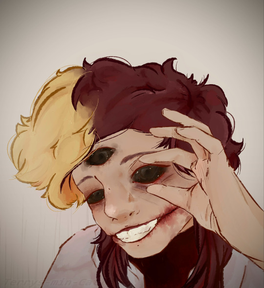
I like to learn different sorts of new things about medicine and how interesting diseases or injuries can be. And one day I came across a post about melanin and cancer. Melanoma can appear on skin or on any mucous membrane. Even in eyes. An eye cancer called eye melanoma. A tumor originating in one particular part of the eye, in the absence of any action, completely affects the eyeball, causing it to turn black. And this type of disease reminded me of Kevin!
So I present you my headcanon: Kevin's eye melanoma.
(In addition, if we imagine that in such an extreme stage of infection, Kevin miraculously remains alive (and unknown in any way to science can see). So: he may also has a mild form of exaphthalmia (bug-eyed), lack of peripheral vision and bad vision in general)
Bonus:
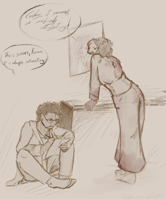
Small fast sketch bc why not. (Sorry for my bad handwriting lol)
#art#welcome to night vale#wtnv#wtnv kevin#kevin#wtnv carlos#carlos the scientist#wtdb#wtdb kevin#sketch#interesting fact#Moles and melanoma are two different things#despite the presence of melanocytes in both cases; one is a malignant tumor and the other is just a harmless cluster of cells#medicine#I love science facts#cw blood#wtnv headcanon#headcanon
34 notes
·
View notes
Text
cancer.
not a dental issue.
#squamous cell carcinoma of the mouth. they did not pull the tooth or do a biopsy bc there's really nothing else to gain from it now#that explains why the whs seemed so strange. it's not the genetic whs. he was likely having balance issues bc of the tumor#they're keeping him on the anti-inflammatory and as long as he's still eating and in good spirits we're just waiting it out#there's not much else left to do. if he stops eating or his quality of life becomes impacted we'll have to discuss putting him down#but until then it's just making sure he's comfortable for as long as possible#i'm just. i don't even know what to think. glad i didn't lose him today but also fucking devastated it's not something that can be fixed
9 notes
·
View notes
Text


Why is this man peak gender tho, like all of him is just a gender, he is pure euphoria.
#maws speaks#seriously#the funniest thing about this is he is literally cancer#hes from cells at work he's literally A CANCEROUS TUMOR#mogai#anti radqueer#actually mogai#mogai blog#mogai community#mogai friendly#xenogender euphoria#xenogender positivity#tw cancer mention#trans#transgender#gender euphoria
7 notes
·
View notes
Text


The Prince and Princess of Wales shared photos on their official shared Instagram account of a meeting with Liz, a 16-year-old aspiring photographer from Harrogate, England, who was diagnosed with cancer in January.
Liz has a desmoplastic small round cell tumor, a rare and aggressive form of cancer. She was given between six months and three years to live, per the BBC.
Prince William invited Liz to photograph the investiture ceremony he was hosting as part of her “photography bucket list.” (Credit: New York Post)
#Princess of Wales#catherine princess of wales#catherine middleton#kate middleton#prince william#prince of wales#british royal family#cancer#desmoplastic small round cell tumor#investiture ceremony#bucket list
9 notes
·
View notes
Text
...
#had an interesting conversation with my sister the other day. odd i guess bc my sister is pretty smart#on paper shes smarter than me. or at least less dyslexic than me#but she didnt seem to kno what cancer is. i mean like how it works. i mean. cancer is a mistake. a confluence of unfortunate accidents#leading to unrestrained cellular growth. when it metastasizes. when it moves to other parts of the body. those same cells continue growing#if u have smooth muscle cancer and it moves to your kidney. you body is trying to grow more smooth muscle on your kidney#at least as i understand it. and she asked why it wants to kill you. it doesnt want anything. it just is. its not a thing of malicious#intent. its neutral. it grows. it takes up resources. it takes up space. and it grows and grows until the organ it grows on stops#functioning properly. like a parasite she said. but no. not like a parasite. it grows like an empty space. a mass of flesh. a constant#obstructive pressure. it grows like only a tumor can. i dunno. it didnt seem to connect with her that this thing didnt want to kill our mom#but it did anyway. and she felt weird about how long she lived after they took her off any support. but thats how cancer kills#it stops an organ from functioning and most of those r important so it only takes one. so her heart kept beating for 12 more hrs bc it was#meant to beat for 40 more years. but not much it could do without working kidneys and without working blood#but that's life. that's death. that's nature. its all nutral even if it feels horrible to the individual.#i dunno. i thought it was interesting. shes 25 and her mother had cancer for 10 years so id think shed kno more#we're at a weird phase now bc its been a week since she died and everything feels normal. we'll see what happens at the wake this week#its been interesting for sure bc she was sick for 10 years but my parents didnt prepare at all for her to die#so my dad is scrambling to put together the pieces shr left behind to make sure that all the bills r paid and whatnot. he had to guess her#computer password. she didnt tell us what she wanted us to have. she didnt tell us the importance of her jewelry and who it belonged to#before her. i dunno. we're seeing the outline of my mothers Pathology in what she left behind. both in the physical objects and in the#feelings she imparted. i dunno. its been weird#unrelated
21 notes
·
View notes
Text
youtube
#Tumor biology#cancer progression#tumor microenvironment#metastasis#oncogenes#tumor suppressor genes#angiogenesis#apoptosis#cell proliferation#cancer biomarkers#tumor heterogeneity#cancer stem cells#immuno-oncology#molecular oncology#cancer metabolism#genomic instability#drug resistance#tumor invasion#epigenetic alterations#cancer immunotherapy.#Youtube
2 notes
·
View notes
Text
Send positive vibes and prayers to any deity that may be listening. Dixie has a little bump that's suddenly behaving like a mast cell tumor and I'm absolutely petrified. My stomach is in knots.
Luckily we have tons of Benadryl and even an itch spray, so hopefully we can keep the histamine response from going out of control.
I know the next steps. Full blood panel work up, and fine needle aspirate. Her vet opens soon, so I'll call and get an appointment just as soon as I can. But holy shit I'm so worried about my best girl.
5 notes
·
View notes
Text
they inject tumors into mice balls????????
#ok so im learning abt stem cells and apparently the best way to check if an obtained cell line is pluripotent#(that is can differentiate into any kind of human body cell)#is to inject them into mice with a severe immune deficiency and kind of??? see what grows out of them???#and like ideally (that is if the cells are actually pluripotent) youll get a tumor with all these different tissue types in it#which is called a teratoma. and its kind of gross but also fascinating#anyway apparently a good injection site for that whole experiment to happen is mouse testes. so. yeah. they inject mice balls with tumors#stem cell biology is kinda crazy ngl#thots
2 notes
·
View notes
Text
vent moment but my health is a bit worse than i let on, which is weird ik since it seems like complain about it all the time here, and apparently i also look sick, because two separate people in their 40s or 50s asked me, 24, if i needed their seat on the bus. kind of them. but humiliating nonetheless.
#medical stuff cw#i sat on the steps instead of taking their seat#vent cw#i have to take five different pills a day excluding birth control which i also take for health reasons but okay#i have to thank italy for its healthcare system because at least i dont have to pay a fuckton for all that stuff. except birthcontrol.#as i may have mentioned they found quite a bit of blood in my piss so im getting tested for ✨️cancer✨️#also because i've been having health issues which might be rated#my blood work is all off but i didnt get tested for tumoral cells specifically because i may have 'just' an autoimmune condition#so im on heavy duty antibiotics too now bc i also developed antibiotic resistance last year. anyway.#i need to take those and then they'll test my peepee again but this time they will also test explicitly for tumoral cells#because something is off and my previous blood work didnt point out what exactly#terrible anemia and other slightly-off numbers that however shouldnt be off considering my lifestyle#i eat almost everything. drink plenty of water. exercise. barely smoke. not even drinking anymore. i'm not too fat nor too skinny.#so. some of the numbers that are off dont really have a reason to be off which is why they are testing my blood and piss for cancer#but like. in 3 weeks because i have to take antibiotics and iron meds (not supplements. meds.) first#so my mind's trying to convince itself that i dont have a tumor. but what if i do? i know i dont. but not knowing makes me go insane#also i have to get tested for heart disease because that motherfucker is not working properly. doesnt pump enough blood to my brain.#i took an ekg and it came back pretty normal except for tachycardia#now i have to go get an holter ekg - but was told to wait until uni starts again bc i need that exam to be done when i have a daily routine#so basically they slap electrodes and shit on me for 24 hrs while i go do my shit around the city and then see how my heart behaved#because i cant stand without struggling to breathe and sometimes it happens when in laying down to.#sometimes i cant fall asleep because i cant breathe#at first the doc thought it might be a reflux issue but not. all good on that front.#so. we'll see. and i mean. i KNOW it's not cancer. like. i'd be dead by now bc i've been having these symptoms for five months#however. i dont know if it's not an autoimmune disease. and if it is? what am i gonna do?
5 notes
·
View notes
Text
Primary dural-based parafalcine diffuse large b-cell lymphoma mimicking meningioma by Amr El Mohamad in Journal of Clinical Case Reports Medical Images and Health Sciences
Abstract
Background: Primary dural-based diffuse large B-cell lymphoma is very rare. Only few cases were reported in the literature. Case presentation: Herein, we present a case of an immunocompetent patient with primary dural-based diffuse large B-cell lymphomas mimicking meningioma associated with ghost tumor phenomenon without any evidence of a systemic lymphoma. Conclusion: Primary central nervous system lymphomas are rare. Clinicians should always consider this lesion as a differential diagnosis if radiological findings are not indicative of typical one meningiomas.
Key words: Dural-based tumor, diffuse large B-cell lymphoma, ghost tumor, MATRIX regimen, central nervous system.
Introduction
Primary central nervous system lymphomas (CNSLs) (PCNSLs) are rare and account for 2%–5% of all brain tumor cases, whereas secondary CNSLs are more common [1,2]. One study has shown that the most common intraparenchymal histological type is diffuse large B-cell lymphoma, as among 26 patients with PCNSL, 25 had diffuse large B-cell lymphoma [3]. Although primary dural-based lymphomas are rare, the most common area of involvement is the cerebral hemispheres. Most dural-based lymphomas are secondary and present as extra-nodal systemic diffuse large B-cell lymphomas. Primary dural-based lymphomas are usually histologically marginal-zone lymphomas, representing a group of lymphomas that have been historically classified together because they appear to arise from post-germinal center and marginal-zone B cells and share a similar immunophenotype, and few cases were reported to be diffuse large B lymphomas [4]. Here, we present a case of an immunocompetent patient with primary dural-based diffuse large B-cell lymphomas mimicking meningioma associated with ghost tumor phenomenon without any evidence of systemic disease.
Case Presentation
A 58-year-old male individual previously healthy and immunocompetent presented with headache, recurrent vomiting, and memory problems lasting for 3 days. No loss of consciousness, seizure, subjective weakness, or fever was observed. On physical examination, the patient’s Glasgow coma scale score was 15; his pupils were 3 mm in diameter, equal, and reactive; and the patient had nominal aphasia without motor and sensory deficit. He had normal cerebellar functions, and cranial nerve exams revealed no deficit. Head computed tomography (CT) (Fig. 1) showed a 2.2 × 3.8 cm (transverse × anteroposterior) iso-dense lesion with internal hypodensity in the left parasagittal frontal region extending to the right frontal region. Extensive perilesional edema was observed with effacement of the sulci and mass effect on bilateral frontal horns, associated with 3-mm midline shift. Head magnetic resonance imaging (MRI) showed an isointense parasagittal lesion on T1 and heterogeneous intense on T2, with redemonstration of perifocal edema (Fig. 2). Head T1-weighted imaging with contrast enhancement (Fig. 3) showed a large, left frontal, parafalcine, irregular-shaped mass located below the superior sagittal sinus level. It measured 4 × 3 × 3.3 cm in anteroposterior, mediolateral, and craniocaudal, respectively. It showed diffusion restriction (Fig. 4). There was central hyperintensity on T2-weighted imaging, without post-contrast enhancement area representing cyst formation. It exerts a mass effect characterized by effacement of the adjacent sulci, compression of the left lateral ventricle, and a 3-mm shift of the midline structures to the right side, and the impression of our neuroradiologist was atypical meningioma. Regarding extensive edema, dexamethasone was started at a dose of 4 mg, thrice a day, and the patient was planned for craniotomy and resection of the tumor. Initially, the patient was reluctant to undergo surgery; however, subsequently, the patient agreed to undergo surgery after approximately 10 days. During surgery, parasagittal craniotomy was performed; however, to our surprise, no definite mass lesion was found at the proposed site, in contrast to the findings described on imaging. The falx was thinned out and partly deficient. A biopsy sample was obtained from this abnormally appearing falx. Moreover, we obtained biopsy samples under neuronavigation guidance from abnormally appearing tissue, which was completely intra-axial, deep down in the lesion visualized on navigation. On postoperative day 1, MRI head with contrast enhancement (Fig. 5) showed that the previously seen lesion had a significant regression in size. Its right frontal extension and adjacent enhanced meningeal tail showed size reduction. Moreover, some regression in the perilesional vasogenic edema was observed. A significant regression in the previously described enhancement was noted at the left-side lentiform nucleus and external capsule. The MR spectroscopy study showed an increased choline/N-acetyl aspartate ratio and elevated lactate level within the lesion.
The histopathology results of the first brain biopsy samples (Figs.6–7) obtained from the falx cerebri showed meningothelial hyperplasia with calcification and focal perivascular lymphocytic infiltrate composed of small and large, atypical lymphocytes. Immunohistochemical staining was performed; however, the area of interest disappeared. The pathology team recommended another fresh biopsy to have the final diagnosis and flowmetry studies. So, the patient underwent redo craniotomy using the same incision, and multiple biopsy samples were taken. The second fresh brain biopsy (Figs. 8–9) showed multiple brain fragments with predominant perivascular atypical lymphoid infiltrates. Most cells were medium to large with moderate cytoplasm, atypical irregular nuclei having vesicular chromatin, variably prominent nucleoli, and several mitoses, including atypical one. Necrotic areas were also seen. Immunohistochemistry of the second biopsy (Fig.10 A-D) showed that atypical perivascular cells were positive for CD45, CD20, CD79a, BCl2, BCl6, MUM1, OCT2, and C-MYC, and negative for CD10, CD21, TDT, ALK1, EBV-LMP1, CD3, and CD5; however, few reactive/residual lymphocytes were positive for these enzymes. Moreover, 80% of lymphoid cellular nuclei were positive for Ki67. These findings were consistent with diffuse large B-cell lymphoma, not otherwise specified.
Whole-body positron emission tomography (PET) showed intense fluorodeoxyglucose (FDG) uptake higher than that in the healthy brain cortex, without evidence of coexisting systemic disease. In addition to PET scan, contrast-enhanced chest, abdomen, pelvis CT did not show any other lesions in the body; furthermore, workup for viral markers and autoimmune conditions were all unremarkable, thus confirming the diagnosis of “primary dural-based diffuse large B-cell lymphoma,” distinguishing it from secondary CNSL. The patient was transferred to the Oncology Department and started on three cycles of the methotrexate, cytarabine, thiotepa, and rituximab (MATRIX) protocol, which is the current standard treatment regimen for PCNSLs [5]. Three months after the diagnosis and after receiving two cycles of the MATRIX protocol, brain MRI with contrast enhancement (Fig. 11A, B) showed regression of the lesion, and PET scan showed complete metabolic resolution in terms of decreased FDG activity of the previously seen PCNSL without signs of lymphoma activity elsewhere. Subsequently, the patient received the third cycle of the MATRIX protocol without specific complications. Two weeks later, autologous stem cell transplantation (50 × 106/kg) was performed as part of the consolidation phase of treatment. Six weeks later, conditioning chemotherapy with carmustine–thiotepa was administered, followed by stem cell infusion (CD34 = 12 million/kg). The post-transplant course was complicated with mucositis, folliculitis, diarrhea, febrile neutropenia, and prolonged thrombocytopenia. Two months after transplantation, PET scan was repeated and showed complete metabolic resolution of initially seen PCNSL involvement. Currently, the patient is being followed by the hematology team; the patient is in good health and remission. The last outpatient follow-up was 8 months after the first surgery. The patient was seen by the vascular surgery (for permcath removal) and oncology teams. At this time, the patient was stable with complete remission; then, the patient was lost to follow-up. Another head MRI was performed and showed almost total regression of the lesion.
Discussion
Lymphomas in CNS are classified as primary, arising de novo from brain parenchyma, leptomeninges, eye, and spinal cord and as secondary to systemic lymphoma, which can be dural-based lesions. Secondary CNSLs are more common than PCNSLs. Most PCNSLs are intraparenchymal diffuse large B-cell lymphomas with a predilection to occur in the frontal lobe and then deep nucleic and periventricular locations; the infratentorial cerebellum is the most common location. However, primary dural-based lymphomas are rare, and even when found, they are histologically marginal-zone lymphomas. Few cases of primary dural-based diffuse large B-cell lymphoma have been reported in the literature [4,6]. Furthermore, PCNSLs are more common in immunocompromised patients with a mean age of 34 years, and they occur in immunocompetent individuals at an older age with a mean of 52 years [7]. The patient in this case report was 58 years old and immunocompetent without significant previous medical conditions. The latest review of the literature on primary dural-based lymphoma has been conducted by Quinn et al., who have found only 24 reported cases of primary dural-based diffuse large B-cell lymphoma, which confirms the rarity of the disease and subsequently the limited knowledge regarding this disease entity [8]. CNSLs have rapid response to steroids with shrinkage in size and initial remission [9]. Moreover, the initial response to steroids is associated with a better response to chemoradiotherapy and good prognosis [9]. In the patient in this case report, there was an unintentional delay of surgery for approximately 10 days, and the patient was on steroids (dexamethasone). In this case report, the failure to identify a discrete lesion of the size expected as perceived on initial imaging, despite proper surgical planning using neuronavigation, was probably due to the rapid regression of the tumor in response to steroids. This phenomenon agrees with the scientific literature reporting about the disappearance of lymphomas in response to steroids (ghost tumors) [10,11]. The pathogenesis of primary dural-based lymphoma remains unknown as there is no lymphoid tissue in the dura. It is hypothesized that it is related to chronic infection, autoimmune disease, or chronic inflammatory condition, which recruits polyclonal lymphocytes resulting in monoclonal lymphomas [6]. In contrast, the patient in this case report did not have any chronic conditions. All workups were negative, including the entire viral panel and autoimmune markers. Basic research is needed to determine the etiology of PCNSL, especially dural-based lymphomas. In the patient in this case report, the initial radiological findings were mimicking those of a meningioma: dural-based and uniformly enhanced. There was significant surrounding edema, significant diffusion restriction, and blooming in susceptibility-weighted image, which goes more with higher-grade meningioma or another high-grade lesion. One review has shown that primary dural-based lymphomas can display the “dural tail “sign, further confusing the preoperative diagnosis with meningioma [12], which did happen in the patient in this case report. Therefore, we suggest that in case of a dural-based lesion that has non-typical features of grade 1 meningioma, clinicians should consider lymphoma in the differential diagnosis and avoid steroids unless necessary due to edema and mass effect keeping in mind the ghost tumor phenomena of lymphoma.
The role of surgery in PCNSLs is limited mainly to histological diagnosis through biopsy or tumor debulking in case of increased intracranial pressure or impending brain herniation. Some studies have shown no benefit of complete surgical resection of PCNSLs; however, a recent systematic review of 244 articles has shown evidence in support of cytoreductive surgery [13]. Previously, whole-brain radiotherapy (WBRT) was the recommended treatment; however, this treatment modality resulted in a high rate of relapse and a decrease in performance status and cognitive impairment, and with the improvement in survival with high-dose methotrexate, WBRT is no longer recommended. Currently, newly diagnosed PCNSLs are initially treated with induction chemotherapy until complete radiological response, followed by consolidation therapy, to prolong the overall survival [14]. The International Extra Nodal Lymphoma Study Group-32 trial has shown that a methotrexate-based MATRIX regimen results in a good outcome and control rate in PCNSL [5], and it is the standard induction chemotherapy. Ferreri AJM, in his article “The role of autologous stem cell transplantation in PCNSL” has compared various consolidation phase treatment modalities, including beam radiation, carmustine–thiotepa regimens, and autologous stem cell transplantation, and the results showed that autologous stem cell transplantation resulted in good outcomes [15]. The patient in this case report showed a good response to treatment with almost total resolution of PCNSL with three cycles of MATRIX chemotherapy, followed by conditioning chemotherapy with stem cell infusion.
Conclusion
PCNSL is a rare entity. Clinicians should always consider it in differential diagnosis of meningioma if the radiological findings are not typical for meningioma. When there is a high index of suspicion of lymphoma, repeating neuroimaging, particularly MRI, before surgery, especially if the surgery is delayed while the patient is on steroids, may help develop a better management plan while dealing with this rare lesion. In case of lesion disappearance, falx biopsy can be an option. The aim of surgery in PCNSL is mainly biopsy or debulking to decrease intracranial pressure in case of significant mass effect.
List of abbreviation:
Primary central nervous system lymphomas (PCNSLs)
Central nervous system lymphomas (CNSLs)
Head computed tomography (CT)
magnetic resonance imaging (MRI)
positron emission tomography (PET)
fluorodeoxyglucose (FDG)
whole-brain radiotherapy (WBRT)
#Dural-based tumor#diffuse large B-cell lymphoma#ghost tumor#MATRIX regimen#central nervous system#jcrmhs#Clinical decision making#Is Journal of Clinical Case Reports Medical Images and Health Sciences PubMed indexed
2 notes
·
View notes