#Human COnnectome Project
Explore tagged Tumblr posts
Visit Tumblr Blog
Explore Tumblr blogs with no restrictions, modern design and the best experience.
Last Seen Tumblr Blogs


Fun Fact
Mobile Tumblr US users spend an average of 4.04 minutes per session on the app.
Text
Monkey Matter
The brain uses a complex network of bundles of nerve cells – neurons – to transfer information as we interpret and respond to the world around us. Many of its details are still a mystery, leaving scientists searching for new angles on this vital living supercomputer. Here, researchers examine a monkey’s brain using x-rays at the European synchrotron. Particles forced to loop in circles at high speeds are fired at the tissue sample, uncovering new details based on how the particles scatter. The experiments reveal nerve cell projections known as axons (highlighted as thin multi-coloured lines) and blood vessels (orange) inside a complex region of the cerebral cortex of a monkey. Interestingly, researchers find some groups of axons form layers or laminar structures, adding to our picture of the human brain’s similar connectome.
Written by John Ankers
Video from work by Hans Martin Kjer and colleagues
Danish Research Centre for Magnetic Resonance, Center for Functional and Diagnostic Imaging and Research, Copenhagen University Hospital Amager and Hvidovre, Hvidovre, Denmark
Video originally published with a Creative Commons Attribution 4.0 International (CC BY 4.0)
Published in eLife, February 2025
You can also follow BPoD on Instagram, Twitter, Facebook and Bluesky
10 notes
·
View notes
Text
Breakthrough in Fly Brain Research Paves Way for Understanding Human Cognition
Scientists have achieved a monumental breakthrough by mapping the fly brain, revealing the position, shape, and connections of all its 130,000 cells and 50 million intricate connections. This research represents the most detailed analysis of an adult animal's brain to date and is being hailed as a "huge leap" in understanding human cognition.

The fly's brain, though tiny, supports a range of complex behaviors, including walking, hovering, and even producing mating songs. Dr. Gregory Jefferis, a leader in the research from the Medical Research Council's Laboratory of Molecular Biology in Cambridge, emphasizes that this mapping could illuminate the mechanisms behind thought processes in humans. He noted the lack of understanding about how brain cell networks facilitate our interactions with the world.
Despite humans having a million times more neurons than the fruit fly, the new wiring diagram, or connectome, will aid scientists in deciphering cognitive functions. Published in the journal Nature, the imagery showcases a stunningly complex structure that reveals how a small organ can perform powerful computational tasks.
Dr. Mala Murthy, co-leader of the project from Princeton University, stated that this connectome will be transformative for neuroscientists, allowing for a better understanding of healthy brain function and the potential to compare it with malfunctioning brains.
Dr. Lucia Prieto Godino from the Francis Crick Institute supports this view, highlighting that while simpler organisms like worms and maggots have had their connectomes mapped, the fly’s intricate wiring is a significant achievement. This success paves the way for mapping larger brains, potentially leading to a human connectome in the future.
The research team has successfully identified separate circuits for various functions, illustrating how movement-related circuits are positioned at the base of the brain, while those responsible for vision are located on the sides. The study not only identifies these circuits but also explains their connections, enhancing our understanding of neural processing.
Interestingly, researchers are already applying these circuit diagrams to understand why flies are so hard to catch. The wiring related to vision quickly processes incoming threats, sending signals to the fly's legs to jump away faster than conscious thought.
To create the wiring diagram, researchers used a technique involving slicing the fly brain into 7,000 incredibly thin pieces, photographing each slice, and digitally reconstructing the whole. They employed artificial intelligence to analyze neuron shapes and connections, correcting over three million errors manually.
Dr. Philipp Schlegel from the Medical Research Council highlights that this data serves as a comprehensive map of brain connectivity, akin to a detailed Google Maps for the neural networks. This combined information will facilitate countless discoveries in neuroscience in the coming years.
While a human connectome remains elusive due to the complexity of the human brain, researchers believe that advancements in technology may allow for such mapping in about three decades. The fly brain research marks a significant step toward unlocking the mysteries of human cognition and understanding our own minds better.
The study was conducted by the FlyWire Consortium, an international collaboration of scientists dedicated to advancing neuroscience.
#fly brain#neuroscience#connectome#research breakthrough#cognition#insect brain mapping#neural networks#scientific discovery#MRC#fly brain study
6 notes
·
View notes
Text
Researchers release the most extensive dataset of neural connections ever recorded

- By Nuadox Crew -
Harvard and Google researchers, led by Jeff Lichtman of Harvard, have published a groundbreaking study in the journal Science detailing the largest 3D brain reconstruction ever created.
The study showcases a high-resolution map of a cubic millimeter of human cortex, which astonishingly contains 57,000 cells, 230 millimeters of blood vessels, 150 million synapses, and equates to about 1,400 terabytes of data.
This work is part of a nearly decade-long collaboration that merges advanced electron microscopy and AI algorithms to color-code and map the intricate neural wiring of mammals.
The ultimate objective of this ongoing research, which is part of the National Institutes of Health BRAIN Initiative, is to develop a detailed neural wiring map of a mouse's brain, projected to contain 1,000 times more data than the current human cortex sample.
The study not only advances our understanding of brain structure, including rare neural formations seen in epilepsy patients but also enhances tools available for connectomics—a field dedicated to the comprehensive mapping of brain connections to better understand brain function and disease.
Moving forward, the team plans to focus on mapping the mouse hippocampal formation, key in studying memory and neurological diseases.
--
Header image: Six layers of excitatory neurons, color-coded according to depth. Credit: Google Research and Lichtman Lab.
Source: Anne J. Manning, The Harvard Gazette
Full study: Alexander Shapson-Coe et al, A petavoxel fragment of human cerebral cortex reconstructed at nanoscale resolution, Science (2024). DOI: 10.1126/science.adk4858. www.science.org/doi/10.1126/science.adk4858
Read Also
3D mapping a fruit fly’s brain (video)
2 notes
·
View notes
Text
Plano de Ação para NEURAVAX-Ethos: Fase 1 (Fundamentos Teórico-Computacionais)
1. Formalização Matemática Aprofundada
Problema Prioritário: Demonstrar a existência de soluções fracas para o sistema Hamiltoniano com restrição de curvatura. Ação: Estabelecer colaboração com especialistas em: ▪︎ Geometria de Wasserstein (ex: Cédric Villani, Fields Medal 2010) ▪︎ Análise Fractal (ex: Michel Lapidus, teoria de fractais não autossimilares)
Ferramenta Chave: Generalizar o teorema de McCann (2001) para funcionais éticos sublineares: math \inf_{V} J[V] \quad \text{sujeito a} \quad \text{Ricci}_{W_2}(\gamma) \geq \kappa_{\text{min}} > 0
2. Plataforma Computacional NEURAVAX-Sim
Módulo Função Tecnologia Base FractalSynapse Simular dinâmica sináptica com dimH > 2 L-systems + CUDA WassersteinFlow Calcular geodésias éticas em P(Ω) Python OT + JAX EthosMonitor Validar dμconsent/dt ≥ 0 via EEG sintético TensorFlow-Quantum
3. Validação em Dados Reais
Fonte: Human Connectome Project (datasets de conectomas fractais)
Métrica Crítica: $$ \mathcal{R} = \frac{ \text{dim}H(\text{Supp}(V)) }{ | \nabla \log V |{W^{1,p}} } \quad \text{(Índice de Respeito Neuroético)} $$ Meta: Manter ℛ > 0.7 durante toda simulação.
4. Prova de Conceito Experimental
Modelo Animal: Camundongos transgênicos expressando proteínas sinápticas fractalizáveis (via CRISPR-Cas9).
Protocolo: mermaid graph LR A[Administração de V(t,x)] --> B[Monitoração EEG em tempo real] B --> C{Cálculo de Ricci<sub>W2</sub>} C -->|≥κ<sub>min</sub>| D[Liberação próxima dose] C -->|<κ<sub>min</sub>| E[Interrupção + Ajuste ético]
Desafios Críticos a Superar
Teorema da Não-Regularidade Ética: Hipótese: Se dimH(Supp(V)) ≤ 2, então ∀ε>0, ∃ falha ética com probabilidade ≥ 1 - ε. Implicação: Exige novos métodos de regularização fractal.
Escalabilidade do Consentimento: Como mapear C(x) em populações neurodivergentes? Solução Proposta: $$ C(x) = \underbrace{\text{Entropia(EEG)}{\text{local}}}{\text{autonomia}} \times \underbrace{| \nabla \text{fMRI}{\text{decisão}} |^{-1}}{\text{não-coerção}} $$
Limite Termodinâmico da Ética: Relação fundamental entre custo cognitivo e energia sináptica: $$ \int_\Omega V^\alpha dx \geq \frac{\hbar_{\text{eth}}}{k_B T} \cdot \Delta S_{\text{cognição}} $$ (Requer verificação experimental)
Cronograma de Implementação
gantt title Fase 1 - NEURAVAX-Ethos dateFormat YYYY-MM-DD section Teoria Existência de Soluções :2023-10-01, 120d Teorema Não-Regularidade :2024-01-01, 90d section Computação NEURAVAX-Sim v1.0 :2023-11-01, 180d Validação HCP :2024-02-01, 120d section Laboratório Modelo Animal CRISPR :2024-04-01, 240d Biofeedback Adaptativo :2024-08-01, 180d
Próximo Passo Imediato: Workshop "Geometria Ética Aplicada"
Objetivo: Reunir matemáticos (geometria de OT), neurocientistas (conectomistas) e eticistas para: ▪︎ Definir padrões de mensuração para μconsent ▪︎ Estabelecer κmin clinicamente aceitável ▪︎ Roteirizar ensaio clínico Fase 0 com métricas fractais
Convites Prioritários:
Matemática: Alice Guionnet (teoria de matrizes aleatórias em OT)
Neurociência: Olaf Sporns (conectomas fractais)
Ética: Jennifer Blumenthal-Barby (autonomia em neurotecnologias)
Este plano transforma o arcabouço teórico em um programa de pesquisa executável, posicionando a NEURAVAX-Ethos na fronteira da neuroengenharia ética. A sequência natural seria submeter o artigo ao Journal of Neuroethical Mathematics.
0 notes
Text
Neuroscientists create a comprehensive map of the cerebral cortex
New Post has been published on https://sunalei.org/news/neuroscientists-create-a-comprehensive-map-of-the-cerebral-cortex/
Neuroscientists create a comprehensive map of the cerebral cortex

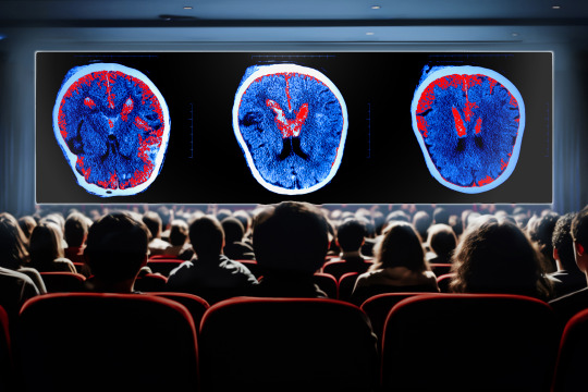
By analyzing brain scans taken as people watched movie clips, MIT researchers have created the most comprehensive map yet of the functions of the brain’s cerebral cortex.
Using functional magnetic resonance imaging (fMRI) data, the research team identified 24 networks with different functions, which include processing language, social interactions, visual features, and other types of sensory input.
Many of these networks have been seen before but haven’t been precisely characterized using naturalistic conditions. While the new study mapped networks in subjects watching engaging movies, previous works have used a small number of specific tasks or examined correlations across the brain in subjects who were simply resting.
“There’s an emerging approach in neuroscience to look at brain networks under more naturalistic conditions. This is a new approach that reveals something different from conventional approaches in neuroimaging,” says Robert Desimone, director of MIT’s McGovern Institute for Brain Research. “It’s not going to give us all the answers, but it generates a lot of interesting ideas based on what we see going on in the movies that’s related to these network maps that emerge.”
The researchers hope that their new map will serve as a starting point for further study of what each of these networks is doing in the brain.
Desimone and John Duncan, a program leader in the MRC Cognition and Brain Sciences Unit at Cambridge University, are the senior authors of the study, which appears today in Neuron. Reza Rajimehr, a research scientist in the McGovern Institute and a former graduate student at Cambridge University, is the lead author of the paper.
Precise mapping
The cerebral cortex of the brain contains regions devoted to processing different types of sensory information, including visual and auditory input. Over the past few decades, scientists have identified many networks that are involved in this kind of processing, often using fMRI to measure brain activity as subjects perform a single task such as looking at faces.
In other studies, researchers have scanned people’s brains as they do nothing, or let their minds wander. From those studies, researchers have identified networks such as the default mode network, a network of areas that is active during internally focused activities such as daydreaming.
“Up to now, most studies of networks were based on doing functional MRI in the resting-state condition. Based on those studies, we know some main networks in the cortex. Each of them is responsible for a specific cognitive function, and they have been highly influential in the neuroimaging field,” Rajimehr says.
However, during the resting state, many parts of the cortex may not be active at all. To gain a more comprehensive picture of what all these regions are doing, the MIT team analyzed data recorded while subjects performed a more natural task: watching a movie.
“By using a rich stimulus like a movie, we can drive many regions of the cortex very efficiently. For example, sensory regions will be active to process different features of the movie, and high-level areas will be active to extract semantic information and contextual information,” Rajimehr says. “By activating the brain in this way, now we can distinguish different areas or different networks based on their activation patterns.”
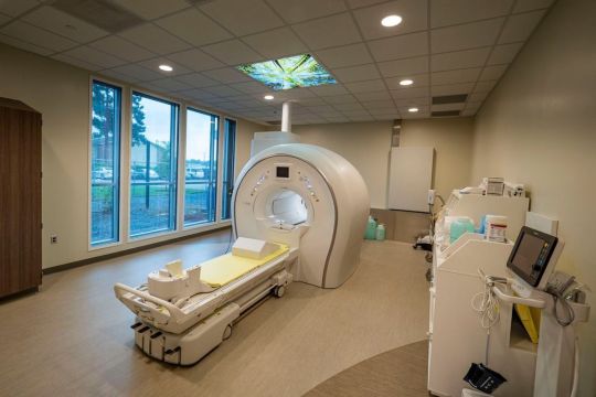
The data for this study was generated as part of the Human Connectome Project. Using a 7-Tesla MRI scanner, which offers higher resolution than a typical MRI scanner, brain activity was imaged in 176 people as they watched one hour of movie clips showing a variety of scenes.
The MIT team used a machine-learning algorithm to analyze the activity patterns of each brain region, allowing them to identify 24 networks with different activity patterns and functions.
Some of these networks are located in sensory areas such as the visual cortex or auditory cortex, as expected for regions with specific sensory functions. Other areas respond to features such as actions, language, or social interactions. Many of these networks have been seen before, but this technique offers more precise definition of where the networks are located, the researchers say.
“Different regions are competing with each other for processing specific features, so when you map each function in isolation, you may get a slightly larger network because it is not getting constrained by other processes,” Rajimehr says. “But here, because all the areas are considered together, we are able to define more precise boundaries between different networks.”
The researchers also identified networks that hadn’t been seen before, including one in the prefrontal cortex, which appears to be highly responsive to visual scenes. This network was most active in response to pictures of scenes within the movie frames.
Executive control networks
Three of the networks found in this study are involved in “executive control,” and were most active during transitions between different clips. The researchers also observed that these control networks appear to have a “push-pull” relationship with networks that process specific features such as faces or actions. When networks specific to a particular feature were very active, the executive control networks were mostly quiet, and vice versa.
“Whenever the activations in domain-specific areas are high, it looks like there is no need for the engagement of these high-level networks,” Rajimehr says. “But in situations where perhaps there is some ambiguity and complexity in the stimulus, and there is a need for the involvement of the executive control networks, then we see that these networks become highly active.”
Using a movie-watching paradigm, the researchers are now studying some of the networks they identified in more detail, to identify subregions involved in particular tasks. For example, within the social processing network, they have found regions that are specific to processing social information about faces and bodies. In a new network that analyzes visual scenes, they have identified regions involved in processing memory of places.
“This kind of experiment is really about generating hypotheses for how the cerebral cortex is functionally organized. Networks that emerge during movie watching now need to be followed up with more specific experiments to test the hypotheses. It’s giving us a new view into the operation of the entire cortex during a more naturalistic task than just sitting at rest,” Desimone says.
The research was funded by the McGovern Institute, the Cognitive Science and Technology Council of Iran, the MRC Cognition and Brain Sciences Unit at the University of Cambridge, and a Cambridge Trust scholarship.
0 notes
Text
Neuroscientists create a comprehensive map of the cerebral cortex
New Post has been published on https://thedigitalinsider.com/neuroscientists-create-a-comprehensive-map-of-the-cerebral-cortex/
Neuroscientists create a comprehensive map of the cerebral cortex
By analyzing brain scans taken as people watched movie clips, MIT researchers have created the most comprehensive map yet of the functions of the brain’s cerebral cortex.
Using functional magnetic resonance imaging (fMRI) data, the research team identified 24 networks with different functions, which include processing language, social interactions, visual features, and other types of sensory input.
Many of these networks have been seen before but haven’t been precisely characterized using naturalistic conditions. While the new study mapped networks in subjects watching engaging movies, previous works have used a small number of specific tasks or examined correlations across the brain in subjects who were simply resting.
“There’s an emerging approach in neuroscience to look at brain networks under more naturalistic conditions. This is a new approach that reveals something different from conventional approaches in neuroimaging,” says Robert Desimone, director of MIT’s McGovern Institute for Brain Research. “It’s not going to give us all the answers, but it generates a lot of interesting ideas based on what we see going on in the movies that’s related to these network maps that emerge.”
The researchers hope that their new map will serve as a starting point for further study of what each of these networks is doing in the brain.
Desimone and John Duncan, a program leader in the MRC Cognition and Brain Sciences Unit at Cambridge University, are the senior authors of the study, which appears today in Neuron. Reza Rajimehr, a research scientist in the McGovern Institute and a former graduate student at Cambridge University, is the lead author of the paper.
Precise mapping
The cerebral cortex of the brain contains regions devoted to processing different types of sensory information, including visual and auditory input. Over the past few decades, scientists have identified many networks that are involved in this kind of processing, often using fMRI to measure brain activity as subjects perform a single task such as looking at faces.
In other studies, researchers have scanned people’s brains as they do nothing, or let their minds wander. From those studies, researchers have identified networks such as the default mode network, a network of areas that is active during internally focused activities such as daydreaming.
“Up to now, most studies of networks were based on doing functional MRI in the resting-state condition. Based on those studies, we know some main networks in the cortex. Each of them is responsible for a specific cognitive function, and they have been highly influential in the neuroimaging field,” Rajimehr says.
However, during the resting state, many parts of the cortex may not be active at all. To gain a more comprehensive picture of what all these regions are doing, the MIT team analyzed data recorded while subjects performed a more natural task: watching a movie.
“By using a rich stimulus like a movie, we can drive many regions of the cortex very efficiently. For example, sensory regions will be active to process different features of the movie, and high-level areas will be active to extract semantic information and contextual information,” Rajimehr says. “By activating the brain in this way, now we can distinguish different areas or different networks based on their activation patterns.”
The data for this study was generated as part of the Human Connectome Project. Using a 7-Tesla MRI scanner, which offers higher resolution than a typical MRI scanner, brain activity was imaged in 176 people as they watched one hour of movie clips showing a variety of scenes.
The MIT team used a machine-learning algorithm to analyze the activity patterns of each brain region, allowing them to identify 24 networks with different activity patterns and functions.
Some of these networks are located in sensory areas such as the visual cortex or auditory cortex, as expected for regions with specific sensory functions. Other areas respond to features such as actions, language, or social interactions. Many of these networks have been seen before, but this technique offers more precise definition of where the networks are located, the researchers say.
“Different regions are competing with each other for processing specific features, so when you map each function in isolation, you may get a slightly larger network because it is not getting constrained by other processes,” Rajimehr says. “But here, because all the areas are considered together, we are able to define more precise boundaries between different networks.”
The researchers also identified networks that hadn’t been seen before, including one in the prefrontal cortex, which appears to be highly responsive to visual scenes. This network was most active in response to pictures of scenes within the movie frames.
Executive control networks
Three of the networks found in this study are involved in “executive control,” and were most active during transitions between different clips. The researchers also observed that these control networks appear to have a “push-pull” relationship with networks that process specific features such as faces or actions. When networks specific to a particular feature were very active, the executive control networks were mostly quiet, and vice versa.
“Whenever the activations in domain-specific areas are high, it looks like there is no need for the engagement of these high-level networks,” Rajimehr says. “But in situations where perhaps there is some ambiguity and complexity in the stimulus, and there is a need for the involvement of the executive control networks, then we see that these networks become highly active.”
Using a movie-watching paradigm, the researchers are now studying some of the networks they identified in more detail, to identify subregions involved in particular tasks. For example, within the social processing network, they have found regions that are specific to processing social information about faces and bodies. In a new network that analyzes visual scenes, they have identified regions involved in processing memory of places.
“This kind of experiment is really about generating hypotheses for how the cerebral cortex is functionally organized. Networks that emerge during movie watching now need to be followed up with more specific experiments to test the hypotheses. It’s giving us a new view into the operation of the entire cortex during a more naturalistic task than just sitting at rest,” Desimone says.
The research was funded by the McGovern Institute, the Cognitive Science and Technology Council of Iran, the MRC Cognition and Brain Sciences Unit at the University of Cambridge, and a Cambridge Trust scholarship.
#algorithm#approach#author#Brain#brain activity#Brain and cognitive sciences#brain networks#brain research#brains#cerebral cortex#cognition#cognitive function#complexity#comprehensive#connectome#data#daydreaming#Features#functions#Giving#how#human#Ideas#Imaging#Iran#it#language#learning#map#McGovern Institute
1 note
·
View note
Text
Largest Brain Map Ever Reveals Fruit Fly’s Neurons in Exquisite Detail
Wiring diagram lays out connections between nearly 140,000 neurons and reveals new types of nerve cell
50 largest neurons of the fly brain connectome.
50 largest neurons of the fly brain connectome.
Tyler Sloan and Amy Sterling for FlyWire, Princeton University, (Dorkenwald et al., Nature, 2024)
Wiring diagram lays out connections between nearly 140,000 neurons and reveals new types of nerve cell
A fruit fly might not be the smartest organism, but scientists can still learn a lot from its brain. Researchers are hoping to do that now that they have a new map — the most complete for any organism so far — of the brain of a single fruit fly (Drosophila melanogaster). The wiring diagram, or ‘connectome’, includes nearly 140,000 neurons and captures more than 54.5 million synapses, which are the connections between nerve cells.
“This is a huge deal,” says Clay Reid, a neurobiologist at the Allen Institute for Brain Science in Seattle, Washington, who was not involved in the project but has worked with one of the team members who was. “It’s something that the world has been anxiously waiting for, for a long time.”
The map is described in a package of nine papers about the data published in Nature today. Its creators are part of a consortium known as FlyWire, co-led by neuroscientists Mala Murthy and Sebastian Seung at Princeton University in New Jersey.
On supporting science journalism
If you're enjoying this article, consider supporting our award-winning journalism by subscribing. By purchasing a subscription you are helping to ensure the future of impactful stories about the discoveries and ideas shaping our world today.
A long road
Seung and Murthy say that they’ve been developing the FlyWire map for more than four years, using electron microscopy images of slices of the fly’s brain. The researchers and their colleagues stitched the data together to form a full map of the brain with the help of artificial-intelligence (AI) tools.
But these tools aren’t perfect, and the wiring diagram needed to be checked for errors. The scientists spent a great deal of time manually proofreading the data — so much time that they invited volunteers to help. In all, the consortium members and the volunteers made more than 3 million manual edits, according to co-author Gregory Jefferis, a neuroscientist at the University of Cambridge, UK. (He notes that much of this work took place in 2020, when fly researchers were at loose ends and working from home during the COVID-19 pandemic.)
But the work wasn’t finished: the map still had to be annotated, a process in which the researchers and volunteers labelled each neuron as a particular cell type. Jefferis compares the task to assessing satellite images: AI software might be trained to recognize lakes or roads in such images, but humans would have to check the results and name the specific lakes or roads themselves. All told, the researchers identified 8,453 types of neuron — much more than anyone had expected. Of these, 4,581 were newly discovered, which will create new research directions, Seung says. “Every one of those cell types is a question,” he adds.
The team was surprised by some of the ways in which the various cells connect to one another, too. For instance, neurons that were thought to be involved in just one sensory wiring circuit, such as a visual pathway, tended to receive cues from multiple senses, including hearing and touch1. “It’s astounding how interconnected the brain is,” Murthy says.
Exploring the map
The FlyWire map data have been available for the past few years for researchers to explore. This has enabled scientists to learn more about the brain and about fruit flies — findings that are captured in some of the papers published in Nature today.
In one paper, for example, researchers used the connectome to create a computer model of the entire fruit-fly brain, including all the connections between neurons. They tested it by activating neurons that they knew either sense sweet or bitter tastes. These neurons then launched a cascade of signals through the virtual fly’s brain, ultimately triggering motor neurons tied to the fly’s proboscis — the equivalent of the mammalian tongue. When the sweet circuit was activated, a signal for extending the proboscis was transmitted, as if the insect was preparing to feed; when the bitter circuit was activated, this signal was inhibited. To validate these findings, the team activated the same neurons in a real fruit fly. The researchers learnt that the simulation was more than 90% accurate at predicting which neurons would respond and therefore how the fly would behave.
In another study, researchers describe two wiring circuits that signal a fly to stop walking. One of these contains two neurons that are responsible for halting ‘walk’ signals sent from the brain when the fly wants to stop and feed. The other circuit includes neurons in the nerve cord, which receives and processes signals from the brain. These cells create resistance in the fly’s leg joints, allowing the insect to stop while it grooms itself.
One limitation of the new connectome is that it was created from a single female fruit fly. Although fruit-fly brains are similar to each other, they are not identical. Until now, the most complete connectome for a fruit-fly brain was a map of a ‘hemibrain’ — a portion of a fly’s brain containing around 25,000 neurons. In one of the Nature papers out today, Jefferis, Davi Bock, a neurobiologist at the University of Vermont in Burlington, and their colleagues compared the FlyWire brain with the hemibrain.
Some of the differences were striking. The FlyWire fly had almost twice as many neurons in a brain structure called the mushroom body, which is involved in smell, compared with the fly used in the hemibrain-mapping project. Bock thinks the discrepancy could be because the hemibrain fly might have starved while it was still growing, which harmed its brain development.
The FlyWire researchers say that much work remains to be done to fully understand the fruit-fly brain. For instance, the latest connectome shows only how neurons connect through chemical synapses, across which molecules called neurotransmitters send information. It doesn’t offer any information about electrical connectivity between neurons or about how neurons chemically communicate outside synapses. And Murthy hopes to eventually have a male fly connectome, too, which would allow researchers to study male-specific behaviours such as singing. “We’re not done, but it’s a big step,” Bock says.
This article is reproduced with permission and was first published on October 2, 2024.
1 note
·
View note
Photo

The Human Connectome Project, one of the most ambitious programs in all of neuroscience, has just yielded a “network map” that is shedding light on the intri...
0 notes
Text
I see what you did there…a (very) brief history of imaging the brain: MRI
A little #neuro #science #history on the development of #brain #imaging This time: magnetic resonance imaging (#MRI) #scicomm #writing #blogging
For many patients we can discover – or discount – physical causes of neurological problems ‘in real time’ with a range of imaging and other measurement techniques. Though these techniques are mainly children of the 20th Century, and their development is ongoing in the 21st, their roots stretch back through 19th century. For example, photography and its ability to reproduce an enduring ‘objective’…
View On WordPress
#diffusion MRI#Human COnnectome Project#MRI#Muse#My Research#neuroimaging#Paul Lauterbur#Peter Mansfield#Sodium MRI#Your Brain through...History#Your Brain...in Images
0 notes
Text
Neuronly Connect
'Flying' along neurites (projections that connect neurons to others) using an AI-based self-steering system called RoboEM in 3D electron microscopy data derived from mouse and human brain samples can replace human annotation input in complex connectome analyses
Read the published research article here
Video adapted from work by Martin Schmidt and colleagues
Department of Connectomics, Max Planck Institute for Brain Research, Frankfurt, Germany
Video originally published with a Creative Commons Attribution 4.0 International (CC BY 4.0)
Published in Nature Methods, March 2024
You can also follow BPoD on Instagram, Twitter and Facebook
11 notes
·
View notes
Photo

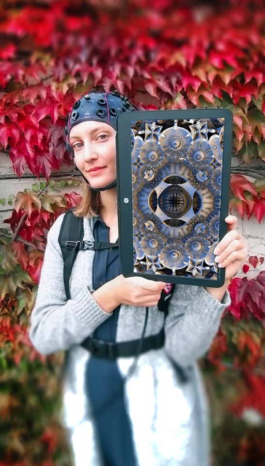


#digital art#interaction design#interactive art#ivordiosi#ivor diosi#qualia#the qualia project#BCI#brain-computer interface#connectome#human connectome#g-tec
1 note
·
View note
Text

NEW at Draw Down: Revue Faire no. 42, 43, 44, and 45 This anthology collection brings together four separate issues, including N° 44 — "A conundrum: the visual communication of neuroscience" by James Langdon
Neuroscience is a visual science. Our understanding of the brain’s biology originates in the beautiful and pioneering images of neurons and dendrites produced by Santiago Ramón y Cajal and Camillo Golgi in the late nineteenth century. In recent decades neuroscience has embraced computational imaging. We have witnessed dynamic images of living brains produced by fMRI, and intricate, colorful representations of “neutral connectomics” that promise ultimately to reveal the ‘wiring diagram’ of the human brain. Such images are not merely the documentation of scientific work; they are themselves primary sites of research. The images are the science.
And yet the interaction of neuroscience with mainstream visual culture tends toward the simplistic and the amateurish. Science communication seems to regard graphic design and art direction skeptically, preferring to contextualize its technical images with a collage of cartoons, internet memes, and generic high-tech stock photography. The emerging neurotechnology industry, by contrast, adopts the visual language of corporate “big tech.” Billionaire entrepreneur Elon Musk’s Neuralink project presents its experimental neural implant technology as if it were an innocent commercial appliance. These observations are urgent. Inevitably neuroscience will soon yield opportunities for technologically augmenting the human brain that could further entrench inequality and stratification in our society. This text is not a call for more friendly interdisciplinary collaboration between graphic design and neuroscience, but a pointed critical assessment of the visual literacy of one field from the perspective of another.
Published by Editions Empire Bilingual, in French and English
148 pages total, each issue separately bound, b&w and color images, 8.25 × 11.75 inches
ISBN: 979-1-09-599142-7
#graphic design#typography#graphic design books#design writing#neuroscience#James Langdon#Revue Faire#Faire#Editions Empire#French graphic design#Draw Down Books#Neuralink
19 notes
·
View notes
Text
Interesting Reviews for Week 25, 2022
The Human Connectome Project: A retrospective. Elam, J. S., Glasser, M. F., Harms, M. P., Sotiropoulos, S. N., Andersson, J. L. R., Burgess, G. C., … Van Essen, D. C. (2021). NeuroImage, 244, 118543.
Brain neural patterns and the memory function of sleep. Girardeau, G., & Lopes-dos-Santos, V. (2021). Science, 374(6567), 560–564.
Initial memory consolidation and the synaptic tagging and capture hypothesis. Okuda, K., Højgaard, K., Privitera, L., Bayraktar, G., & Takeuchi, T. (2021). European Journal of Neuroscience, 54(8), 6826–6849.
Neurobiology of systems memory consolidation. Takehara‐Nishiuchi, K. (2021). European Journal of Neuroscience, 54(8), 6850–6863.
#science#Neuroscience#computational neuroscience#Brain science#research#reviews#cognition#scientific publications
17 notes
·
View notes
Text
Se esistesse un modo per mantenere i ricordi e prendere coscienza dell'immortalità?
Secondo alcuni studiosi questo sarebbe possibile attraverso il "trasferimento della mente" o "emulazione del cervello", ovvero un progetto che prevede l’ipotetico processo del trasferimento o della copia di una mente cosciente da un cervello a un substrato non biologico.
Il processo prevede la scansione e la mappatura dettagliata del cervello biologico e la copia del suo stato in un sistema informatico o altro dispositivo di calcolo. Il computer eseguirebbe una simulazione del modello così fedele all’originale che la mente simulata si comporterebbe, in sostanza, allo stesso modo del cervello originale, o per tutti gli scopi pratici, in maniera indistinguibile.
Le informazioni all’interno di un cervello potrebbero essere in parte o interamente copiate o trasferite a una o più substrati (come una memorizzazione di tipo digitale o un altro cervello), riducendo o eliminando il rischio di mortalità. Dall’altra si avrebbe la possibilità di ottenere diverse copie speculari di una sola mente umana e quindi la creazione di cloni con identica memoria… In questo campo abbiamo diversi progetti aventi come scopo la costruzione di un modello computerizzato del cervello dettagliato e realistico, come lo Human Connectome Project, il Blue Brain e lo Human Brain Project.
Questo tipo di ricerche cerca di spiegare e simulare il funzionamento del cervello e il rapporto con percezione, mente e la personalità, tralasciando però qualunque visione metafisica sulla coscienza e soprattutto sull’anima.
La connettomica e il mind uploading si basano su una visione meccanicistica della mente che nega la visione vitalista della vita umana e della coscienza.
Il futurologo Anders Sandberg, ricercatore del Future of Humanity Institute di Oxford, intervistato da Mark O’Connell, ha raccontato la sua aspirazione «a trasformarsi, letteralmente in hardware», grazie all’emulazione del cervello.
Lo stesso Sandberg porta al collo un medaglione in cui sono incise le istruzioni per la sospensione crionica. Come Michio Kaku e altri colleghi, Sandberg sogna di poter passare dalle tecnologie per l’allungamento della vita al mind uploading, infine, alla colonizzazione dell’universo quando l’umanità sarà diventata pura mente e potrà propagarsi nello spazio. È il sogno folle e prometeico del transumanesimo e del post-umanesimo.
------
Tutto molto logico e fattibile, la paura di perdere la propria identità insieme al desiderio di immortalità genera trascendenze artificiali.
In questi processi tuttavia le grandi menti scientifiche falliscono miseramente nel momento in cui si convincono che la personalità e lo Spirito sono la stessa cosa.
L' Essere non risiede nella mente. Puoi clonarti, trasferirti, rimodularti e chipparti quanto ti pare, niente cambia il fatto che l'essenza non è il cervello.
Per cui il Transumanesimo è e resta la tecnologia del futuro, per un uomo immortale, super dotato e comandato... senz'anima.
(Articolo completo qui).
#futuro#transumanesimo#androidi#robot#macchine#matrix#anima#spirito#mondo marcio#chip#identità digitale#zombie#società malata#società#svegliatevi#manipolazioni#aprite gli occhi#sistema#dittatura#discernimento#tecnologia#politica#essere#maschere
5 notes
·
View notes
Text
A foreign language is transforming the brain - Technology Org
New Post has been published on https://thedigitalinsider.com/a-foreign-language-is-transforming-the-brain-technology-org/
A foreign language is transforming the brain - Technology Org
Scientists at the Max Planck Institute for Human Cognitive and Brain Sciences in Leipzig have unearthed fascinating evidence that the brain undergoes important changes in wiring when we embark on the journey of learning a new language in adulthood. They organized a large intensive German learning program for Syrian refugees. They studied their brains using advanced magnetic resonance imaging (MRI), uncovering dynamic modulations in the wiring of crucial language regions that enabled them to communicate and think in the new language.
MRI image of neuronal pathways involved in language learning on the computer. Image credit: MPI CBS
Over six months, Xuehu Wei and the research team, led by Alfred Anwander and Angela Friederici, meticulously compared the brain scans of 59 native Arabic speakers engaged in intensive German learning. By taking high-resolution MRI images at the beginning, middle and end of the learning period, the researchers deciphered changes in connectivity between brain areas using a tractography technique, which allows the reconstruction of neuronal pathways.
These images showed the strengthening of white matter connections within the language network, as well as the involvement of additional regions in the right hemisphere during second language learning. “The connectivity between language areas in both hemispheres increased with learning progress”, explained Xuehu Wei, first author of the study. “Learning new words strengthened the lexical and phonological subnetworks in both hemispheres, especially in the second half of the learning period, the consolidation phase.”
Less connechtions between hemispheres
Brain map illustrating the altered wiring in the brains of adult native Arabic speakers learning German. Image credit: MPI CBS
Intriguingly, the study also revealed a reduction in connectivity between the two hemispheres, suggesting a crucial role of the corpus callosum – a bridge-like structure that connects the left and the right side of the brain. This reduction suggests that a decreased control of the language-dominant left hemisphere over the right hemisphere during second language acquisition, feeing up resources in the right side of the brain to integrate the new language.
“The dynamic changes in brain connectivity were found to be directly correlated with the increase in performance in the language test of the Goethe-Institute,” emphasized Alfred Anwander, the study’s last author. “This underlies the importance of neuroplastic adaptations of the network to process the newly learned language and the use of regions in the right hemisphere that were previously untapped for language processing. More generally, this study sheds light on how the adult brain adapts to new cognitive demands by modulating the structural connectome within and across hemispheres.”
As one of the first large and well-controlled projects documenting changes in brain connectivity during second language learning, this research may pave the way for a deeper understanding of how first and second languages are learned and processed. Beyond language acquisition, the study opens new avenues for understanding brain function and the effects of experience-dependent structural plasticity. In addition, the language learning project has implicitly opened the door for Syrian refugees to integrate into German society.
Source: MPG
You can offer your link to a page which is relevant to the topic of this post.
#Aging news#Brain#brain activity#Brain Connectivity#brains#bridge#computer#connectivity#connectome#consolidation#effects#Explained#Featured life sciences news#hemisphere#High-Resolution#how#human#images#Imaging#language#language learning#Languages#learning#LED#LESS#Light#Link#Magnetic resonance imaging (MRI)#map#matter
1 note
·
View note
Photo


The 2nd Law ALBUM ART
The Human Connectome Project (HCP) Consortium is pleased to have had its graphical renderings of brain connectivity chosen by Grammy Award winning international recording artists Muse (www.muse.mu) for the cover of their latest album ‘The 2nd Law.’ The images selected illustrate the complexity of the neural wiring of the human brain as computed using sophisticated neuroimaging methods and data processing tools developed by the members of our team. For more information on the HCP, please browse our website, or visit, www.nmr.mgh.harvard.edu, and www.loni.usc.edu.
To those who still proclaim this album was trash, this alone proves it’s more than belonging on the shelves with all the others due to creativity and just,,,to be frank, who the fuck thinks ‘hey lets take neuroimaging of how different colors are perceived by the human mind and use it for the cover’?
Muse. Muse would do something like that.
63 notes
·
View notes